29 Nisan 2014 Salı

Research shows brain’s predictive nature when listening

from

Bioengineer.org http://bioengineer.org/research-shows-brains-predictive-nature-listening/


Our brain activity is more similar to that of speakers we are listening to when we can predict what they are going to say, a team of neuroscientists has found. The study, which appears in the Journal of Neuroscience, provides fresh evidence on the brain’s role in communication.


Research shows brain's predictive nature when listening


Photo Credits: iStockPhoto.com/arekmalang


“Our findings show that the brains of both speakers and listeners take language predictability into account, resulting in more similar brain activity patterns between the two,” says Suzanne Dikker, the study’s lead author and a post-doctoral researcher in New York University’s Department of Psychology and Utrecht University. “Crucially, this happens even before a sentence is spoken and heard.”


“A lot of what we’ve learned about language and the brain has been from controlled laboratory tests that tend to look at language in the abstract—you get a string of words or you hear one word at a time,” adds Jason Zevin, an associate professor of psychology and linguistics at the University of Southern California and one of the study’s co-authors. “They’re not so much about communication, but about the structure of language. The current experiment is really about how we use language to express common ground or share our understanding of an event with someone else.”


The study’s other authors were Lauren Silbert, a recent PhD graduate from Princeton University, and Uri Hasson, an assistant professor in Princeton’s Department of Psychology.


Traditionally, it was thought that our brains always process the world around us from the “bottom up”—when we hear someone speak, our auditory cortex first processes the sounds, and then other areas in the brain put those sounds together into words and then sentences and larger discourse units. From here, we derive meaning and an understanding of the content of what is said to us.


However, in recent years, many neuroscientists have shifted to a “top-down” view of the brain, which they now see as a “prediction machine”: We are constantly anticipating events in the world around us so that we can respond to them quickly and accurately. For example, we can predict words and sounds based on context—and our brain takes advantage of this. For instance, when we hear “Grass is…” we can easily predict “green.”


What’s less understood is how this predictability might affect the speaker’s brain, or even the interaction between speakers and listeners.


In the Journal of Neuroscience study, the researchers collected brain responses from a speaker while she described images that she had viewed. These images varied in terms of likely predictability for a specific description. For instance, one image showed a penguin hugging a star (a relatively easy image in which to predict a speaker’s description). However, another image depicted a guitar stirring a bicycle tire submerged in a boiling pot of water—a picture that is much less likely to yield a predictable description: Is it “a guitar cooking a tire,” “a guitar boiling a wheel,” or “a guitar stirring a bike”?


Then, another group of subjects listened to those descriptions while viewing the same images. During this period, the researchers monitored the subjects’ brain activity.


When comparing the speaker’s brain responses directly to the listeners’ brain responses, they found that activity patterns in brain areas where spoken words are processed were more similar between the listeners and the speaker when the listeners could predict what the speaker was going to say.


When listeners can predict what a speaker is going to say, the authors suggest, their brains take advantage of this by sending a signal to their auditory cortex that it can expect sound patterns corresponding to predicted words (e.g., “green” while hearing “grass is…”). Interestingly, they add, the speaker’s brain is showing a similar effect as she is planning what she will say: brain activity in her auditory language areas is affected by how predictable her utterance will be for her listeners.


“In addition to facilitating rapid and accurate processing of the world around us, the predictive power of our brains might play an important role in human communication,” notes Dikker, who conducted some of the research as a post-doctoral fellow at Weill Cornell Medical College’s Sackler Institute for Developmental Psychobiology. “During conversation, we adapt our speech rate and word choices to each other—for example, when explaining science to a child as opposed to a fellow scientist—and these processes are governed by our brains, which correspondingly align to each other.”


Story Source:


The above story is based on materials provided by New York University, James Devitt.


The post Research shows brain’s predictive nature when listening appeared first on Bioengineer.org.


28 Nisan 2014 Pazartesi

Scientists create circuit board modeled on the human brain

from

Bioengineer.org http://bioengineer.org/scientists-create-circuit-board-modeled-human-brain/


Scientists have developed faster, more energy-efficient microchips based on the human brain – 9,000 times faster and using significantly less power than a typical PC. This offers greater possibilities for advances in robotics and a new way of understanding the brain. For instance, a chip as fast and efficient as the human brain could drive prosthetic limbs with the speed and complexity of our own actions.


neurogrid


Stanford scientists have developed a new circuit board modeled on the human brain, possibly opening up new frontiers in robotics and computing.


For all their sophistication, computers pale in comparison to the brain. The modest cortex of the mouse, for instance, operates 9,000 times faster than a personal computer simulation of its functions.


Not only is the PC slower, it takes 40,000 times more power to run, writes Kwabena Boahen, associate professor of bioengineering at Stanford, in an article for the Proceedings of the IEEE.


“From a pure energy perspective, the brain is hard to match,” says Boahen, whose article surveys how “neuromorphic” researchers in the United States and Europe are using silicon and software to build electronic systems that mimic neurons and synapses.


Boahen and his team have developed Neurogrid, a circuit board consisting of 16 custom-designed “Neurocore” chips. Together these 16 chips can simulate 1 million neurons and billions of synaptic connections. The team designed these chips with power efficiency in mind. Their strategy was to enable certain synapses to share hardware circuits. The result was Neurogrid – a device about the size of an iPad that can simulate orders of magnitude more neurons and synapses than other brain mimics on the power it takes to run a tablet computer.


The National Institutes of Health funded development of this million-neuron prototype with a five-year Pioneer Award. Now Boahen stands ready for the next steps – lowering costs and creating compiler software that would enable engineers and computer scientists with no knowledge of neuroscience to solve problems – such as controlling a humanoid robot – using Neurogrid.


Its speed and low power characteristics make Neurogrid ideal for more than just modeling the human brain. Boahen is working with other Stanford scientists to develop prosthetic limbs for paralyzed people that would be controlled by a Neurocore-like chip.


“Right now, you have to know how the brain works to program one of these,” said Boahen, gesturing at the $40,000 prototype board on the desk of his Stanford office. “We want to create a neurocompiler so that you would not need to know anything about synapses and neurons to able to use one of these.”


Brain ferment


In his article, Boahen notes the larger context of neuromorphic research, including the European Union’s Human Brain Project, which aims to simulate a human brain on a supercomputer. By contrast, the U.S. BRAIN Project – short for Brain Research through Advancing Innovative Neurotechnologies – has taken a tool-building approach by challenging scientists, including many at Stanford, to develop new kinds of tools that can read out the activity of thousands or even millions of neurons in the brain as well as write in complex patterns of activity.


Zooming from the big picture, Boahen’s article focuses on two projects comparable to Neurogrid that attempt to model brain functions in silicon and/or software.


One of these efforts is IBM’s SyNAPSE Project – short for Systems of Neuromorphic Adaptive Plastic Scalable Electronics. As the name implies, SyNAPSE involves a bid to redesign chips, code-named Golden Gate, to emulate the ability of neurons to make a great many synaptic connections – a feature that helps the brain solve problems on the fly. At present a Golden Gate chip consists of 256 digital neurons each equipped with 1,024 digital synaptic circuits, with IBM on track to greatly increase the numbers of neurons in the system.


Heidelberg University’s BrainScales project has the ambitious goal of developing analog chips to mimic the behaviors of neurons and synapses. Their HICANN chip – short for High Input Count Analog Neural Network – would be the core of a system designed to accelerate brain simulations, to enable researchers to model drug interactions that might take months to play out in a compressed time frame. At present, the HICANN system can emulate 512 neurons each equipped with 224 synaptic circuits, with a roadmap to greatly expand that hardware base.


Each of these research teams has made different technical choices, such as whether to dedicate each hardware circuit to modeling a single neural element (e.g., a single synapse) or several (e.g., by activating the hardware circuit twice to model the effect of two active synapses). These choices have resulted in different trade-offs in terms of capability and performance.


In his analysis, Boahen creates a single metric to account for total system cost – including the size of the chip, how many neurons it simulates and the power it consumes.


Neurogrid was by far the most cost-effective way to simulate neurons, in keeping with Boahen’s goal of creating a system affordable enough to be widely used in research.


Speed and efficiency


But much work lies ahead. Each of the current million-neuron Neurogrid circuit boards cost about $40,000. Boahen believes dramatic cost reductions are possible. Neurogrid is based on 16 Neurocores, each of which supports 65,536 neurons. Those chips were made using 15-year-old fabrication technologies.


By switching to modern manufacturing processes and fabricating the chips in large volumes, he could cut a Neurocore’s cost 100-fold – suggesting a million-neuron board for $400 a copy. With that cheaper hardware and compiler software to make it easy to configure, these neuromorphic systems could find numerous applications.


For instance, a chip as fast and efficient as the human brain could drive prosthetic limbs with the speed and complexity of our own actions – but without being tethered to a power source. Krishna Shenoy, an electrical engineering professor at Stanford and Boahen’s neighbor at the interdisciplinary Bio-X center, is developing ways of reading brain signals to understand movement. Boahen envisions a Neurocore-like chip that could be implanted in a paralyzed person’s brain, interpreting those intended movements and translating them to commands for prosthetic limbs without overheating the brain.


A small prosthetic arm in Boahen’s lab is currently controlled by Neurogrid to execute movement commands in real time. For now it doesn’t look like much, but its simple levers and joints hold hope for robotic limbs of the future.


Of course, all of these neuromorphic efforts are beggared by the complexity and efficiency of the human brain.


In his article, Boahen notes that Neurogrid is about 100,000 times more energy efficient than a personal computer simulation of 1 million neurons. Yet it is an energy hog compared to our biological CPU.


“The human brain, with 80,000 times more neurons than Neurogrid, consumes only three times as much power,” Boahen writes. “Achieving this level of energy efficiency while offering greater configurability and scale is the ultimate challenge neuromorphic engineers face.”


Tom Abate writes about the students, faculty and research of the School of Engineering. Amy Adams of Stanford University Communications contributed to this report.


Story Source:


The above story is based on materials provided by Stanford University, Tom ABETE.


The post Scientists create circuit board modeled on the human brain appeared first on Bioengineer.org.


First disease-specific human embryonic stem cell line by nuclear transfer

from

Bioengineer.org http://bioengineer.org/first-disease-specific-human-embryonic-stem-cell-line-nuclear-transfer/


Using somatic cell nuclear transfer, a team of scientists led by Dr. Dieter Egli at the New York Stem Cell Foundation (NYSCF) Research Institute and Dr. Mark Sauer at Columbia University Medical Center has created the first disease-specific embryonic stem cell line with two sets of chromosomes.


First disease-specific human embryonic stem cell line by nuclear transfer


Blastocyst derived after somatic cell nuclear transfer. The green fluorescence originates from the somatic cell genome and marks the inner cell mass from which embryonic stem cells are derived. Photo Credit: Dieter Egli, NYSCF


As reported today in Nature, the scientists derived embryonic stem cells by adding the nuclei of adult skin cells to unfertilized donor oocytes using a process called somatic cell nuclear transfer (SCNT). Embryonic stem cells were created from one adult donor with type 1 diabetes and a healthy control. In 2011, the team reported creating the first embryonic cell line from human skin using nuclear transfer when they made stem cells and insulin-producing beta cells from patients with type 1 diabetes. However, those stem cells were triploid, meaning they had three sets of chromosomes, and therefore could not be used for new therapies.


The investigators overcame the final hurdle in making personalized stem cells that can be used to develop personalized cell therapies. They demonstrated the ability to make a patient-specific embryonic stem cell line that has two sets of chromosomes (a diploid state), the normal number in human cells. Reports from 2013 showed the ability to reprogram fetal fibroblasts using SCNT; however, this latest work demonstrates the first successful derivation by SCNT of diploid pluripotent stem cells from adult and neonatal somatic cells.


“From the start, the goal of this work has been to make patient-specific stem cells from an adult human subject with type 1 diabetes that can give rise to the cells lost in the disease,” said Dr. Egli, the NYSCF scientist who led the research and conducted many of the experiments. “By reprograming cells to a pluripotent state and making beta cells, we are now one step closer to being able to treat diabetic patients with their own insulin-producing cells.”


“I am thrilled to say we have accomplished our goal of creating patient-specific stem cells from diabetic patients using somatic cell nuclear transfer,” said Susan L. Solomon, CEO and co-founder of NYSCF. “I became involved with medical research when my son was diagnosed with type 1 diabetes, and seeing today’s results gives me hope that we will one day have a cure for this debilitating disease. The NYSCF laboratory is one of the few places in the world that pursues all types of stem cell research. Even though many people questioned the necessity of continuing our SCNT work, we felt it was critical to advance all types of stem-cell research in pursuit of cures. We don’t have a favorite cell type, and we don’t yet know what kind of cell is going to be best for putting back into patients to treat their disease.”


Dieter Egli 2


Blastocyst derived after somatic cell nuclear transfer. The green fluorescence originates from the somatic cell genome and marks nuclei. Credit: Dieter Egli, NYSCF


The research is the culmination of an effort begun in 2006 to make patient-specific embryonic stem cell lines from patients with type 1 diabetes. Ms. Solomon opened NYSCF’s privately funded laboratory on March 1, 2006, to facilitate the creation of type 1 diabetes patient-specific embryonic stem cells using SCNT. Initially, the stem cell experiments were done at Harvard and the skin biopsies from type 1 diabetic patients at Columbia; however, isolation of the cell nuclei from these skin biopsies could not be conducted in the federally funded laboratories at Columbia, necessitating a safe-haven laboratory to complete the research. NYSCF initially established its lab, now the largest independent stem cell laboratory in the nation, to serve as the site for this research.


In 2008, all of the research was moved to the NYSCF laboratory when the Harvard scientists determined they could no longer move forward, as restrictions in Massachusetts prevented their obtaining oocytes. Dr. Egli left Harvard University and joined NYSCF; at the same time, NYSCF forged a collaboration with Dr. Sauer who designed a unique egg-donor program that allowed the scientists to obtain oocytes for the research.


“This project is a great example of how enormous strides can be achieved when investigators in basic science and clinical medicine collaborate. I feel fortunate to have been able to participate in this important project,” said Dr. Sauer. Dr. Sauer is vice chair of the Department of Obstetrics and Gynecology, professor of obstetrics and gynecology, and chief of reproductive endocrinology at Columbia University Medical Center and program director of assisted reproduction at the Center for Women’s Reproductive Care.


Patients with type 1 diabetes lack insulin-producing beta cells, resulting in insulin deficiency and high blood-sugar levels. Therefore, producing beta cells from stem cells for transplantation holds promise as a treatment and potential cure for type 1 diabetes. Because the stem cells are made using a patient’s own skin cells, the beta cells for replacement therapy would be autologous, or from the patient, matching the patient’s DNA.


Generating autologous beta cells using SCNT is only the first step in developing a complete cell replacement therapy for type 1 diabetes. In type 1 diabetes, the body’s immune system attacks its own beta cells; therefore, further work is underway at NYSCF, Columbia, and other institutions to develop strategies to protect existing and therapeutic beta cells from attack by the immune system, as well as to prevent such attack.


The technique described in the report published today can also be translated for use in the development of personalized autologous cell therapies for many other diseases and conditions including Parkinson’s disease, macular degeneration, multiple sclerosis, and liver diseases and for replacing or repairing damaged bones.


As part of the work, the scientists systematically analyzed the factors that affect stem-cell derivation after SCNT. The reprogramming of skin cells from a type 1 diabetes patient by SCNT has long been sought, but has been challenging to achieve because of logistical difficulties in obtaining human oocytes for research, as well as an incomplete understanding of the biology of human oocytes.

The scientists found that the addition of specific chemicals, called histone deacetylase inhibitors, and an efficient protocol for human oocyte activation were critical to achieving development to the stage at which embryonic stem cells are derived. These findings are consistent with the 2013 report by Tachibana and colleagues that used fetal cells. Though the authors of the 2013 paper also performed studies with cells of an infant with Leigh syndrome, they did not demonstrate that diploid pluripotent stem cells could be derived from these cells. Because fetal cells are less mature than the cells after birth, it was critical to determine if diploid pluripotent stem cells could be derived from the cells of both infants and adults.


As an additional optimization of the SCNT protocol, the scientists found that it was important to maintain the integrity of the plasma membrane during manipulation, and that to do so, the agent used in the manipulations had to be at a low dose. The scientists applied this optimized protocol to skin cells of a male newborn and the cells of the adult patient with type 1 diabetes. From these two cell lines, the scientists produced a total of four SCNT-derived embryonic stem cell lines. All cell lines were diploid and could give rise to neurons, pancreatic cells, and cartilage, as well as various other cell types, demonstrating their pluripotency. Importantly, the cells of the type 1 diabetes patient also gave rise to insulin-producing beta cells.


Therefore, this is the first report of the derivation of diploid pluripotent stem cells from a patient. And together with a paper published this month in Cell Stem Cell by Chung et al., it is also the first report of diploid embryonic stem cell lines derived from a human after birth.


Dr. Nissim Benvenisty and his laboratory at Hebrew University of Jerusalem collaborated on this report by demonstrating that the cells produced were, in fact, embryonic stem cells by using microarrays to perform gene expression analysis of the cells.


Dr. Rudolph Leibel, a co-author and co-director with Dr. Robin Goland of the Naomi Berrie Diabetes Center, where aspects of these studies were conducted, said, “This accomplishment is the product of an ongoing inter-institutional collaboration across scientific and clinical disciplines, supported by thoughtful philanthropy. The resulting technical and scientific insights bring closer the promise of cell replacement for a wide range of human disease.”


NYSCF continues pursuing SCNT research despite many scientific obstacles and in light of the advent of induced pluripotent stem (iPS) cells, as it is not yet clear which type of stem cells will prove best for personalized treatments. Many thought that iPS cells, first created from human cells in 2007, would replace the need for patient-specific embryonic stem cells because they allow patient- and disease-specific stem cell lines to be generated by genetically reprogramming adult cells into becoming pluripotent cells. However, it is not clear how similar iPS cells are to naturally occurring embryonic stem cells, which remain the gold standard, and what will be the preferred cell type for therapies.


Though it is now possible to derive stem cell lines with a patient’s genotype using iPS technology, the generation of stem cells using oocytes may have an advantage for use in cell replacement for diseases such as type 1 diabetes. The generation of pluripotent stem cell lines by SCNT uses human oocytes, while iPS cells use recombinant DNA, RNA, or chemicals, each of which requires its own safety testing and approval for clinical use. Human oocytes are already used routinely around the world to generate clinically relevant cells. The generation of pluripotent stem cell lines using human oocytes may therefore be particularly suitable for the development of cell-replacement therapies. Therefore, this work brings the scientists a significant step closer to this goal.


The post First disease-specific human embryonic stem cell line by nuclear transfer appeared first on Bioengineer.org.


Researchers identify potential new strategy to treat ovarian cancer

from

Bioengineer.org http://bioengineer.org/researchers-identify-potential-new-strategy-treat-ovarian-cancer/


Scientists studying cancerous tumour tissues in a laboratory believe they have identified a potential new strategy to treat ovarian cancer.


Researchers identify potential new strategy to treat ovarian cancer


Recently developed drugs have increased patient survival rates by targeting a tumour’s blood vessels that supply essential nutrients and oxygen to cancer cells.


However, many patients go on to develop resistance to these therapies and grow new blood vessels that spread the cancer again.


A team from The University of Manchester – part of the Manchester Cancer Research Centre – say blocking several avenues that tumour cells use to escape eradication at the same time is now the way forward rather than current drugs, which target only one molecule.


The research gives scientists the opportunity to develop new anticancer drugs that target ovarian tumour growth through the inhibition of the development of new tumour blood vessels.


Ovarian cancer is the deadliest of all gynaecological cancers, and since the majority of patients are diagnosed when the disease is at an advanced stage, prognosis is generally poor. Currently 7,000 women are diagnosed with the disease in the UK each year. Of those, more than 4,000 are not expected to survive but if women are diagnosed earlier 90% of those cases could beat the disease.


Scientists looked at the role of a particular set of molecules in controlling the activity of growth factors, proteins that are responsible for the stimulation of blood vessel growth.


Dr Egle Avizienyte, who co-led the research with Professor Gordon Jayson, said: “We know that a molecule called heparan sulphate (HS) is involved in blood vessel growth through facilitating interactions between the growth factors and their receptors that induce the development of new blood vessels. This is controlled by proteins known as HS6STs which regulate HS structure. By knocking down these proteins – reducing their levels in cancer cells – we were able to reduce activity of growth factors and stop ovarian cancer cells inducing the development of new blood vessels.”


The studies in tumour tissue in the laboratory showed that reducing HS6STs led to a reduction of tumour growth.


Professor Gordon Jayson, who leads the research group, said: “This knowledge gives us the opportunity to develop new anticancer drugs aimed against these growth factors. Targeting multiple factors and blocking several avenues that tumour cells use to escape eradication at the same time may be a better strategy than current drugs, which target only one molecule.”


Story Source:


The above story is based on materials provided by The University of Manchester, Alison Barbuti.


The post Researchers identify potential new strategy to treat ovarian cancer appeared first on Bioengineer.org.


27 Nisan 2014 Pazar

Biotech to the rescue

from

Bioengineer.org http://bioengineer.org/biotech-rescue/


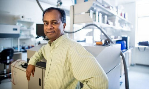
“It’s about the impact we can have on patient care,” says Sasisekharan, the Alfred H. Caspary Professor of Biological Engineering and Health Sciences and Technology. “Whether it’s monitoring for disease, or diagnosing, or treating — that’s the common element.”


Biotech to the rescue


Each company was born from Sasisekharan’s MIT lab, and each is now developing technologies to make stronger therapeutics, battle cancer and infectious diseases, and improve overall global health.

The most established of the three, founded in 2001, is Momenta Pharmaceuticals, which uses technology invented by Sasisekharan to sequence and engineer complex molecules — including proteins, polypeptides, and polysaccharides — to create powerful drugs from these molecules. In his 12 years on Momenta’s board, Sasisekharan helped the now multimillion-dollar company market its first commercial drug — a low-cost, highly potent version of the blood-thinner Lovenox that’s being used today by hundreds of thousands of patients worldwide.


Sasisekharan has since left Momenta to focus on his younger startups — Cerulean, founded in 2006, and Visterra, founded in 2008 — both of which are developing drugs that are now in advanced clinical trials. Cerulean uses “nanopharmaceuticals” that act like Trojan horses, invading tumors and then slowly releasing highly potent chemotherapeutics. Visterra is developing a vaccine that intervenes early in influenza A’s infection cycle, inhibiting the virus’ fusion to host cells — and possibly laying the groundwork for a universal vaccine for influenza.


Thriving today in Cambridge — “all within a 10-minute walk from MIT” — these companies owe their success, Sasisekharan says, to his leveraging of the novel scientific ideas, entrepreneurial ecosystem, and disparate scientific fields found at MIT. “The convergence of biology, analytics, computation, and engineering is a critical ingredient to solving the problems that are part of the Momenta, Cerulean, and Visterra stories,” he says.


Tackling the complex


Momenta’s story dates to 1999, when Sasisekharan and an MIT team “pieced together a toolkit” to sequence complex sugars (or polysaccharides), much as scientists had already done with DNA and proteins.


It was a massive undertaking: Compared with DNA, which has four building blocks, and proteins, which have 20, polysaccharides have 32 building blocks — and, potentially, a million sequences per sample. “Everyone told me to avoid them,” Sasisekharan says.


The team coded each building block of a polysaccharide sample by its mass and, using computational tools, determined all possible sequences of a sample. Using custom enzymes, they then cut the sample at the edge of each building block — so they knew the beginning and ending block — and, in so doing, began eliminating unviable sequences.


But the tool’s true value was in its speed, Sasisekharan says. “Previously, it would almost take an entire PhD thesis to solve the structure of a very small carbohydrate,” Sasisekharan says. “This was something that very rapidly allowed us to solve important sequencing puzzles of large chains in a matter of days.”


Among other things, this method — described in papers published in Science (1999) and the Proceedings of the National Academy of Sciences (2000) — could lead to better understanding of the role polysaccharides play in viral infections and tissue development.


There were commercial applications, too. But entrepreneurship “took me out of my comfort zone,” Sasisekharan says. “That’s where the MIT ecosystem becomes important. We had interactions with people with business backgrounds, clinical backgrounds, which gave us very different perspectives on commercial applications for the first time.”


One thing that became very clear, Sasisekharan says, was the tool’s broad use in understanding complex molecules that make up commercial drugs — especially a molecule called heparin. Heparin-based drugs are created by chopping the molecule randomly, creating pieces with varying sizes and active sites and disparate strengths from batch to batch. Momenta’s technology could identify and remove heparin’s active ingredient, separating it from the junk to build a more efficient drug.


In 2001, Sasisekharan co-founded Momenta (then Mimeon) to apply the technology to the U.S. regulatory pathway for drug approval, “where it was generally considered impossible to make these complex molecules,” Sasisekharan says.


“Once you know you can correct these things, we knew we could use this technology in a way to make more of these complex medicines more accessible to the world,” he says.


Using the technology, Momenta has since grown a pipeline of therapeutics, including its widely used generic Lovenox product, numerous novel drug candidates, various biogenics, and a generic version of Copaxone, a drug for multiple sclerosis, that is now ready for potential launch. Apart from the therapeutic benefits, Momenta’s lower-cost drugs have the potential to save millions of dollars, according to the company.


Detecting the undetectable


But while seeing MIT research find practical application and earn millions in industry is rewarding, Sasisekharan says, the technology may have best demonstrated its real-world value two years before Momenta’s products even hit the market — during a heparin contamination crisis of 2008.

That year, contaminated batches of heparin slipped past the U.S. Food and Drug Administration. Supplies were put under quarantine, leading to a massive shortage. Needing to rapidly identify the contaminant, the FDA called on Sasisekharan.


Using Momenta’s core technology, Sasisekharan and a team of MIT and international researchers, within weeks, identified the contaminant as oversulfated chondroitin sulfate, a sugar chain very similar to heparin (rendering it undetectable) that caused allergic reactions in patients. Batches were tested and recalled, and the crisis ended. Sasisekharan published these findings with the FDA in Nature Biotechnology and the New England Journal of Medicine.


“This was one key piece of the Momenta story, where the technology became extremely valuable and useful in the real world,” says Sasisekharan, now Momenta’s scientific advisor. “It was a very humbling application of the technology that saved lives.”


Nanotechnology and “Napoleon strategy”


Back in 2005 — before the heparin crisis, but years after the launch of Momenta — Sasisekharan found himself with a new batch of grad students, itching to start another venture. (Many of his students had joined Momenta — a recurring theme among all of Sasisekharan’s startups.)


At the time, nanotechnology was on the rise, especially at MIT. “There was a big interest in ‘going nano’ in regard to drug delivery,” Sasisekharan says. And there was the application of this concept in anti-angiogenesis, which involves cutting tumors’ blood supply to starve them to death — “what is called a ‘Napoleon strategy’ of cutting the supply off from the enemy,” Sasisekharan explains.

Combine the rise of anti-angiogenesis with Sasisekharan’s wife’s career as an oncologist — “who inspired me to focus on cancer treatment,” he says — and you have the ingredients for the scientific core of Cerulean.


Building on groundwork laid by Institute Professor Robert Langer, Sasisekharan led a team from MIT in engineering nanoparticles that could carry anti-angiogenic drugs on their outer membranes and highly potent chemotherapeutic agents inside.


When sucked into a tumor’s pores, the nanoparticles’ outer membrane disintegrates, rapidly deploying the anti-angiogenic drug — causing blood vessels feeding the tumor to collapse, and trapping the loaded nanoparticle. Inside the tumor, the nanoparticles slowly release a chemotherapeutic agent, such as camptothecin and docetaxel, while leaving healthy cells unscathed. This avoids a major challenge of chemotherapy: its toxicity to the healthy cells surrounding cancerous ones. This platform was described in a paper published in 2005 in Nature.


“It’s basically a one-two punch,” Sasisekharan says, “cutting off the supply and releasing chemotherapeutics.”


The following year, in 2006, Sasisekharan co-founded Cerulean to commercialize the technology; today, it remains one of the few companies using nanotechnology to treat cancer. But because nanotechnology is still relatively new, Cerulean is working on ways to improve the platform. “The field is moving fast, and some of the things we’re still learning,” Sasisekharan says.


Still, the company has raised $85 million and partnered with cancer centers and hospitals around the nation to further refine its technology; its first drug candidate, CRLX101, has entered clinical trials. “With the clinical trials, we’re past some of the safety issues that were of concern for nanoparticles, and are beginning to see efficacy,” Sasisekharan says. “In a few years we may see an approval of ‘nanodrugs’ for oncology applications.” Cerulean was one of a few Boston-area biotech companies to go public recently.


Fighting flu and dengue


While growing Momenta and Cerulean, Sasisekharan slowly assembled the pieces for his most recent venture, Visterra, which focuses on a separate global health issue: influenza and other infectious diseases.


In 2003, during a trip with his wife to Thailand (where Sasisekharan spends most summers teaching), he found himself in the middle of the country’s H5N1 epidemic. “I remember we couldn’t even order eggs in our hotel — that’s how severe it was,” he says: The flu ravaged the poultry industry in Thailand.

Nudged by the princess of Thailand to address a global health issue, Sasisekharan worked with an MIT team to determine how and when bird flu may make the jump from birds to humans.


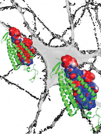
Sasisekharan and his MIT team ultimately found, five years later, that H5N1’s hemagglutinin, a protein on the virus surface, must bind to our umbrella-shaped receptors in order to infect humans. Published in 2008 in Nature Biotechnology, this discovery could help scientists monitor the virus’ evolution and develop vaccines against a deadly flu pandemic. Sasisekharan and his team applied this approach more recently to the emerging N7N9 influenza virus, with results published in 2013 in Cell.


Visterra grew from the novel technology Sasisekharan and his team invented for this research — which combined computation and bioengineering.


Using algorithms, the technology builds a 3-D model of key viral proteins and identifies optimal hierotopes — sites where antibodies bind — on the viral hemagglutinin. These sites are found across all 15 influenza A subtypes, but don’t mutate — meaning they can’t develop resistance to vaccines. Visterra scientists build and tweak antibodies, using bioengineering tools, to specifically target these hierotopes.


Visterra’s first commercial antibody, called VIS410, is now in its first phase of clinical trials; it has the potential to vaccinate against all influenza A subtypes.


In 2012, the Bill and Melinda Gates Foundation partnered with Visterra — which has raised nearly $40 million in venture capital — to help grow its infectious disease product pipeline. Next year, that pipeline may include a second therapeutic candidate, for an equally deadly virus: the mosquito-borne dengue.


In a 2009 visit to Singapore as part of the Singapore-MIT Alliance for Research and Technology, Sasisekharan saw that the country was “ground zero” for dengue. Now Visterra is working to develop an antibody that broadly neutralizes all four dengue virus serotypes — and other viruses, including the West Nile virus, which is familiar to many in the United States.


“Apart from the flu, dengue is the biggest global health agent,” he says. “We’re trying to target diseases that are broadly prevalent in the world, but that a lot of people don’t really know about.”


Biotech entrepreneurship, here and there


Having found success with biotech startups, Sasisekharan has been working in developing countries with little venture capital — such as Thailand and Singapore — to help people start companies.


“In a lot of Asia, there’s this ‘valley of death,’ where angels and venture capitalists are only now beginning to fall in place,” he says. “We’ve come up with pragmatic ways to help people start companies in such a constrained context.”


Among other things, this includes promoting academic institutions as key players in biotech innovation and working with governments and pharmaceuticals to offer support.


Back home, however, the biotech industry in Kendall Square “has exploded,” Sasisekharan says, with advanced technology and unprecedented access to venture capital funding. “We’re experiencing a unique window for biotech companies to go public. That’s thanks, in part, to the venture capital community and MIT. It’s a melting pot of people, ideas, opportunities,” he says. “And fundamentally it’s the mindset: solving problems and focusing on things that have some inherent value to make a difference in the world.”


Story Source:


The above story is based on materials provided by MIT News.


The post Biotech to the rescue appeared first on Bioengineer.org.


26 Nisan 2014 Cumartesi

Bioengineering to Switch off Neurons

from

Bioengineer.org http://bioengineer.org/bioengineering-switch-neurons/


Researchers discovered how to switch brain cells on or off with light pulses by using special proteins from microbes to pass electrical current into neurons.


opsin


In the excitatory opsin (left), the red depicts a channel of negatively charged amino acids that draws positive sodium ions into the neuron to turn it on. The new bioengineered inhibitory opsin (right) is more blue to denote its channel of positively charged amino acids that draws in negative chloride ions to turn the neuron off. Photo Credit: Deisseroth lab


Since then, research teams around the world have used the technique that this scientist, Karl Deisseroth, dubbed “optogenetics” to study not just brain cells but also heart cells, stem cells and the vast array of cell types across biology that can be regulated by electrical signals – the movement of ions across cell membranes.


Optogenetics gave researchers a powerful investigational technique to deepen their understanding of biological system design and function in animal models. But first-generation optogenetics had a shortcoming: Its light-sensitive proteins were potent at switching cells on but less effective at turning them off.


Now in a paper culminating years of effort, Deisseroth’s team has re-engineered its light-sensitive proteins to switch cells off far more efficiently than before. The paper will be published April 25 in Science.


“This is something we and others in the field have sought for a very long time,” said Deisseroth, senior author of the paper and professor of bioengineering and of psychiatry and behavioral sciences.


Thomas Insel, director of the National Institute of Mental Health, which funded the study, said this improved “off” switch will help researchers to better understand the brain circuits involved in behavior, thinking and emotion.


“This latest discovery by the Deisseroth team is the kind of neurotechnology envisioned by President Obama when he launched the BRAIN Initiative a year ago,” Insel said. “It creates a powerful tool that allows neuroscientists to apply a brake in any specific circuit with millisecond precision, beyond the power of any existing technology.”


Behind this development lay many years of foundational experiments in the Deisseroth lab to discover how a key optogenetic “on” switch worked so efficiently to stimulate cells, followed by a systematic effort to redesign this molecular machine to turn it into a fundamentally new kind of “off” switch.


“This is a complex story at the interface of science and engineering,” said Deisseroth, who is also the D.H. Chen Professor at Stanford and a Howard Hughes Medical Institute investigator.


Proteins are the machinery of life. These gigantic molecules are built out of smaller molecules known as amino acids. Like so many Lego parts, amino acids join to form proteins, and those proteins then interact to perform every marvelous task of life from controlling the muscles in our fingertips to firing the neurons in our brains. With optogenetics, Deisseroth’s lab turned a family of proteins known as microbial opsins into a research tool. That story has been told elsewhere, but the result was that optogenetics allowed scientists to fire laser pulses through thin fiberoptics inserted into the brains of animal models. These light pulses triggered the opsins to deliver a stimulating flow of positive ions or an inhibiting pulse of negative ions to control cell behavior with high precision and specificity.


As more researchers embraced optogenetics, many discoveries were made with both excitatory and inhibitory opsins. But Deisseroth and other scientists began to notice that stimulation was more effective than inhibition. He therefore sought to lay the groundwork for developing new inhibitory tools by delving more deeply into the mechanism by which the excitatory opsins delivered only positive ions.


Conceptually, the mechanism by which the excitatory opsins operate is simple. Light activation causes the opsin to open a channel through the cell membrane; positive ions then flow through this channel like water through a garden hose. Taking advantage of this channel mechanism, Deisseroth and his collaborators made modified versions of the excitatory opsins in 2008. They called these step-function opsins. A single pulse of light was all it took to flip these switches on and keep the channel open. This allowed the ions to continue flowing, which kept the cell in an excited state even after the light was turned off. The continuous flow of ions enabled by these step-function opsins also made the cell many times more light-sensitive. This allowed scientists to excite neurons deep within the brains of animal models without penetrating the tissue with fiberoptics.


In contrast, none of the inhibitory opsins acted as channels. They were all “pumps” moving only a single ion across the membrane for every incoming photon – acting more like a squirt gun than a garden hose. Each incoming photon was like pulling the trigger to deliver a negative ion. But it took another photon, or trigger pull, to deliver the next negative ion, and so on. This pump mechanism was less efficient than a channel mechanism in terms of ions moved per photon. It was also impossible to engineer the inhibitory opsins to stay open. Therefore it took more intrusive use of light to perform experiments. Finally, the inhibitory pump did not work in the same way as the normal mechanisms that inhibit brain cells. The normal mechanisms make neuron membranes leaky – like water balloons with pinpricks – and more resistant to firing. All these factors made opsin pumps less efficient as inhibitors. Ideally, bioengineers wanted an inhibitory opsin that functioned like a channel.


In a 2012 article in Nature, Deisseroth’s team (with collaborator Osamu Nureki, director of biological sciences at the University of Tokyo) completed the first big step along the path toward improving optogenetic inhibition when it revealed the detailed structure of the on-switch channel protein. This work showed that the amino acids inside the channel pore created a lining of negative charges. This negative lining attracted positive ions to flow through the membrane in order to excite the cell. This discovery pointed to a potential strategy to create an inhibitory channel: bioengineer the opsin to create an inner lining of positive amino acids to draw a flow of negative (inhibitory) ions, such as chloride, into the cell.


From that point, it took roughly two more years for Deisseroth’s team, led by Andre Berndt and Soo Yeun Lee, postdoctoral scholars in bioengineering and lead authors of the paper, to complete this process. In order to create this new lining, the Stanford team re-engineered the excitatory channel opsin from its 2012 experiment to change nine of the protein’s roughly 300 amino acids. When triggered by light, this newly bioengineered protein opened a channel lined by more positively charged amino acids and thereby attracted a flow of negative (chloride) ions to inhibit activity. This created a microbial opsin capable of delivering a powerful channel effect for inhibition.


Finally, fulfilling one of the most anticipated goals of the project, the researchers changed a 10th amino acid, which gave them the ability to keep this new negative channel open – making the negative channel more like the positive channel. As hoped, this created a vastly more light-sensitive inhibitory channel opsin. The targeted neurons remained off for several minutes after a single, brief, blue light pulse, in a manner that was instantaneously reversible with red light. Deisseroth called this new negative channel opsin SwiChR.


As the Stanford team continues a 10-year journey that began with its first microbial opsin experiments in neurons, Deisseroth anticipates that the long-acting and stable responses of SwiChR will open new realms of opportunities for optogenetics.


His enthusiasm was echoed by Merab Kokaia, a professor at Lund University Hospital in Sweden who has used optogenetics to study epilepsy, among other conditions, in rodent models. Kokaia, who was not involved in the Deisseroth team’s new study, noted the new opsin’s main advantages: more effective at inhibiting neuronal activity based on chloride conductance, greater sensitivity to light, and the ability to stay open for longer periods and maintain inhibition even without light for longer periods.


“These features could be much more useful for behavioral studies in animals but could also become an effective treatment alternative for neurological conditions where drugs do not work, such as some cases of severe epilepsy and other hyper-excitability disorders,” Kokaia said. “The novel approach of channel engineering used by Deisseroth’s lab opens unprecedented perspectives for developing new optogenetic tools for system neuroscience studies that will help us to understand better how the brain works.”


Story Source:


The above story is based on materials provided by Stanford University, Tom Abate.


The post Bioengineering to Switch off Neurons appeared first on Bioengineer.org.


25 Nisan 2014 Cuma

Researchers Generate Immunity Against Tumor Vessel Protein

from

Bioengineer.org http://bioengineer.org/researchers-generate-immunity-tumor-vessel-protein/


Sometimes a full-on assault isn’t the best approach when dealing with a powerful enemy. A more effective approach, in the long run, may be to target the support system replenishing the supplies that keep your foe strong and ready for battle. A group of researchers from the Abramson Cancer Center and the Perelman School of Medicine at the University of Pennsylvania is pursuing this strategy by employing a novel DNA vaccine to kill cancer, not by attacking tumor cells, but targeting the blood vessels that keep them alive. The vaccine also indirectly creates an immune response to the tumor itself, amplifying the attack by a phenomenon called epitope spreading. The results of the study were published this month in the Journal of Clinical Investigation.


Researchers Generate Immunity Against Tumor Vessel Protein


Photo Credit: University of Pennsylvania School of Medicine


Previous studies have targeted tumor angiogenesis (the formation of new blood vessels that feed the tumor cells). However, this approach can also interfere with normal processes involved in wound healing and development. Penn researchers avoided this pitfall by designing a DNA vaccine that specifically targets TEM1 (tumor endothelial marker 1), a protein that is overexpressed in tumors and poorly expressed in normal tissues.


“We demonstrated that by targeting TEM1, our vaccine can decrease tumor vascularization, increase hypoxia of the tumor and reduce tumor growth,” says Andrea Facciabene, PhD, research assistant professor of Obstetrics and Gynecology and a faculty member in the Ovarian Cancer Research Center at Penn Medicine. “Our results confirm that we were directly targeting the tumor vasculature and also indirectly killing tumor cells through epitope spreading.”


The Penn team injected mice with a DNA fusion vaccine called TEM1-TT, created by fusing TEM1 complementary DNA with a fragment of the tetanus toxoid (TT). In mouse models of three cancer types (breast, colon, and cervical), tumor formation was delayed or prevented in mice vaccinated with the TEM1-TT DNA vaccine. Specifically, they found that the mouse tumors had suppressed growth, decreased tumor vessel formation, and increased infiltration of immune cells into tumors.


The researchers found that the DNA vaccine, after killing the endothelial cells that make up the tumor vessels (vasculature), also resulted in epitope spreading, meaning that the immune cells of the mice gathered pieces of dead tumor cells (due to hypoxia) to create a secondary immune response against the tumor itself. The vaccine induced specific T cells to fight other tumor cells expression other proteins, in addition to TEM1, thus increasing its therapeutic efficacy.


The new DNA vaccine approach to fight cancer is showing great potential compared to previous studies that focused on tumor cells rather than the blood vessels that allow tumor cells to thrive.


“Until now there have been a lot of clinical trials using DNA vaccines to target tumors themselves, but unfortunately the results have been disappointing,” Facciabene notes. “This is a different approach which should heighten optimism for cancer vaccines in general. Moreover, based on what we’ve seen in our mouse studies, this vaccine doesn’t seem to show any significant side effects.”


The prevalence of TEM1 in a wide range of tumor types coupled with its scarcity in normal vessels makes it a suitable target both for a prophylactic defense against cancer and a complement to other therapies such as radiotherapy and chemotherapy. “Using this vaccine simultaneously with radiation may eventually have a double synergy,” Facciabene says. “Both treatments affect the tumor endothelium, radiotherapy could help the phenomenon of epitope spreading induced by the TEM1-TT vaccine.” In addition to ongoing pre-clinical work with human TEM1, Facciabene and colleagues are planning to move on to Phase I human clinical trials.


The authors suggest that TEM1 may also be an excellent target as a prophylactic cancer vaccine for individuals that have a high risk of developing ovarian cancer, such as carriers of the BRCA1/2 mutations, predominant in breast and ovarian cancer. Research to develop those types of strategies is a key goal of Penn’s Basser Research Center for BRCA. As a bonafide vaccine, TEM-TT DNA vaccine generates a memory immune response, which Facciabene says is an ideal attribute for high risk populations.


Story Source:


The above story is based on materials provided by University of Pennsylvania School of Medicine.


The post Researchers Generate Immunity Against Tumor Vessel Protein appeared first on Bioengineer.org.


Biologists discover a key regulator in the pacemakers of our brain and heart

from

Bioengineer.org http://bioengineer.org/biologists-discover-key-regulator-pacemakers-brain-heart/


Biologists have discovered how an outer shield over T-type channels change the electrochemical signaling of heart and brain cells. Understanding how these shields work will help researchers eventually develop a new class of drugs for treating epilepsy, cardiovascular disease and cancer.


Biologists discover a key regulator in the pacemakers of our brain and heart


T-type channels in pond snails and other invertebrates are similar to those found in humans. Biologists have discovered how an outer shield over T-type channels change the electrochemical signaling of heart and brain cells. Photo Credit: University of Waterloo


The study from the University of Waterloo is published in the Journal of Biological Chemistry today and is featured as the “Paper of the Week” for its significance.


The researchers discovered T-type channels in the pond snail, Lymnaea stagnalis, can shift from using calcium ions to using sodium ions to generate the electrical signal because of an outer shield of amino acids called a turret situated above the channel’s entrance.


Low voltage T-type channels generate tiny pulses of current at regular intervals by selectively passing positively charged cations across the cell’s membrane through a gate-like channel. The channels are normally extremely selective, allowing just one sodium ion to pass for every 10,000 calcium ions.


The resulting rhythmic signals produced by this transfer of cations are what support the synchronous contraction of our heart muscles and neuronal firing in parts of the brain, like the thalamus, which helps regulate our sleep-wake cycle, or circadian rhythm.


In addition to their published findings, the researchers also found the shield-like turrets in pond snails restrict access of therapeutic drugs to the channel.


T-type channels in pond snails and other invertebrates are similar to those found in humans. Although pond snails reach only 7 cm in length, its simple neural network and physiology make it a popular model organism with neurobiologists.


Over-active T-type channels are linked to epilepsy, cardiac problems, neuropathic pain, as well as the spreading of several kinds of cancer. Drugs that could quench out-of-control T-type channel activity are unable to bind to the channels themselves.


“We wanted to understand the molecular structures of T-type channels,” said Spafford. “How they pass ionic currents to generate electrical activity, and to identify drug binding sites, and the drugs which may block these channels to treat neurological disease or heart complications.”


The group is currently investigating how dismantling this extracellular turret will improve drug access and binding in T-type channels.


Story Source:


The above story is based on materials provided by University of Waterloo.


The post Biologists discover a key regulator in the pacemakers of our brain and heart appeared first on Bioengineer.org.


Skin layer grown from human stem cells could replace animals in drug and cosmetics testing

from

Bioengineer.org http://bioengineer.org/skin-layer-grown-human-stem-cells-replace-animals-drug-cosmetics-testing/


An international team led by King’s College London and the San Francisco Veteran Affairs Medical Center (SFVAMC) has developed the first lab-grown epidermis – the outermost skin layer – with a functional permeability barrier akin to real skin. The new epidermis, grown from human pluripotent stem cells, offers a cost-effective alternative lab model for testing drugs and cosmetics, and could also help to develop new therapies for rare and common skin disorders.


Skin layer grown from human stem cells could replace animals in drug and cosmetics testing


Photo Credit: King’s College London


The epidermis, the outermost layer of human skin, forms a protective interface between the body and its external environment, preventing water from escaping and microbes and toxins from entering. Tissue engineers have been unable to grow epidermis with the functional barrier needed for drug testing, and have been further limited in producing an in vitro (lab) model for large-scale drug screening by the number of cells that can be grown from a single skin biopsy sample.


The new study, published in the journal Stem Cell Reports, describes the use of human induced pluripotent stem cells (iPSC) to produce an unlimited supply of pure keratinocytes – the predominant cell type in the outermost layer of skin – that closely match keratinocytes generated from human embryonic stem cells (hESC) and primary keratinocytes from skin biopsies. These keratinocytes were then used to manufacture 3D epidermal equivalents in a high-to-low humidity environment to build a functional permeability barrier, which is essential in protecting the body from losing moisture, and preventing the entry of chemicals, toxins and microbes.


A comparison of epidermal equivalents generated from iPSC, hESC and primary human keratinocytes (skin cells) from skin biopsies showed no significant difference in their structural or functional properties compared with the outermost layer of normal human skin.


Dr Theodora Mauro, leader of the SFVAMC team, says: “The ability to obtain an unlimited number of genetically identical units can be used to study a range of conditions where the skin’s barrier is defective due to mutations in genes involved in skin barrier formation, such as ichthyosis (dry, flaky skin) or atopic dermatitis. We can use this model to study how the skin barrier develops normally, how the barrier is impaired in different diseases and how we can stimulate its repair and recovery.”


Dr Dusko Ilic, leader of the team at King’s College London, says: “Our new method can be used to grow much greater quantities of lab-grown human epidermal equivalents, and thus could be scaled up for commercial testing of drugs and cosmetics. Human epidermal equivalents representing different types of skin could also be grown, depending on the source of the stem cells used, and could thus be tailored to study a range of skin conditions and sensitivities in different populations.”


Story Source:


The above story is based on materials provided by King’s College London.


The post Skin layer grown from human stem cells could replace animals in drug and cosmetics testing appeared first on Bioengineer.org.


Researchers trace HIV evolution in North America

from

Bioengineer.org http://bioengineer.org/researchers-trace-hiv-evolution-north-america/


A study tracing the evolution of HIV in North America involving researchers at Simon Fraser University has found evidence that the virus is slowly adapting over time to its human hosts. However, this change is so gradual that it is unlikely to have an impact on vaccine design.


researchers trace HIV evolution in North America


Photo Credit: Wayne Leidenfrost


“Much research has focused on how HIV adapts to antiviral drugs—we wanted to investigate how HIV adapts to us, its human hosts, over time,” says lead author Zabrina Brumme, an assistant professor in SFU’s Faculty of Health Sciences.


The study, published today in PLOS Genetics, was led by Brumme’s lab in collaboration with scientists at the BC Centre for Excellence in HIV/AIDS, UBC, and sites across the U.S. including Harvard University, the New York Blood Center and the San Francisco Department of Public Health.


“HIV adapts to the immune response in reproducible ways. In theory, this could be bad news for host immunity—and vaccines—if such mutations were to spread in the population,” says Brumme. “Just like transmitted drug resistance can compromise treatment success, transmitted immune escape mutations could erode our ability to naturally fight HIV.”


Researchers characterized HIV sequences from patients dating from 1979, the beginning of the North American HIV epidemic, to the modern day.


The team reconstructed the epidemic’s ancestral HIV sequence and from there, assessed the spread of immune escape mutations in the population.


“Overall, our results show that the virus is adapting very slowly in North America,” says Brumme. “In parts of the world harder hit by HIV though, rates of adaptation could be higher.”


The study ends with a message of hope, Brumme adds. “We already have the tools to curb HIV in the form of treatment—and we continue to advance towards a vaccine and a cure. Together, we can stop HIV/AIDS before the virus subverts host immunity through population-level adaptation.”


Numerous SFU researchers contributed to the analysis, which required the careful recovery of viral RNA from historic specimens followed by laboratory culture. A trio of SFU graduate students, including health sciences student Laura Cotton, shared the lead author role.


“It was painstaking work,” says Cotton, “but it was fascinating to study these isolates in the lab, knowing that they had played an important role in the history of HIV on our continent.” Numerous undergraduate students, graduate students, postdocs and faculty including adjunct professor Art Poon and associate professor Mark Brockman were among other co-authors.


Story Source:


The above story is based on materials provided by Simon Fraser University.


The post Researchers trace HIV evolution in North America appeared first on Bioengineer.org.


3D printing cancer tumors

from

Bioengineer.org http://bioengineer.org/3d-printing-cancer-tumors/


Drexel’s Wei Sun, PhD, Albert Soffa chair professor in the College of Engineering, has devised a method for 3D printing tumors that could soon be taking cancer research out of the petri dish.


3D printing cancer tumors


Photo Credit: Drexel University


Using a mixture of cervical cancer cells and a hydrogel substance that resembles an ointment balm, Sun can print out a tumor model that can be used for studying their growth and response to treatment. This living model will give cancer researchers a better look at how tumors behave and a more accurate measure of how they respond to treatment.


“This is the first time to report that one can build a 3D in vitro tumor model through 3D Printing technology,” said Sun, the director of Drexel’s research center at the Shanghai Advanced Research Institute. “This may lead to a new paradigm for cancer research and for individual cancer therapies. We have developed a technological platform and would like to work with biologists and pathologists to encourage them to use the developed platform for 3D biology and disease studies.”


While researchers have been able to make cell models and tissues using rapid prototyping methods for some time, Sun’s lab, is the first to produce a living 3D tumor model through additive manufacturing –also known as 3D printing. In a study published in the journal Biofabrication in April, Sun reports a procedure his team developed for growing research-grade models of cervical cancer tumors.


Cancer researchers are aware that working with two-dimensional samples comes with inherent limitations. For example, tumors in the body have a much different surface area, shape and cellular composition than samples grown in a lab, thus data from tests of cancer treatments will differ from the reaction of an actual tumor to the drugs. But until now, these in vitro cell cultures were their best option.


3dprintingca


This is an illustration of the process developed by Drexel University researcher Dr. Wei Sun to 3D print cancer tumors. Photo Credit: Drexel University


“Two-dimensional cell culture models are traditionally used for biology study and drug screening,” Sun said. “However, two-dimensional culture models can not represent true 3D physiological tissues so it lacks the microenvironment characteristics of natural 3D tissues in vivo. This inherent inadequacy leads to shortcomings in cancer research and anti-tumor drug development. On the other hand, 3D tumor models can represent true tumor 3D pathological organizations and will lead to a new paradigm for cancer study.”


As part of the National Science Foundation-funded study, Sun tested his tumor model against a two-dimensional culture sample using a common anti-cancer drug. Sun’s 3D printed tumors showed more resistance to chemical treatment than the same cancer cells grown in a petri dish –an illustration of the disparity that exists between test results and success rates of cancer treatments.


Sun, who is a mechanical engineering researcher with a focus on biomodeling, designed and patented a special 3D printer in 2002 so his lab, the Drexel Biofabrication Laboratory, could make tissue samples and bone scaffolds as part of their research. As his work progressed, it became apparent that the next step was to figure out a way to print living 3D models of tissues and organs.


With a significant background in the extrusion, or additive, modeling process, Sun and his team were able to control for the main variables: diameter of nozzle, speed and pressure of extrusion, pattern and size of deposition, and viscosity and temperature of substrate materials.


For the undertaking, Sun’s team, composed of researchers at Drexel University, Tsinghua University in Beijing, China, and Drexel-Shanghai Advanced Research Center in Shanghai, China, used a multi-nozzle printer to extrude a gelatinous mixture of hydrogels and living cervical cancer cells. The result was a cellular deposition in which 90 percent of the cancer cells survived the process and within eight days had grown into spheroid-shaped tumors.


“The keys to keeping the cells alive were controlling the temperature of the nozzle and using a hearty strain of cancer cells,” Sun said. “We chose the Hela cell, which is a robust form of cervical cancer that has been used in research for many years. Because of this, we had a good idea as to how it would behave under certain conditions. This allowed us to control the variables of the extrusion process until we were able to successfully create a model.”


Sun’s team plans to continue its research in hopes of creating tumors that are even more similar to those that grow in the body. They will work to print tumors composed of multiple different cells –a trait often found in those removed from cancer patients. In addition, the group is working on ways to attach the models to tissues and vasculature that they’ve printed, which would recreate the way tumors grow in their bodily habitat.


“We will try to understand the cell-cell and cell-substrate communication and immune responses for the printed tumor-like models,” Sun said. “Our goal is to take this tumor-like model and make it into a more of an in vivo simulation. And to apply it to study the development, invasion and metastasis of cancer, to test the efficacy and safety of new cancer drugs, as well as the specific therapy for individual cancer patient”


Story Source:


The above story is based on materials provided by Drexel University, Britt Faulstick.


The post 3D printing cancer tumors appeared first on Bioengineer.org.


24 Nisan 2014 Perşembe

Hearing Quality Restored With Bionic Ear Tech

from

Bioengineer.org http://bioengineer.org/hearing-quality-restored-bionic-ear-tech/


Researchers at UNSW Australia have for the first time used electrical pulses delivered from a cochlear implant to deliver gene therapy, thereby successfully regrowing auditory nerves.


Auditory Nerves after Gene Therapy


This photo shows regenerated auditory nerves after gene therapy (top) compared with no treatment (below). Photo Credit: UNSW Translational Neuroscience Facility


The research also heralds a possible new way of treating a range of neurological disorders, including Parkinson’s disease, and psychiatric conditions such as depression through this novel way of delivering gene therapy.


The research is published today (Thursday 24 April) in the journal Science Translational Medicine.

“People with cochlear implants do well with understanding speech, but their perception of pitch can be poor, so they often miss out on the joy of music,” says UNSW Professor Gary Housley, who is the senior author of the research paper.


“Ultimately, we hope that after further research, people who depend on cochlear implant devices will be able to enjoy a broader dynamic and tonal range of sound, which is particularly important for our sense of the auditory world around us and for music appreciation,” says Professor Housley, who is also the Director of the Translational Neuroscience Facility at UNSW Medicine.


The research, which has the support of Cochlear Limited through an Australian Research Council Linkage Project grant, has been five years in development.


The work centres on regenerating surviving nerves after age-related or environmental hearing loss, using existing cochlear technology. The cochlear implants are “surprisingly efficient” at localised gene therapy in the animal model, when a few electric pulses are administered during the implant procedure.


“This research breakthrough is important because while we have had very good outcomes with our cochlear implants so far, if we can get the nerves to grow close to the electrodes and improve the connections between them, then we’ll be able to have even better outcomes in the future,” says Jim Patrick, Chief Scientist and Senior Vice-President, Cochlear Limited.


It has long been established that the auditory nerve endings regenerate if neurotrophins — a naturally occurring family of proteins crucial for the development, function and survival of neurons — are delivered to the auditory portion of the inner ear, the cochlea.


But until now, research has stalled because safe, localised delivery of the neurotrophins can’t be achieved using drug delivery, nor by viral-based gene therapy.


Professor Housley and his team at UNSW developed a way of using electrical pulses delivered from the cochlear implant to deliver the DNA to the cells close to the array of implanted electrodes. These cells then produce neurotrophins.


“No-one had tried to use the cochlear implant itself for gene therapy,” says Professor Housley. “With our technique, the cochlear implant can be very effective for this.”


While the neurotrophin production dropped away after a couple of months, Professor Housley says ultimately the changes in the hearing nerve may be maintained by the ongoing neural activity generated by the cochlear implant.


“We think it’s possible that in the future this gene delivery would only add a few minutes to the implant procedure,” says the paper’s first author, Jeremy Pinyon, whose PhD is based on this work. “The surgeon who installs the device would inject the DNA solution into the cochlea and then fire electrical impulses to trigger the DNA transfer once the implant is inserted.”


Integration of this technology into other ‘bionic’ devices such as electrode arrays used in deep brain stimulation (for the treatment of Parkinson’s disease and depression, for example) could also afford opportunities for safe, directed gene therapy of complex neurological disorders.


“Our work has implications far beyond hearing disorders,” says co-author Associate Professor Matthias Klugmann, from the UNSW Translational Neuroscience Facility research team. “Gene therapy has been suggested as a treatment concept even for devastating neurological conditions and our technology provides a novel platform for safe and efficient gene transfer into tissues as delicate as the brain.”


Story Source:


The above story is based on materials provided by University of New South Wales, Susi Hamilton.


The post Hearing Quality Restored With Bionic Ear Tech appeared first on Bioengineer.org.


Stem cells in circulating blood affect cardiovascular health

from

Bioengineer.org http://bioengineer.org/stem-cells-in-circulating-blood-affect-cardiovascular-health/


New research suggests that attempts to isolate an elusive adult stem cell from blood to understand and potentially improve cardiovascular health – a task considered possible but very difficult – might not be necessary.


stem cells


Instead, scientists have found that multiple types of cells with primitive characteristics circulating in the blood appear to provide the same benefits expected from a stem cell, including the endothelial progenitor cell that is the subject of hot pursuit.


“There are people who still dream that the prototypical progenitors for several components of the cardiovascular tree will be found and isolated. I decided to focus the analysis on the whole nonpurified cell population – the blood as it is,” said Nicanor Moldovan, senior author of the study and a research associate professor of cardiovascular medicine at The Ohio State University.


“Our method determines the contributions of all blood cells that serve the same function that an endothelial progenitor cell is supposed to. We can detect the presence of those cells and their signatures in a clinical sample without the need to isolate them.”


The study is published in the journal PLOS ONE.


Stem cells, including the still poorly understood endothelial progenitor cells, are sought-after because they have the potential to transform into many kinds of cells, suggesting that they could be used to replace damaged or missing cells as a treatment for multiple diseases.


By looking at gene activity patterns in blood, Moldovan and colleagues concluded that many cell types circulating throughout the body may protect and repair blood vessels – a key to keeping the heart healthy.


The scientists also found that several types of blood cells retain so-called “primitive” properties. In this context, primitive is positive because these cells are the first line of defense against an injury and provide a continuous supply of repair tissue either directly or by telling local cells what to do.


By comparing gene activation patterns with the study blood donors’ health status, the research showed which genes in blood are associated with such problems as high blood pressure and inflexible blood vessels.


In physicians’ hands, this analysis could be used to diagnose certain diseases, monitor the effects of some treatments and determine a cardiovascular patient’s prognosis. Further analysis also could help explain how primitive properties in cells, which decrease as humans age, reduce the body’s protective and repairing resources.


At the start of this work, the research group proposed a method intended to physically isolate an endothelial progenitor cell candidate. When the evidence didn’t support their hypothesis, they adjusted to a systems biology approach and decided to “keep the whole soup” of blood for analysis instead, said Moldovan, an investigator in the Davis Heart and Lung Research Institute and a member of the Center for Regenerative Medicine and Cell-Based Therapies at Ohio State.


The researchers analyzed gene activity, or expression, in the blood cells at an early point in the process – at the messenger RNA (mRNA) level, before proteins are made. Based on previous studies that have helped identify markers that indicate whether cells are primitive or mature, they narrowed the search to detect mRNA for 45 genes in the blood cells.


Using the bioinformatics principle of “guilt-by-association” to analyze the massive amounts of data, “we let those genes that participate in a common function or that share a common structure to show themselves up objectively in the data,” he said.


The sorting produced clusters of genes that were further analyzed to identify their purpose. The genes aggregated into two modules, and the researchers zeroed in on a module in which a total of 15 primitive and cardiovascular genes showed a clear connection.


An initial finding countered conventional wisdom about embryonic-level primitive cells: It’s thought by some scientists that these types of cells cannot exist in bone marrow, and thus in the adult blood. But this analysis detected embryonic stemness genes “constantly and in all samples we looked at,” Moldovan said.


The blood samples came from two groups of human research participants: 26 healthy volunteers and 20 patients with a diagnosis of high blood pressure. Blood from healthy people helped define the cardiovascular-relevant module – a gene profile of cardiac health that was then compared to characteristics of gene behavior in patients with hypertension.


To determine the physiological significance of the clustered primitive and cardiovascular genes, researchers compared their expression patterns to four patient measures: age, a measure of vascular stiffness (healthy vessels are flexible, not stiff), blood pressure and body mass index (BMI). The correlation pattern suggested that higher expression of these genes was linked to younger age, more flexible vessels and lower blood pressure.


“This means the genes in this module are protective against high blood pressure and vascular stiffness, which are related,” Moldovan said. “And they reflect a property of blood that is being lost in time, as you would expect from progenitor cells. They have a protective and presumably repairing function, which diminishes with age.”


In women, a higher BMI was associated with higher expression of these genes, which Moldovan said might help explain the overweight paradox – the apparently protective role of a few extra pounds in a variety of medical conditions.


Scientists also compared this gene module to images representing stiffness of human aortas and found similar connections – low expression of these genes was associated with more stiffness in the body’s largest artery.


“This is an example of how we intend to apply this in a clinical setting,” Moldovan said.


He said that with this comprehensive knowledge about expression patterns of 45 genes in the blood, scientists can now search for the molecules produced by those genes to identify which kinds of blood cells resemble adult stem cells – namely, those cells whose genes show that they retain primitive characteristics. He added that, based on other preliminary data, he and colleagues are confident that this gene module can be expanded using the same bioinformatics approach to add new candidate members.


“Our goal is to assess the status of the system of progenitors in the bloodstream in its natural complexity, to understand and anticipate the prognosis of what’s going to happen with the patient,” Moldovan said. “It requires letting go of the old paradigm of ‘cell type’ and embracing the more abstract notion of a cluster of genes – a ‘metagene’– that associates with blood and changes as the condition of a patient changes.”


Story Source:

The above story is based on materials provided by Ohio State University, Emily Caldwell.


The post Stem cells in circulating blood affect cardiovascular health appeared first on Bioengineer.org.


Enzymes to Help Fix Cancer-Causing DNA Defects

from

Bioengineer.org http://bioengineer.org/enzymes-help-fix-cancer-causing-dna-defects/


Purdue University researchers have identified an important enzyme pathway that helps prevent new cells from receiving too many or too few chromosomes, a condition that has been directly linked to cancer and other diseases.


Enzymes to Help Fix Cancer-Causing DNA Defects


Mark Hall is also exploring the possibility of using Cdc14 inhibitors to combat deadly fungal diseases in crops.


Mark Hall, associate professor of biochemistry, found that near the end of cell division, the enzyme Cdc14 activates Yen1, an enzyme that ensures any breaks in DNA are fully repaired before the parent cell distributes copies of the genome to daughter cells. This process helps safeguard against some of the most devastating genome errors, including the loss of chromosomes or chromosome segments.

“It only takes one cell to start a tumor,” Hall said. “This study gives us a platform for figuring out exactly what these enzymes are doing in human cells and how they impact genome stability and the avoidance of cancer.”


Cdc14 has been linked to DNA damage repair in humans, but exactly how the enzyme helps preserve the genome and which proteins it regulates in this process have not been known.


Hall and his fellow researchers developed a novel method of identifying the protein substrates upon which Cdc14 acts. Cdc14 regulates the function of other proteins by removing phosphate, a small chemical group, from them. Using Cdc14 in baker’s yeast — which is very similar to human Cdc14 — the team studied the activity of the enzyme on a wide variety of synthetic substrate molecules, looking for similar features among the molecules most preferred by Cdc14.


“We were basically trying different keys in the lock to see which would fit the best,” Hall said.

The team identified the most common structural features on molecules targeted by Cdc14 and used bioinformatics tools to pinpoint matching features in yeast proteins. Yen1 proved to be the best match, and further tests confirmed its role as a substrate of Cdc14. Yen1 is the first Cdc14 substrate involved in DNA repair to be identified.


Hall said the remarkable similarity of these enzymes in yeast and humans makes it likely that this method could be used to identify targets of Cdc14 in humans as well.


“Despite belonging to extremely different species, the ‘lock’ in yeast and human Cdc14 enzymes is exactly the same,” he said. “That gives us confidence that we can use this strategy to identify substrates of human CDC14 and how they work to control DNA repair processes and prevent cancer.”

Hall said understanding Cdc14′s role in DNA repair and how the enzyme binds to its substrates could be used to develop more effective chemotherapeutic weapons against cancer. Many chemotherapeutic drugs work by producing such extensive DNA damage in cancer cells that they kill themselves. Designing a chemical that mimics the features of a Cdc14 substrate would help block Cdc14 from repairing damaged DNA in cancer cells, speeding their death.


“Developing Cdc14 inhibitory compounds could make certain cancer treatments more specific and potent,” Hall said. “You could think of Cdc14 inhibitors as kryptonite to cancer cells, potentially weakening their ability to heal themselves and making them more vulnerable to chemotherapy treatment.”

Hall also is exploring the possibility of using Cdc14 inhibitors to combat deadly fungal diseases in crops.


Story Source:


The above story is based on materials provided by Purdue University.


The post Enzymes to Help Fix Cancer-Causing DNA Defects appeared first on Bioengineer.org.


Brain Circuits Involved in Emotion Discovered

from

Bioengineer.org http://bioengineer.org/brain-circuits-involved-in-emotion-discovered/


Neuroscientists have discovered a brain pathway that underlies the emotional behaviours critical for survival.


Brain circuits involved in emotion


New research by the University of Bristol, published in the Journal of Physiology, has identified a chain of neural connections which links central survival circuits to the spinal cord, causing the body to freeze when experiencing fear.


Understanding how these central neural pathways work is a fundamental step towards developing effective treatments for emotional disorders such as anxiety, panic attacks and phobias.


An important brain region responsible for how humans and animals respond to danger is known as the PAG (periaqueductal grey), and it can trigger responses such as freezing, a high heart rate, increase in blood pressure and the desire for flight or fight.


This latest research has discovered a brain pathway leading from the PAG to a highly localised part of the cerebellum, called the pyramis. The research went on to show that the pyramis is involved in generating freezing behaviour when central survival networks are activated during innate and learnt threatening situations.


The pyramis may therefore serve as an important point of convergence for different survival networks in order to react to an emotionally challenging situation.


Dr Stella Koutsikou, first author of the study and Research Associate in the School of Physiology and Pharmacology at the University of Bristol, said: “There is a growing consensus that understanding the neural circuits underlying fear behaviour is a fundamental step towards developing effective treatments for behavioural changes associated with emotional disorders.”


Professor Bridget Lumb, Professor of Systems Neuroscience, added: “Our work introduces the novel concept that the cerebellum is a promising target for therapeutic strategies to manage dysregulation of emotional states such as panic disorders and phobias.”


The researchers involved in this work are all members of Bristol Neuroscience which fosters interactions across one of the largest communities of neuroscientists in the UK.

Professor Richard Apps said “This is a great example of how Bristol Neuroscience brings together expertise in different fields of neuroscience leading to exciting new insights into brain function.”


Story Source:


The above story is based on materials provided by University of Bristol.


The post Brain Circuits Involved in Emotion Discovered appeared first on Bioengineer.org.


Y Chromosome Appeared 180 Million Years Ago

from

Bioengineer.org http://bioengineer.org/y-chromosome-appeared-180-million-years-ago/


Man or woman? Male or female? In humans and other mammals, the difference between sexes depends on one single element of the genome: the Y chromosome. It is present only in males, where the two sexual chromosomes are X and Y, whereas women have two X chromosomes. Thus, the Y is ultimately responsible for all the morphological and physiological differences between males and females.


Y Chromosome Appeared 180 Million Years Ago


But this has not always been the case. A very long time ago, the X and Y were identical, until the Y started to differentiate from the X in males. It then progressively shrank to such an extent that, nowadays, it only contains about 20 genes (the X carries more than one thousand genes). When did the Y originate and which genes have been kept? The answer has just been brought to light by the team of Henrik Kaessmann, Associate Professor at the CIG (UNIL) and group leader at the SIB Swiss Institute of Bioinformatics, and their collaborators in Australia. They have established that the first ” sex genes ” appeared concomitantly in mammals around 180 million years ago.


4,3 billion genetic sequences


By studying samples from several male tissues — in particular testicles — from different species, the researchers recovered the Y chromosome genes from the three major mammalian lineages: placentals (which include humans, apes, rodents and elephants), marsupials (such as opossums and kangaroos) and monotremes (egg-laying mammals, such as the platypus and the echidna, a kind of Australian porcupine). In total, the researchers worked with samples from 15 different mammals, representing these three lineages, as well as the chicken, which they included for comparison.


Instead of sequencing all Y chromosomes, which would have been a ” colossal task ” according to Diego Cortez, researcher at CIG and SIB and main author of the study, the scientists ” opted for a shortcut .” By comparing genetic sequences from male and female tissues, they eliminated all sequences common to both sexes in order to keep only those sequences corresponding to the Y chromosome. By doing so, they established the largest gene atlas of this ” male ” chromosome to date.


This study required more than 29,500 computing hours! A gigantic task, which could not have been performed without important technical means: the high-throughput DNA sequencers of the genomics platform at the Center for Integrative Genomics, for the generation of the genetic sequences, and the calculation means of Vital-IT, SIB’s high-performance computing centre, for the biological analyses.


Two independent sex-determining genes


The study shows that the same sex-determining gene, named SRY, in placentals and marsupials had formed in the common ancestor of both lineages around 180 million years ago. Another gene, AMHY, is responsible for the emergence of Y chromosomes in monotremes and appeared some 175 million years ago. Both genes, which according to Henrik Kaessmann are “involved in testicular development ,” have thus emerged ” nearly at the same time but in a totally independent way .”


The nature of the sex-determination system present in the common ancestor of all mammals remains unclear, given that mammalian Y chromosomes did not yet exist at that time — at least not those discovered in this study. So what triggered back then that an individual was born male or female? Was this determination linked to other sex chromosomes, or even environmental factors such as the temperature? The latter is not an unreasonable scenario, given that temperature determines sex in present-day crocodiles. As far as mammals are concerned, “the question remains open ,” concludes Diego Cortez.


Story Source:


The above story is based on materials provided by Swiss Institute of Bioinformatics, Henrik Kaessmann.


The post Y Chromosome Appeared 180 Million Years Ago appeared first on Bioengineer.org.