23 Haziran 2014 Pazartesi

Seeing the inner workings of brain made easier by new technique

from

Bioengineer.org http://bioengineer.org/seeing-inner-workings-brain-made-easier-new-technique/


Last year Karl Deisseroth, a Stanford professor of bioengineering and of psychiatry and behavioral sciences, announced a new way of peering into a brain – removed from the body – that provided spectacular fly-through views of its inner connections. Since then laboratories around the world have begun using the technique, called CLARITY, with some success, to better understand the brain’s wiring.


A three-dimensional rendering of clarified brain imaged from below


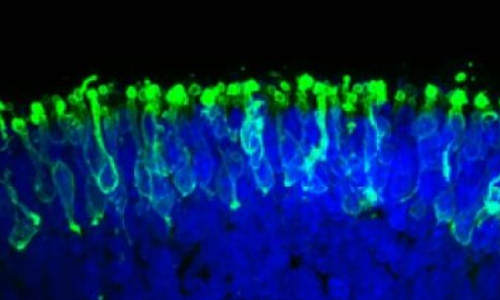
A three-dimensional rendering of clarified brain imaged from below. Photo Credit: Deisseroth lab


However, Deisseroth said that with two technological fixes CLARITY could be even more broadly adopted. The first problem was that laboratories were not set up to reliably carry out the CLARITY process. Second, the most commonly available microscopy methods were not designed to image the whole transparent brain. “There have been a number of remarkable results described using CLARITY,” Deisseroth said, “but we needed to address these two distinct challenges to make the technology easier to use.”


In a Nature Protocols paper published June 19, Deisseroth presented solutions to both of those bottlenecks. “These transform CLARITY, making the overall process much easier and the data collection much faster,” he said. He and his co-authors, including postdoctoral fellows Raju Tomer and Li Ye and graduate student Brian Hsueh, anticipate that even more scientists will now be able to take advantage of the technique to better understand the brain at a fundamental level, and also to probe the origins of brain diseases.


This paper may be the first to be published with support of the White House BRAIN Initiative, announced last year with the ambitious goal of mapping the brain’s trillions of nerve connections and understanding how signals zip through those interconnected cells to control our thoughts, memories, movement and everything else that makes us us.


“This work shares the spirit of the BRAIN Initiative goal of building new technologies to understand the brain – including the human brain,” said Deisseroth, who is also a Stanford Bio-X affiliated faculty member.


Eliminating fat


When you look at the brain, what you see is the fatty outer covering of the nerve cells within, which blocks microscopes from taking images of the intricate connections between deep brain cells. The idea behind CLARITY was to eliminate that fatty covering while keeping the brain intact, complete with all its intricate inner wiring.


The way Deisseroth and his team eliminated the fat was to build a gel within the intact brain that held all the structures and proteins in place. They then used an electric field to pull out the fat layer that had been dissolved in an electrically charged detergent, leaving behind all the brain’s structures embedded in the firm water-based gel, or hydrogel. This is called electrophoretic CLARITY.


The electric field aspect was a challenge for some labs. “About half the people who tried it got it working right away,” Deisseroth said, “but others had problems with the voltage damaging tissue.” Deisseroth said that this kind of challenge is normal when introducing new technologies. When he first introduced optogenetics, which allows scientists to control individual nerves using light, a similar proportion of labs were not initially set up to easily implement the new technology, and ran into challenges.


To help expand the use of CLARITY, the team devised an alternate way of pulling out the fat from the hydrogel-embedded brain – a technique they call passive CLARITY. It takes a little longer, but still removes all the fat, is much easier and does not pose a risk to the tissue. “Electrophoretic CLARITY is important for cases where speed is critical, and for some tissues,” said Deisseroth, who is also the D.H. Chen Professor. “But passive CLARITY is a crucial advance for the community, especially for neuroscience.” Passive CLARITY requires nothing more than some chemicals, a warm bath and time.


Many groups have begun to apply CLARITY to probe brains donated from people who had diseases like epilepsy or autism, which might have left clues in the brain to help scientists understand and eventually treat the disease. But scientists, including Deisseroth, had been wary of trying electrophoretic CLARTY on these valuable clinical samples with even a very low risk of damage. “It’s a rare and precious donated sample, you don’t want to have a chance of damage or error,” Deisseroth said. “Now the risk issue is addressed, and on top of that you can get the data very rapidly.”


Fast CLARITY imaging in color


The second advance had to do this rapidity of data collection. In studying any cells, scientists often make use of probes that will go into the cell or tissue, latch onto a particular molecule, then glow green, blue, yellow or other colors in response to particular wavelengths of light. This is what produces the colorful cellular images that are so common in biology research. Using CLARITY, these colorful structures become visible throughout the entire brain, since no fat remains to block the light.


But here’s the hitch. Those probes stop working, or get bleached, after they’ve been exposed to too much light. That’s fine if a scientist is just taking a picture of a small cellular structure, which takes little time. But to get a high-resolution image of an entire brain, the whole tissue is bathed in light throughout the time it takes to image it point by point. This approach bleaches out the probes before the entire brain can be imaged at high resolution.


The second advance of the new paper addresses this issue, making it easier to image the entire brain without bleaching the probes. “We can now scan an entire plane at one time instead of a point,” Deisseroth said. “That buys you a couple orders of magnitude of time, and also efficiently delivers light only to where the imaging is happening.” The technique is called light sheet microscopy and has been around for a while, but previously didn’t have high enough resolution to see the fine details of cellular structures. “We advanced traditional light sheet microscopy for CLARITY, and can now see fine wiring structures deep within an intact adult brain,” Deisseroth said. His lab built their own microscope, but the procedures are described in the paper, and the key components are commercially available. Additionally, Deisseroth’s lab provides free training courses in CLARITY, modeled after his optogenetics courses, to help disseminate the techniques.


Brain imaging to help soldiers


The BRAIN Initiative is being funded through several government agencies including the Defense Advanced Research Projects Agency (DARPA), which funded Deisseroth’s work through its new Neuro-FAST program. Deisseroth said that like the National Institute of Mental Health (NIMH, another major funder of the new paper), DARPA “is interested in deepening our understanding of brain circuits in intact and injured brains to inform the development of better therapies.” The new methods Deisseroth and his team developed will accelerate both human- and animal-model CLARITY; as CLARITY becomes more widely used, it will continue to help reveal how those inner circuits are structured in normal and diseased brains, and perhaps point to possible therapies.


Other arms of the BRAIN Initiative are funded through the National Science Foundation (NSF) and the National Institutes of Health (NIH). A working group for the NIH arm was co-led by William Newsome, professor of neurobiology and director of the Stanford Neurosciences Institute, and also included Deisseroth and Mark Schnitzer, associate professor of biology and of applied physics. That group recently recommended a $4.5 billion investment in the BRAIN Initiative over the next 12 years, which NIH Director Francis Collins approved earlier this month.


Story Source:


The above story is based on materials provided by Stanford University, Amy Adams.


The post Seeing the inner workings of brain made easier by new technique appeared first on Bioengineer.org.


15 Haziran 2014 Pazar

‘Trust hormone’ oxytocin helps old muscle work like new

from

Bioengineer.org http://bioengineer.org/trust-hormone-oxytocin-helps-old-muscle-work-like-new/


UC Berkeley researchers have discovered that oxytocin — a hormone associated with maternal nurturing, social attachments, childbirth and sex — is indispensable for healthy muscle maintenance and repair, and that in mice it declines with age.


‘Trust hormone’ oxytocin helps old muscle work like new


The new study, published today (June 10) in the journal Nature Communications, presents oxytocin as the latest treatment target for age-related muscle wasting, or sarcopenia.


A few other biochemical factors in blood have been connected to aging and disease in recent years, but oxytocin is the first anti-aging molecule identified that is approved by the Food and Drug Administration for clinical use in humans, the researchers said. Pitocin, a synthetic form of oxytocin, is already used to help with labor and to control bleeding after childbirth. Clinical trials of an oxytocin nasal spray are also underway to alleviate symptoms associated with mental disorders such as autism, schizophrenia and dementia.


“Unfortunately, most of the molecules discovered so far to boost tissue regeneration are also associated with cancer, limiting their potential as treatments for humans,” said study principal investigator Irina Conboy, associate professor of bioengineering. “Our quest is to find a molecule that not only rejuvenates old muscle and other tissue, but that can do so sustainably long-term without increasing the risk of cancer.”


Conboy and her research team say that oxytocin, secreted into the blood by the brain’s pituitary gland, is a good candidate because it is a broad range hormone that reaches every organ, and it is not known to be associated with tumors or to interfere with the immune system.


A happy hormone


Oxytocin is sometimes referred to as the “trust hormone” because of its association with romance and friendship. It is released with a warm hug, a grasped hand or a loving gaze, and it increases libido. The hormone kicks into high gear during and after childbirth, helping new mothers bond with and breastfeed their new babies.


“This is the hormone that makes your heart melt when you see kittens, puppies and human babies,” said Conboy, who is also a member of the Berkeley Stem Cell Center and of the California Institute for Quantitative Biosciences (QB3). “There is an ongoing joke among my research team that we’re all happy, friendly and trusting because oxytocin permeates the lab.”


The researchers pointed out that while oxytocin is found in both young boys and girls, it is not yet known when levels of the hormone start to decline in humans, and what levels are necessary for maintaining healthy tissues.


Christian Elabd and Wendy Cousin, both senior scientists in Conboy’s lab, were co-lead authors on this study.


Previous research by Elabd found that administering oxytocin helped prevent the development of osteoporosis in mice that had their ovaries removed to mimic menopause.


Extra oxytocin more beneficial for the old


The new study determined that in mice, blood levels of oxytocin declined with age. They also showed that there are fewer receptors for oxytocin in muscle stem cells in old versus young mice.


To tease out oxytocin’s role in muscle repair, the researchers injected the hormone under the skin of old mice for four days, and then for five days more after the muscles were injured. After the nine-day treatment, they found that the muscles of the mice that had received oxytocin injections healed far better than those of a control group of mice without oxytocin.


“The action of oxytocin was fast,” said Elabd. “The repair of muscle in the old mice was at about 80 percent of what we saw in the young mice.”


Interestingly, giving young mice an extra boost of oxytocin did not seem to cause a significant change in muscle regeneration.


“This is good because it demonstrates that extra oxytocin boosts aged tissue stem cells without making muscle stem cells divide uncontrollably,” Cousin added.


The researchers also found that blocking the effects of oxytocin in young mice rapidly compromised their ability to repair muscle, which resembled old tissue after an injury.


The researchers also studied mice whose gene for oxytocin was disabled, and compared them with a group of control mice. At a young age, there was no significant difference between the two groups in muscle mass or repair efficiency after an injury. It wasn’t until the mice with the disabled oxytocin gene reached adulthood that signs of premature aging began to appear.


“When disabling other types of genes associated with tissue repair, defects appear right away either during embryonic development, or early in life,” said Conboy. “To our knowledge, the oxytocin gene is the only one whose impact is seen later in life, suggesting that its role is closely linked to the aging process.”


Future treatment options


Cousin noted that oxytocin could become a viable alternative to hormone replacement therapy as a way to combat the symptoms of both female and male aging, and for long-term health. Hormone therapy did not show improvements in agility or muscle regeneration ability, and it is no longer recommended for disease prevention because research has found that the therapy’s benefits did not outweigh its health risks.


In addition to healthy muscle, oxytocin is predicted to improve bone health, and it might be important in combating obesity.


Conboy said her lab plans to examine oxytocin’s role in extending a healthy life in animals, and in conserving its beneficial anti-aging effects in humans.


She noted that there is a growing circle of scientists who believe that aging is the underlying cause of a number of chronic diseases, including Parkinson’s and Type 2 diabetes.


“If you target processes associated with aging, you may be tackling those diseases at the same time,” said Conboy. “Aging is a natural process, but I believe that we can meaningfully intervene with age-imposed organ degeneration, thereby slowing down the rate at which we become progressively unhealthy.”


Story Source:


The above story is based on materials provided by University of California – Berkeley.


The post ‘Trust hormone’ oxytocin helps old muscle work like new appeared first on Bioengineer.org.


Unexpected origin for important parts of the nervous system

from

Bioengineer.org http://bioengineer.org/unexpected-origin-important-parts-nervous-system/


A new study from Karolinska Institutet shows that a part of the nervous system, the parasympathetic nervous system, is formed in a way that is different from what researchers previously believed. In this study, which is published in the journal Science, a new phenomenon is investigated within the field of developmental biology, and the findings may lead to new medical treatments for congenital disorders of the nervous system.


Unexpected origin for important parts of the nervous system


Almost all of the body’s functions are controlled by the autonomous, involuntary nervous system, for example the heart and blood vessels, liver and gastrointestinal system. At rest, the body is set up for energy saving functions, which is regulated by the parasympathetic part of the autonomous nervous system.


Current understanding is that many important types of cells, including the parasympathetic nerve cells in various organs, originate in early progenitor cells that move short distances while the embryo is still small. But this model does not explain how many of our organs – which develop relatively late, when the embryo is large – are furnished with cells that form the parasympathetic neurons.


This study alters a fundamental principal of our understanding of how the peripheral nervous system develops in the body. Researchers at Karolinska Institutet have made three-dimensional reconstructions of mouse embryos. These show that the parasympathetic neurons are formed from immature glial cells known as Schwann cell precursors that travel along the peripheral nerves out to the body’s tissues and organs. The immature cells have the properties of stem cells and may be the origin of several different types of cells. For example, the researchers behind this new study have previously demonstrated that the majority of our melanocytes (pigment cells) are born from these cells.


New principal of developmental biology


“Our study focuses on a new principal of developmental biology, a targeted recruitment of cells that are probably also used in the reconstruction of tissue. Despite the elegance, simplicity and beauty of this principal, it is still unclear how the number of parasympathetic neurons is controlled and why only some of the cells transported by nerves are transformed into that which becomes an important part of the nervous system”, says Igor Adameyko at the Department of Physiology and Pharmacology who, together with Patrik Ernfors at the Department of Medical Biochemistry and Biophysics, is responsible for the study.


Somewhat surprisingly, the researchers found that the entire parasympathetic nervous system arises from these progenitor cells that travel along the peripheral nerves. The researchers hope that this discovery will open up the possibility of new ways to treat congenital disorders of the autonomous nervous system using regenerative medicine.


The study has been financed with funds from, among others, the Swedish Research Council, the Swedish Cancer Society, the European Research Council and Hjärnfonden.


Story Source:


The above story is based on materials provided by Karolinska Institutet.


The post Unexpected origin for important parts of the nervous system appeared first on Bioengineer.org.


New computer program aims to teach itself everything about anything

from

Bioengineer.org http://bioengineer.org/new-computer-program-aims-teach-everything-anything/


In today’s digitally driven world, access to information appears limitless. But when you have something specific in mind that you don’t know, like the name of that niche kitchen tool you saw at a friend’s house, it can be surprisingly hard to sift through the volume of information online and know how to search for it. Or, the opposite problem can occur – we can look up anything on the Internet, but how can we be sure we are finding everything about the topic without spending hours in front of the computer?


New computer program aims to teach itself everything about anything


Some of the many variations the new program has learned for three different concepts. Photo Credit: Image courtesy of University of Washington


Computer scientists from the University of Washington and the Allen Institute for Artificial Intelligence in Seattle have created the first fully automated computer program that teaches everything there is to know about any visual concept. Called Learning Everything about Anything, or LEVAN, the program searches millions of books and images on the Web to learn all possible variations of a concept, then displays the results to users as a comprehensive, browsable list of images, helping them explore and understand topics quickly in great detail.


“It is all about discovering associations between textual and visual data,” said Ali Farhadi, a UW assistant professor of computer science and engineering. “The program learns to tightly couple rich sets of phrases with pixels in images. This means that it can recognize instances of specific concepts when it sees them.”


The research team will present the project and a related paper this month at the Computer Vision and Pattern Recognition annual conference in Columbus, Ohio.


The program learns which terms are relevant by looking at the content of the images found on the Web and identifying characteristic patterns across them using object recognition algorithms. It’s different from online image libraries because it draws upon a rich set of phrases to understand and tag photos by their content and pixel arrangements, not simply by words displayed in captions.


Users can browse the existing library of roughly 175 concepts. Existing concepts range from “airline” to “window,” and include “beautiful,” “breakfast,” “shiny,” “cancer,” “innovation,” “skateboarding,” “robot,” and the researchers’ first-ever input, “horse.”


If the concept you’re looking for doesn’t exist, you can submit any search term and the program will automatically begin generating an exhaustive list of subcategory images that relate to that concept. For example, a search for “dog” brings up the obvious collection of subcategories: Photos of “Chihuahua dog,” “black dog,” “swimming dog,” “scruffy dog,” “greyhound dog.” But also “dog nose,” “dog bowl,” “sad dog,” “ugliest dog,” “hot dog” and even “down dog,” as in the yoga pose.


The technique works by searching the text from millions of books written in English and available on Google Books, scouring for every occurrence of the concept in the entire digital library. Then, an algorithm filters out words that aren’t visual. For example, with the concept “horse,” the algorithm would keep phrases such as “jumping horse,” “eating horse” and “barrel horse,” but would exclude non-visual phrases such as “my horse” and “last horse.”


Once it has learned which phrases are relevant, the program does an image search on the Web, looking for uniformity in appearance among the photos retrieved. When the program is trained to find relevant images of, say, “jumping horse,” it then recognizes all images associated with this phrase.


“Major information resources such as dictionaries and encyclopedias are moving toward the direction of showing users visual information because it is easier to comprehend and much faster to browse through concepts. However, they have limited coverage as they are often manually curated. The new program needs no human supervision, and thus can automatically learn the visual knowledge for any concept,” said Santosh Divvala, a research scientist at the Allen Institute for Artificial Intelligence and an affiliate scientist at UW in computer science and engineering.


The research team also includes Carlos Guestrin, a UW professor of computer science and engineering. The researchers launched the program in March with only a handful of concepts and have watched it grow since then to tag more than 13 million images with 65,000 different phrases.


Right now, the program is limited in how fast it can learn about a concept because of the computational power it takes to process each query, up to 12 hours for some broad concepts. The researchers are working on increasing the processing speed and capabilities.


The team wants the open-source program to be both an educational tool as well as an information bank for researchers in the computer vision community. The team also hopes to offer a smartphone app that can run the program to automatically parse out and categorize photos.


Story Source:


The above story is based on materials provided by University of Washington.


The post New computer program aims to teach itself everything about anything appeared first on Bioengineer.org.


13 Haziran 2014 Cuma

New insight into how brain regulates its blood flow

from

Bioengineer.org http://bioengineer.org/new-insight-into-how-brain-regulates-its-blood-flow/


In a new study published online in the Journal of the American Heart Association June 12, 2014, researchers at Columbia Engineering report that they have identified a new component of the biological mechanism that controls blood flow in the brain. Led by Elizabeth M. C. Hillman, associate professor of biomedical engineering, the team has demonstrated, for the first time, that the vascular endothelium plays a critical role in the regulation of blood flow in response to stimulation in the living brain.


Blush of the brain


The blush of the brain. Red hue shows the increase in the amount of blood in the brain (total hemoglobin concentration) in response to stimulation. This response extends up along surface arteries in a wave of dilation that travels over a millimeter in less than half a second. This ‘brain blush’ provides the contrast seen in fMRI images, and is essential for normal brain function. The green vessel crossing the responding region is a draining vein. Veins exhibit very little diameter change in response to normal stimulation, acting as passive drains of increased blood flow. Photo Credit: Elizabeth Hillman


“We think we’ve found a missing link in our understanding of how the brain dynamically tunes its blood flow to stay in sync with the activity of neurons,” says Hillman, who has a joint appointment in Radiology. She is also a member of the Zuckerman Mind Brain Behavior Institute and the Kavli Institute for Brain Science at Columbia. Hillman has spent more than 10 years using advanced imaging tools to study how blood flow is controlled in the brain. “Earlier studies identified small pieces of the puzzle, but we didn’t believe they formed a cohesive ‘big picture’ that unified everybody’s observations. Our new finding seems to really connect the dots.”


Understanding how and why the brain regulates its blood flow could provide important clues to understanding early brain development, disease, and aging. The brain increases local blood flow when neurons fire, and this increase is what is detected by a functional magnetic resonance imaging (fMRI) scan. Hillman found that the vascular endothelium, the inner layer of blood vessels, plays a critical role in propagating and shaping the blood flow response to local neuronal activity. While the vascular endothelium is known to do this in other areas of the body, until now the brain was thought to use a different, more specialized mechanism and researchers in the field were focused on the cells surrounding the vessels in the brain.


“Once we realized the importance of endothelial signaling in the regulation of blood flow in the brain,” Hillman adds, “we wondered whether overlooking the vascular endothelium might have led researchers to misinterpret their results.”


“As we identified this pathway, so many things fell into place,” she continues, “We really hope that our work will encourage others to take a closer look at the vascular endothelium in the brain. So far, we think that our findings have far-reaching and really exciting implications for neuroscience, neurology, cardiovascular medicine, radiology, and our overall understanding of how the brain works.”


This research was carried out in Hillman’s Laboratory for Functional Optical Imaging, led by PhD student and lead author on the study, Brenda Chen. Other lab members who assisted with the study included PhD and MD/PhD students from Columbia Engineering, Neurobiology and Behavior, and Columbia University Medical Center. The group combined their engineering skills with their expertise in neuroscience, biology, and medicine to understand this new aspect of brain physiology.


To tease apart the role of endothelial signaling in the living brain, they had to develop new ways to both image the brain at very high speeds, and also to selectively alter the ability of endothelial cells to propagate signals within intact vessels. The team achieved this through a range of techniques that use light and optics, including imaging using a high-speed camera with synchronized, strobed LED illumination to capture changes in the color, and thus the oxygenation level of flowing blood. Focused laser light was used in combination with a fluorescent dye within the bloodstream to cause oxidative damage to the inner endothelial layer of blood brain arterioles, while leaving the rest of the vessel intact and responsive. The team showed that, after damaging a small section of a vessel using their laser, the vessel no longer dilated beyond the damaged point. When the endothelium of a larger number of vessels was targeted in the same way, the overall blood flow response of the brain to stimulation was significantly decreased.


“Our finding unifies what is known about blood flow regulation in the rest of the body with how it is regulated in the brain,” Hillman explains. “This has wider reaching implications since there are many disease states known to affect blood flow regulation in the rest of the body that, until now, were not expected to directly affect brain health.” For instance, involvement of the endothelium might explain neural deficits in diabetics; a clue that could lead to new diagnostics tests and treatments for neurological conditions associated with broader cardiovascular problems.


“Improving our fundamental understanding of how and why the brain regulates its blood flow is key to understanding how and when this mechanism could be altered or broken,” she says. “We think this could extend to studies of early brain development, aging, and diseases such as Alzheimer’s and dementia.”


The team’s research findings may also explain the effects of some drugs on the brain, and on the fMRI response to stimulation, since the vascular endothelium is exposed to chemicals in the bloodstream. “Overall, this work could dramatically improve our ability to interpret fMRI data collected in humans, perhaps making it a better tool for doctors to understand brain disease,” she adds. Hillman’s work in this area is also featured in an upcoming review in the 2014 edition of the Annual Review of Neuroscience, as well as an article in Scientific American MIND (July/August 2014).


Hillman plans next to address the broad range of implications her latest finding may have. She wants to explore the effects of drugs and disease states on the coupling of blood flow to neuronal activity in the brain, and is now starting studies to explore fMRI data from a range of different disease states to see whether she can find signs of neurovascular dysfunction. She is also working on characterizing the co-evolution of neuronal and hemodynamic activity during brain development and is beginning to develop new imaging tools that will enable non-invasive, inexpensive monitoring of brain hemodynamics in infants and children who cannot be imaged within an MRI scanner.


“Our latest finding gives us a new way of thinking about brain disease—that some conditions assumed to be caused by faulty neurons could actually be problems with faulty blood vessels,” Hillman adds. “This gives us a new target to focus on to explore treatments for a wide range of disorders that have, until now, been thought of as impossible to treat. The brain’s vasculature is a critical partner in normal brain function. We hope that we are slowly getting closer to untangling some of the mysteries of the human brain.”


Story Source:


The above story is based on materials provided by Columbia University School of Engineering and Applied Science.


The post New insight into how brain regulates its blood flow appeared first on Bioengineer.org.


Synchronized brain waves enable rapid learning

from

Bioengineer.org http://bioengineer.org/synchronized-brain-waves-enable-rapid-learning/


The human mind can rapidly absorb and analyze new information as it flits from thought to thought. These quickly changing brain states may be encoded by synchronization of brain waves across different brain regions, according to a new study from MIT neuroscientists.


Synchronized brain waves enable rapid learning


MIT neuroscientists found that brain waves originating from the striatum (red) and from the prefrontal cortex (blue) become synchronized when an animal learns to categorize different patterns of dots. Photo Credits: Jose-Luis Olivares/MIT


The researchers found that as monkeys learn to categorize different patterns of dots, two brain areas involved in learning — the prefrontal cortex and the striatum — synchronize their brain waves to form new communication circuits.


“We’re seeing direct evidence for the interactions between these two systems during learning, which hasn’t been seen before. Category-learning results in new functional circuits between these two areas, and these functional circuits are rhythm-based, which is key because that’s a relatively new concept in systems neuroscience,” says Earl Miller, the Picower Professor of Neuroscience at MIT and senior author of the study, which appears in the June 12 issue of Neuron.


There are millions of neurons in the brain, each producing its own electrical signals. These combined signals generate oscillations known as brain waves, which can be measured by electroencephalography (EEG). The research team focused on EEG patterns from the prefrontal cortex —the seat of the brain’s executive control system — and the striatum, which controls habit formation.


The phenomenon of brain-wave synchronization likely precedes the changes in synapses, or connections between neurons, believed to underlie learning and long-term memory formation, Miller says. That process, known as synaptic plasticity, is too time-consuming to account for the human mind’s flexibility, he believes.


“If you can change your thoughts from moment to moment, you can’t be doing it by constantly making new connections and breaking them apart in your brain. Plasticity doesn’t happen on that kind of time scale,” says Miller, who is a member of MIT’s Picower Institute for Learning and Memory. “There’s got to be some way of dynamically establishing circuits to correspond to the thoughts we’re having in this moment, and then if we change our minds a moment later, those circuits break apart somehow. We think synchronized brain waves may be the way the brain does it.”


The paper’s lead author is former Picower Institute postdoc Evan Antzoulatos, who is now at the University of California at Davis.


Humming together


Miller’s lab has previously shown that during category-learning, neurons in the striatum become active early, followed by slower activation of neurons in the prefrontal cortex. “The striatum learns very simple things really quickly, and then its output trains the prefrontal cortex to gradually pick up on the bigger picture,” Miller says. “The striatum learns the pieces of the puzzle, and then the prefrontal cortex puts the pieces of the puzzle together.”


In the new study, the researchers wanted to investigate whether this activity pattern actually reflects communication between the prefrontal cortex and striatum, or if each region is working independently. To do this, they measured EEG signals as monkeys learned to assign patterns of dots into one of two categories.


At first, the animals were shown just two different examples, or “exemplars,” from each category. After each round, the number of exemplars was doubled. In the early stages, the animals could simply memorize which exemplars belonged to each category. However, the number of exemplars eventually became too large for the animals to memorize all of them, and they began to learn the general traits that characterized each category.


By the end of the experiment, when the researchers were showing 256 novel exemplars, the monkeys were able to categorize all of them correctly.


As the monkeys shifted from rote memorization to learning the categories, the researchers saw a corresponding shift in EEG patterns. Brain waves known as “beta bands,” produced independently by the prefrontal cortex and the striatum, began to synchronize with each other. This suggests that a communication circuit is forming between the two regions, Miller says.


“There is some unknown mechanism that allows these resonance patterns to form, and these circuits start humming together,” he says. “That humming may then foster subsequent long-term plasticity changes in the brain, so real anatomical circuits can form. But the first thing that happens is they start humming together.”


A little later, as an animal nailed down the two categories, two separate circuits formed between the striatum and prefrontal cortex, each corresponding to one of the categories.


“This is the first paper that provides data suggesting that coupling in the beta-band between prefrontal cortex and striatum may play a key role in category-formation. In addition to revealing a novel mechanism involved in category-learning, the results also contribute to better understanding of the significance of coupled beta-band oscillations in the brain,” says Andreas Engel, a professor of physiology at the University Medical Center Hamburg-Eppendorf in Germany.


“Expanding your knowledge”


Previous studies have shown that during cognitively demanding tasks, there is increased synchrony between the frontal cortex and visual cortex, but Miller’s lab is the first to show specific patterns of synchrony linked to specific thoughts.


Miller and Antzoulatos also showed that once the prefrontal cortex learns the categories and sends them to the striatum, they undergo further modification as new information comes in, allowing more expansive learning to take place. This iteration can occur over and over.


“That’s how you get the open-ended nature of human thought. You keep expanding your knowledge,” Miller says. “The prefrontal cortex learning the categories isn’t the end of the game. The cortex is learning these new categories and then forming circuits that can send the categories down to the striatum as if it’s just brand-new material for the brain to elaborate on.”


In follow-up studies, the researchers are now looking at how the brain learns more abstract categories, and how activity in the striatum and prefrontal cortex might reflect that type of abstraction.


Story Source:


The above story is based on materials provided by Massachusetts Institute of Technology.


The post Synchronized brain waves enable rapid learning appeared first on Bioengineer.org.


11 Haziran 2014 Çarşamba

Scientists use stem cells to create HIV resistance

from

Bioengineer.org http://bioengineer.org/scientists-use-stem-cells-create-hiv-resistance/


Yuet Wai Kan of the University of California, San Francisco and colleagues have created HIV-resistant white blood cells by editing the genomes of induced pluripotent stem cells. The researchers inserted genes with a mutation that confers resistance to HIV into stem cells. White blood cells grown from these stem cells were HIV resistant. The research appears in the Proceedings of the National Academy of Sciences.


A false-color scanning electron micrograph of a T cell


A false-color scanning electron micrograph of a T cell. Photo Credit: NIH


The HIV virus attacks CD+4 T cells, a type of white blood cell, by locking onto a protein called CCR5. A small number of people of European descent have a mutation in the gene that codes for CCR5. HIV infection progresses more slowly in people with one copy of this mutation, known as CCR5Δ32, than in people without the mutation. Those who are homozygous for CCR5Δ32 are resistant to HIV infection.


Inserting genes with the CCR5Δ32 mutation into cells of people suffering from HIV infection could cure them of the virus. Previously, scientists tried doing this by transplanting stem cells from people with natural HIV resistance into people with HIV. In a well known case, Timothy Ray Brown, an HIV patient, received stem cells from the bone marrow of someone with the mutation. After the procedure, signs of HIV infection disappeared.


Unfortunately, because so few people naturally carry the CCR5Δ32 mutation, finding enough donors to treat all HIV patients would virtually be impossible. It would be better if scientists could create the mutation in people with the infection. Researchers have tried disrupting normal copies of the gene that codes for CCR5, so the virus could not latch on to the protein. However, this might not be a good idea, as completely destroying the gene could have an unknown harmful effect.


Kan and his team thought a preferable solution would be to recreate the CCR5Δ32 mutation in pluripotent stem cells. People with this mutation usually are healthy, so the team didn’t think this would cause any problems. They generated stem cells homozygous for CCR5Δ32, using a new method of genome editing that relies on the CRISPR-Cas9 system, a bacterial immune system that works by splicing DNA from invading viruses into the bacteria’s own DNA.


White blood cells derived from the stem cells were HIV resistant. These white blood cells were not CD+4 T-cells. However, previous attempts to modify the CCR5 gene in CD+4 T cells of HIV-infected patients, using an older genome editing method, show that patients receiving this treatment would require repeated T-cell transplants. The researchers suggest creating HIV resistant stem cells, which would later develop into all kinds of blood cells.


Story Source:


The above story is based on materials provided by the University of California.


The post Scientists use stem cells to create HIV resistance appeared first on Bioengineer.org.


Researchers Discover New Form of Cancer

from

Bioengineer.org http://bioengineer.org/researchers-discover-new-form-cancer/


This is the story of two perfectly harmless genes. By themselves, PAX3 and MAML3 don’t cause any problems. However, when they combine during an abnormal but recurring chromosomal mismatch, they can be dangerous.


The result is a chimera — a gene that is half of each — and that causes biphenotypic sinonasal sarcoma. The tumor usually begins in the nose and may infiltrate the rest of the face, requiring disfiguring surgery to save the individual. Because Mayo Clinic pathology researchers have now described the molecular makeup of the rare tumor, several existing cancer drugs may be targeted against it. The findings appear in the current issue of Nature Genetics.


In 2004, Mayo Clinic pathologists Andre Oliveira, M.D., Ph.D., and Jean Lewis, M.D., first noticed something unusual about a tumor sample they were analyzing under the microscope. By 2009, they had seen the same pathology several times and had begun collecting data. In 2012, they and a team of Mayo collaborators published their discovery and defined a new class of tumor not previously described. Now, less than two years later, they are Closeup of lab worker with test tubes and doing researchinforming the medical community of the “nature of the beast” — the genetic structure and molecular signature of this seldom-recognized type of cancer, which seems to strike women 75 percent of the time. It is rare, but how rare no one knows as most of the cases they examined were initially diagnosed as various other types of cancer. They were able to first identify and then characterize it because Mayo Clinic is considered one of the world’s largest referral centers for sarcoma diagnosis and treatment.


“It’s unusual that a condition or disease is recognized, subsequently studied in numerous patients, and then genetically characterized all at one place,” says Dr. Oliveira, who subspecializes in the molecular genetics of sarcomas. “Usually these things happen over a longer period of time and involve separate investigators and institutions. Because of Mayo’s network of experts, patient referrals, electronic records, bio repositories, and research scientists, it all happened here. And this is only the tip of the iceberg. Who knows what is in our repositories waiting to be discovered.”


First Seen Nearly 60 Years Earlier


While the cancer wasn’t formally identified until 2009, a subsequent search of Mayo Clinic’s medical records showed that a Mayo patient had the cancer in 1956. The identical description was found in physician notes within Mayo’s computerized database and confirmed with careful microscopic analysis. Dr. Oliveira took his investigation one step further and located that patient’s original tumor samples kept all those years in Mayo’s bio repositories. His analysis confirmed that the tumor possessed this same genetic chimera. In a way, Mayo Clinic had discovered the same rare cancer twice. The notes from the original physician added to the findings of the more recent discovery. For Dr. Oliveira, it was a surprising but not unheard of “collaboration” with a Mayo colleague from two generations ago.


Significance of the Discovery


Other than the increased knowledge about this rare cancer, its mechanisms and the potential for a treatment drug, researchers also are interested in the discovery because of its potential as a disease model.


“The PAX3-MAML3 chimera we identified in this cancer has some similarities to a unique protein found in alveolar rhabdomyosarcoma, a common cancer found in children,” says Mayo Clinic molecular biologist and co-author Jennifer Westendorf, Ph.D. “Our findings may also lead to a better understanding of this pediatric disease for which, unfortunately, there is no specific treatment.”


Story Source:


The above story is based on materials provided by Mayo Clinic, Bob Nellis.


The post Researchers Discover New Form of Cancer appeared first on Bioengineer.org.


Experts unlock key to blood vessel repair

from

Bioengineer.org http://bioengineer.org/experts-unlock-key-blood-vessel-repair/


Scientists from the University of Leeds have found a way to restore the function of damaged blood vessel repairing cells, in a potentially important step for the future treatment of heart disease.


Experts unlock key to blood vessel repair


Dr. Richard Cubbon Photo Credit: University of Leeds


The research, part-funded by the British Heart Foundation (BHF), could also pave the way for new targets for drug development in the fight against heart disease.


The findings have also identified a potential reason why South Asian men in the UK experience an increased risk of heart disease.


Led by Dr Richard Cubbon in the School of Medicine, the research team studied cells which can be grown in test tubes from routine blood samples (called outgrowth endothelial cells, or OEC), that can repair damaged blood vessels, or even form new ones.


Using a variety of experimental models which mimicked cardiovascular diseases, the researchers showed that cells grown from apparently healthy young South Asian men were effectively unable to repair damaged blood vessels or form new blood vessels in damaged tissues, compared to a matched control group of white European men.


The team then identified a protein, called Akt, known to be important in blood vessel formation, which was much less active in the South Asian men’s cells. By adding active Akt back into their OEC using specially-designed viruses, it was possible to completely restore the ability of these cells to repair vessels.


Dr Cubbon said: “Our work provides a proof of principle that it is possible to restore the blood vessel healing properties of cells, which might be suitable for use as a treatment.


“By understanding why Akt is less active in these cells, we may gain a better understanding of why South Asian men are prone to cardiovascular disease, and possibly find new targets for drug development.”


Dr Shannon Amoils, Senior Researcher Advisor at the BHF, which helped to fund the study, said: “We already know that people of South Asian ethnicity are at a higher risk of cardiovascular disease. In their latest study Dr Cubbon and his colleagues have found one possible reason for this increased risk. The study showed that cells involved in the repair of blood vessels may not work as well in some South Asian people because of low levels of proteins involved in the healing process.


“This research gives hope that in the future it may be possible to enhance blood vessel repair by targeting these proteins. Knowing how to improve blood supply could one day help to mend hearts damaged after a heart attack – the focus of our Mending Broken Hearts Appeal.”


Story Source:


The above story is based on materials provided by University of Leeds, Ben JONES.


The post Experts unlock key to blood vessel repair appeared first on Bioengineer.org.


10 Haziran 2014 Salı

3-D Light-Sensitive Retina Made in the Lab

from

Bioengineer.org http://bioengineer.org/3-d-light-sensitive-retina-made-lab/


Using a type of human stem cell, Johns Hopkins researchers say they have created a three-dimensional complement of human retinal tissue in the laboratory, which notably includes functioning photoreceptor cells capable of responding to light, the first step in the process of converting it into visual images.


retina


These are rod photoreceptors (in green) within a “mini retina” derived from human iPS cells in the lab. Photo Credit: Image courtesy of Johns Hopkins Medicine


“We have basically created a miniature human retina in a dish that not only has the architectural organization of the retina but also has the ability to sense light,” says study leader M. Valeria Canto-Soler, Ph.D., an assistant professor of ophthalmology at the Johns Hopkins University School of Medicine. She says the work, reported online June 10 in the journal Nature Communications, “advances opportunities for vision-saving research and may ultimately lead to technologies that restore vision in people with retinal diseases.”


Like many processes in the body, vision depends on many different types of cells working in concert, in this case to turn light into something that can be recognized by the brain as an image. Canto-Soler cautions that photoreceptors are only part of the story in the complex eye-brain process of vision, and her lab hasn’t yet recreated all of the functions of the human eye and its links to the visual cortex of the brain. “Is our lab retina capable of producing a visual signal that the brain can interpret into an image? Probably not, but this is a good start,” she says.


The achievement emerged from experiments with human induced pluripotent stem cells (iPS) and could, eventually, enable genetically engineered retinal cell transplants that halt or even reverse a patient’s march toward blindness, the researchers say.


The iPS cells are adult cells that have been genetically reprogrammed to their most primitive state. Under the right circumstances, they can develop into most or all of the 200 cell types in the human body. In this case, the Johns Hopkins team turned them into retinal progenitor cells destined to form light-sensitive retinal tissue that lines the back of the eye.


Using a simple, straightforward technique they developed to foster the growth of the retinal progenitors, Canto-Soler and her team saw retinal cells and then tissue grow in their petri dishes, says Xiufeng Zhong, Ph.D., a postdoctoral researcher in Canto-Soler’s lab. The growth, she says, corresponded in timing and duration to retinal development in a human fetus in the womb. Moreover, the photoreceptors were mature enough to develop outer segments, a structure essential for photoreceptors to function.


Retinal tissue is complex, comprising seven major cell types, including six kinds of neurons, which are all organized into specific cell layers that absorb and process light, “see,” and transmit those visual signals to the brain for interpretation. The lab-grown retinas recreate the three-dimensional architecture of the human retina. “We knew that a 3-D cellular structure was necessary if we wanted to reproduce functional characteristics of the retina,” says Canto-Soler, “but when we began this work, we didn’t think stem cells would be able to build up a retina almost on their own. In our system, somehow the cells knew what to do.”


When the retinal tissue was at a stage equivalent to 28 weeks of development in the womb, with fairly mature photoreceptors, the researchers tested these mini-retinas to see if the photoreceptors could in fact sense and transform light into visual signals.


They did so by placing an electrode into a single photoreceptor cell and then giving a pulse of light to the cell, which reacted in a biochemical pattern similar to the behavior of photoreceptors in people exposed to light.


Specifically, she says, the lab-grown photoreceptors responded to light the way retinal rods do. Human retinas contain two major photoreceptor cell types called rods and cones. The vast majority of photoreceptors in humans are rods, which enable vision in low light. The retinas grown by the Johns Hopkins team were also dominated by rods.


Canto-Soler says that the newly developed system gives them the ability to generate hundreds of mini-retinas at a time directly from a person affected by a particular retinal disease such as retinitis pigmentosa. This provides a unique biological system to study the cause of retinal diseases directly in human tissue, instead of relying on animal models.


The system, she says, also opens an array of possibilities for personalized medicine such as testing drugs to treat these diseases in a patient-specific way. In the long term, the potential is also there to replace diseased or dead retinal tissue with lab-grown material to restore vision.


Story Source:


The above story is based on materials provided by Johns Hopkins Medicine.


The post 3-D Light-Sensitive Retina Made in the Lab appeared first on Bioengineer.org.


9 Haziran 2014 Pazartesi

Fasting triggers stem cell regeneration of damaged, old immune system

from

Bioengineer.org http://bioengineer.org/fasting-triggers-stem-cell-regeneration-damaged-old-immune-system/


In the first evidence of a natural intervention triggering stem cell-based regeneration of an organ or system, a study in the June 5 issue of the Cell Stem Cell shows that cycles of prolonged fasting not only protect against immune system damage — a major side effect of chemotherapy — but also induce immune system regeneration, shifting stem cells from a dormant state to a state of self-renewal.


Fasting for two days could regenerate the immune system


During fasting the number of hematopoietic stem cells increases but the number of the normally much more abundant white blood cells decreases. In young or healthy mice undergoing multiple fasting/re-feeding cycles, the population of stem cells increases in size although the number of white blood cells remain normal. In mice treated with chemotherapy or in old mice, the cycles of fasting reverse the immunosuppression and immunosenescence, respectively. Photo Credit: Cell Stem Cell, Cheng et al.


In both mice and a Phase 1 human clinical trial, long periods of not eating significantly lowered white blood cell counts. In mice, fasting cycles then “flipped a regenerative switch,” changing the signaling pathways for hematopoietic stem cells, which are responsible for the generation of blood and immune systems, the research showed.


The study has major implications for healthier aging, in which immune system decline contributes to increased susceptibility to disease as people age. By outlining how prolonged fasting cycles — periods of no food for two to four days at a time over the course of six months — kill older and damaged immune cells and generate new ones, the research also has implications for chemotherapy tolerance and for those with a wide range of immune system deficiencies, including autoimmunity disorders.


“We could not predict that prolonged fasting would have such a remarkable effect in promoting stem cell-based regeneration of the hematopoietic system,” said corresponding author Valter Longo, Edna M. Jones Professor of Gerontology and the Biological Sciences at the USC Davis School of Gerontology and director of the USC Longevity Institute. Longo has a joint appointment at the USC Dornsife College of Letters, Arts and Sciences.


“When you starve, the system tries to save energy, and one of the things it can do to save energy is to recycle a lot of the immune cells that are not needed, especially those that may be damaged,” Longo said. “What we started noticing in both our human work and animal work is that the white blood cell count goes down with prolonged fasting. Then when you re-feed, the blood cells come back. So we started thinking, well, where does it come from?”


Fasting cycles

Prolonged fasting forces the body to use stores of glucose, fat and ketones, but it also breaks down a significant portion of white blood cells. Longo likens the effect to lightening a plane of excess cargo.


During each cycle of fasting, this depletion of white blood cells induces changes that trigger stem cell-based regeneration of new immune system cells. In particular, prolonged fasting reduced the enzyme PKA, an effect previously discovered by the Longo team to extend longevity in simple organisms and which has been linked in other research to the regulation of stem cell self-renewal and pluripotency — that is, the potential for one cell to develop into many different cell types. Prolonged fasting also lowered levels of IGF-1, a growth-factor hormone that Longo and others have linked to aging, tumor progression and cancer risk.


“PKA is the key gene that needs to shut down in order for these stem cells to switch into regenerative mode. It gives the OK for stem cells to go ahead and begin proliferating and rebuild the entire system,” explained Longo, noting the potential of clinical applications that mimic the effects of prolonged fasting to rejuvenate the immune system. “And the good news is that the body got rid of the parts of the system that might be damaged or old, the inefficient parts, during the fasting. Now, if you start with a system heavily damaged by chemotherapy or aging, fasting cycles can generate, literally, a new immune system.”


Prolonged fasting also protected against toxicity in a pilot clinical trial in which a small group of patients fasted for a 72-hour period prior to chemotherapy, extending Longo’s influential past research.


“While chemotherapy saves lives, it causes significant collateral damage to the immune system. The results of this study suggest that fasting may mitigate some of the harmful effects of chemotherapy,” said co-author Tanya Dorff, assistant professor of clinical medicine at the USC Norris Comprehensive Cancer Center and Hospital. “More clinical studies are needed, and any such dietary intervention should be undertaken only under the guidance of a physician.”


“We are investigating the possibility that these effects are applicable to many different systems and organs, not just the immune system,” said Longo, whose lab is in the process of conducting further research on controlled dietary interventions and stem cell regeneration in both animal and clinical studies.


The study was supported by the National Institute of Aging of the National Institutes of Health (grant numbers AG20642, AG025135, P01AG34906). The clinical trial was supported by the V Foundation and the National Cancer Institute of the National Institutes of Health (P30CA014089).


Chia Wei-Cheng of USC Davis was first author of the study. Gregor Adams, Xiaoying Zhou and Ben Lam of the Eli and Edythe Broad Center for Regenerative Medicine and Stem Cell Research at USC; Laura Perin and Stefano Da Sacco of the Saban Research Institute at Children’s Hospital Los Angeles; Min Wei of USC Davis; Mario Mirisola of the University of Palermo; Dorff and David Quinn of the Keck School of Medicine of USC; and John Kopchick of Ohio University were co-authors of the study.


Story Source:


The above story is based on materials provided by University of Southern California.


The post Fasting triggers stem cell regeneration of damaged, old immune system appeared first on Bioengineer.org.


Stem cells are a soft touch for nano-engineered biomaterials

from

Bioengineer.org http://bioengineer.org/stem-cells-soft-touch-nano-engineered-biomaterials/


Scientists from Queen Mary University of London have shown that stem cell behaviour can be modified by manipulating the nanoscale properties of the material they are grown on – improving the potential of regenerative medicine and tissue engineering as a result.


Stem cells are a soft touch for nano-engineered biomaterials


Stem cells are special because they are essential to the normal function of our organs and tissues. Previous research shows stem cells grown on hard substrates go on to multiply but do not differentiate: a process by which the cells specialise to perform specific functions in the body. In contrast, stem cells grown on softer surfaces do go on to differentiate.


In this new study, published in the journal Nano Letters, the researchers used tiny material patches known as nanopatches to alter the surface of the substrate and mimic the properties of a softer material.


“By changing the surface properties like the shape of the substrate at the nanoscale level, we tricked the stem cells to behave differently,” explains co-author Dr Julien Gautrot, from QMUL’s School of Engineering and Materials Science and the Institute of Bioengineering.


The team tested different sizes of nanopatches – from 3 microns to 100 nanometres (about one thousandth of the diameter of a hair). The stem cells behaved as if they were on a soft surface when in contact with the smallest patches because they can’t firmly grip them.


Dr Gautrot added: “This development will be useful when there’s a need to create a rigid implant to be inserted into the body. Potentially, such nanopatches could provide a soft touch to the surface of the implant so that cells from the neighbouring tissues are not perturbed by such a hard material.”


Story Source:


The above story is based on materials provided by Queen Mary, University of London.


The post Stem cells are a soft touch for nano-engineered biomaterials appeared first on Bioengineer.org.


Discipline is the bridge between goals and accomplishment.

from

Bioengineer.org http://bioengineer.org/discipline-bridge-goals-accomplishment/


“Discipline is the bridge between goals and accomplishment.” by #JimRohn


Discipline is the bridge between goals and accomplishment


The post Discipline is the bridge between goals and accomplishment. appeared first on Bioengineer.org.


Man’s greatness lies in his power of thought.

from

Bioengineer.org http://bioengineer.org/mans-greatness-lies-power-thought/


“Man’s greatness lies in his power of thought.” – Blaise Pascal


Man's greatness lies in his power of thought - Blaise Pascal


The post Man’s greatness lies in his power of thought. appeared first on Bioengineer.org.


Researchers pinpoint new role for enzyme in DNA repair, kidney cancer

from

Bioengineer.org http://bioengineer.org/researchers-pinpoint-new-role-enzyme-dna-repair-kidney-cancer/


Twelve years ago, UNC School of Medicine researcher Brian Strahl, PhD, found that a protein called Set2 plays a role in how yeast genes are expressed – specifically how DNA gets transcribed into messenger RNA. Now his lab has found that Set2 is also a major player in DNA repair, a complicated and crucial process that can lead to the development of cancer cells if the repair goes wrong.


Researchers pinpoint new role for enzyme in DNA repair, kidney cancer


The enzyme SET2 is needed for DNA repair, a critical step that keeps cells from mutating into cancer cells. When mutated, the human version of the enzyme–SETD2–has been implicated in kidney cancer. Photo Credit: Max Englund, UNC Health Care


“We found that if Set2 is mutated, DNA repair does not properly occur” said Strahl, a professor of biochemistry and biophysics. “One consequence could be that if you have broken DNA, then loss of this enzyme could lead to downstream mutations from inefficient repair. We believe this finding helps explain why the human version of Set2 – which is called SETD2 – is frequently mutated in cancer.”


The finding, published online June 9 in the journal Nature Communications, is the first to show Set2′s role in DNA repair and paves the way for further inquiry and targeted approaches to treating cancer patients.


In previous studies, including recent genome sequencing of cancer patients, human SETD2 has been implicated in several cancer types, especially in renal cell carcinoma – the most common kind of kidney cancer. SETD2 plays such a critical role in DNA transcription and repair that Strahl is now teaming up with fellow UNC Lineberger Comprehensive Cancer Center members Stephen Frye, PhD, director of the UNC Center for Integrative Chemical Biology and Drug Discovery (CICBDD), Jian Jin, PhD, also with the CICBDD, and Kim Rathmell, MD, PhD, an associate professor in the department of genetics. Their hope is to find compounds that can selectively kill cells that lack SETD2. Such personalized medicine is a goal of cancer research at UNC and elsewhere.


In recent years, scientists have discovered the importance of how DNA is packaged inside nuclei. It is now thought that the “mis-regulation” of this packaging process can trigger carcinogenesis. This realm of research is called epigenetics, and at the heart of it is chromatin – the nucleic acids and proteins that package DNA to fit inside cells.


Proper packaging allows for proper DNA replication, prevents DNA damage, and controls how genes are expressed. Typically, various proteins tightly regulate how these complex processes happen, including how specific enzyme modifications occur during these processes. Some proteins are involved in turning “on” or turning “off” these modifications. For instance, protein and DNA modifications involved in gene expression in kidneys must at some point be turned off.


In 2002, Strahl found that Set2 in yeast played a role as an off switch in gene expression – particularly when DNA is copied to make RNA. Now, Strahl’s team found that Set2 also regulates how the broken strands of DNA – the most severe form of DNA damage in cells – are repaired. If DNA isn’t repaired correctly, then that can result in disastrous consequences for cells, one of them being increased mutation that can lead to cancer.


Through a series of biochemical and genetic experiments, Deepak Jha, a graduate student in Strahl’s lab, was able to see what happens when cells experience a break in the double-strand of DNA.


“We found that Set2 is required when cells decide how to repair the break in DNA,” said Jha, the first author of the Nature Communications paper. He said that the loss of Set2 keeps the chromatin in a more open state – not as compact as normal. This, Strahl said, leaves the DNA at greater risk of mutation. “This sort of genetic instability is a hallmark of cancer biology,” Jha said.


Strahl and Jha said they still don’t know the exact mechanism by which Set2 becomes mutated or why its mutation affects its function. But that’s the subject of their next inquiry. They are now collaborating with Rathmell and Ian Davis, also members of UNC Lineberger Comprehensive Cancer Center, to study how the human protein SETD2 is regulated and how its mutation contributes to cancer.


Strahl said, “We think this work will lead to a greater understanding of cancer biology, and open the door to future therapeutic approaches for patients in need of better treatment options.”


Story Source:


The above story is based on materials provided by University of North Carolina Health Care.


The post Researchers pinpoint new role for enzyme in DNA repair, kidney cancer appeared first on Bioengineer.org.


8 Haziran 2014 Pazar

Compact and Extremely Small-Scale Incubator Microscope to Examine Cells in Time Lapse

from

Bioengineer.org http://bioengineer.org/compact-extremely-small-scale-incubator-microscope-examine-cells-time-lapse/


Biologists and doctors rely heavily on incubators and microscopes. Now the Fraunhofer Institute for Biomedical Engineering IBMT has come up with a novel solution that combines the functions of both these tools in a compact and extremely small-scale system.


Compact and Extremely Small-Scale Incubator Microscope to Examine Cells in Time Lapse


No bigger than a soda can, the small-scale incubator microscope is a space-saving and cost effective solution for time-lapse observation of cell cultures. Photo Credit:Fraunhofer IBMT


It is ideally suited for time-lapse examination over a number of weeks and for automatic observation of cell cultures. The incubator microscope is no bigger than a soda can and costs 30 times less than buying an incubator and a microscope separately. It will be on display for the first time at MEDTEC in Stuttgart (Hall 7, Booth B10).


Cells play a prominent role in biology and in medicine. Just like humans, they need nutrients to survive. Cultivating human and animal cells requires parameters such as temperature and humidity to be specified with absolute precision and maintained at an even level over long periods of time. Time-lapse observation over a period of some weeks can be particularly valuable, since a lot happens in that time in terms of cell reproduction and differentiation. Until now, the usual technique to make these sorts of observations has been to use small incubators in combination with conventional microscopes. This takes up about one square meter of space, making operating several such systems alongside each other an inefficient process. There is a need for innovative solutions that will significantly reduce the space needed and the costs involved – without compromising the quality of the cultivation and of the microscope images recorded.


At MEDTEC researchers from the Fraunhofer Institute for Biomedical Engineering IBMT in St. Ingbert will be showcasing their small-scale incubator microscope solution. This system can be used for time lapse observation of cell cultures as well as to gather fluorescent images at different wavelengths. It includes a small-scale incubation chamber and control electronics to ensure defined cell culture parameters. Cells grow on the floor of the miniaturized incubation chamber on a thin, replaceable glass plate and are supplied with a constant stream of nutrients. The only parameters that need to be kept constant within the incubator are the temperature and the nutrient supply flow rate. All in all, the small-scale incubator microscope is extremely good value and allows for many units to be operated in parallel in a very compact space. And despite its space-saving design, the system yields images that are almost as good as those of the big microscopes.


Prototype versions are already in use in a variety of research projects. “The system is stable and can be used for time-lapse observation spanning several weeks,” says Dr. Thomas Velten, head of the Biomedical Microsystems department. The device continuously gathers data and saves them to a computer. Images can be accessed at any time and analyzed using the appropriate image processing software.


“Our customers get a biomedical analysis tool of the highest quality – well priced, space-saving and tailored to their needs,” says Velten. The incubator microscope is suited to a wide variety of applications, for instance examining the reaction of cells to nanoparticles or toxic agents in the environment. Another current application is stem cell research. “The system is compact, mobile, extremely efficient and fully automatic in operation,” concludes Velten.


Story Source:


The above story is based on materials provided by Fraunhofer-Gesellschaft.


The post Compact and Extremely Small-Scale Incubator Microscope to Examine Cells in Time Lapse appeared first on Bioengineer.org.


New Test Predicts If Breast Cancer Will Spread

from

Bioengineer.org http://bioengineer.org/new-test-predicts-breast-cancer-will-spread/


The study was led by researchers at the National Cancer Institute (NCI)─designated Albert Einstein Cancer Center of Albert Einstein College of Medicine of Yeshiva University and Montefiore Einstein Center for Cancer Care and was published online June 03 in the Journal of the National Cancer Institute (JNCI).


New Test Predicts If Breast Cancer Will Spread


Metastasis requires the presence of three cells in direct contact on a blood vessel wall: a tumor cell that produces the protein MENA; a peri-vascular macrophage (cells that guide tumor cells to blood vessels); and a blood-vessel endothelial cell. The presence of three such cells in contact with each other is called a tumor microenvironment of metastasis, or TMEM, which is depicted within the box in this illustration. Photo Credit: Albert Einstein College of Medicine


“Tests assessing metastatic risk can help doctors identify which patients should receive aggressive therapy and which patients should be spared,” said Dr. Thomas Rohan, the lead and corresponding author of the study and professor and chair of epidemiology & population health at Einstein and Montefiore.


To measure the test’s effectiveness, the researchers used it on about 500 breast tumour specimens that had been collected over a 20-year period. The test proved more accurate in predicting the risk of distant tumour spread than a test closely resembling the leading breast cancer prognostic indicator on the market.


According to the NCI, 232,340 American women developed breast cancer last year and 39,620 women died from the disease. It is the most common cancer among women in the United States. Death from breast cancer is mainly due to distant metastasis, when cancer cells in the primary tumour invade blood vessels and travel via the bloodstream to form tumours elsewhere in the body.


Observing How Cancer Cells Metastasize


“Currently marketed tests assess risk for breast cancer metastasis by looking for changes in gene expression or in levels of proteins associated with growth of tumour cells,” said Dr. Joan Jones, senior author of the JNCI paper, professor of pathology of anatomy and structural biology and of epidemiology & population health at Einstein and attending pathologist at Montefiore Medical Center. “But those changes don’t reflect the mechanism by which individual tumour cells invade blood vessels, a necessary step for metastasis. By contrast, our test is based on what Einstein researchers learned from intravital imaging, which reveals biological processes deep within the tissues of a living animal. Using this technology, they determined how breast cancer tumour cells spread in rodents.”


Those observations showed that primary breast cancers metastasize when a specific trio of cells is present together in the same microanatomic site: an endothelial cell (a type of cell that lines the blood vessels), a perivascular macrophage (a type of immune cell found near blood vessels) and a tumour cell that produces high levels of Mena, a protein that enhances a cancer cell’s ability to spread. A site where these three cells touch is where tumour cells can enter blood vessels. That site is called a tumour microenvironment of metastasis, or TMEM (see accompanying figure).


Dr. Jones, who is also director of clinical imaging applications in Einstein’s Integrated Imaging Program (IIP), led a team of pathologists who applied the intravital imaging observations in living rodents to identify TMEMs in human breast biopsy specimens. The scientists developed a test that uses a triple immunostain containing antibodies to the three cell types that make up a TMEM. The test was then optimized using resources in the IIP, established with the support of benefactor Dr. Evelyn Gruss Lipper, an Einstein alumna (’71) and honorary Einstein Overseer.


The IIP combines the attributes of several different imaging technologies to reveal in great detail how cancer and other complex diseases get started and progress in the body, permitting the translation of basic-science observations into relevant clinical applications. The pioneering intravital imaging that made these discoveries possible was developed in the Gruss Lipper Biophotonics Center at Einstein.


Assessing Risk in Breast Cancer Patients


The JNCI paper describes results from a case-control study that evaluated tumour samples from a subset of women in the Kaiser Permanente Northwest health plan who were diagnosed with invasive ductal carcinoma of the breast between 1980 and 2000. TMEM testing was carried out on specimens from 259 women who later developed a distant metastasis (the cases) and on specimens from women who were alive and had not developed a distant metastasis (the controls). Controls were individually matched with cases so that women in each pair were the same age and were diagnosed with breast cancer in the same year. The pathology team applied the triple immunostain to specimens and counted the TMEMs. The team members did not know whether the specimens came from breast tumours that remained localized or from those that had metastasized to distant sites. The final TMEM score for each specimen was calculated by counting the total number of TMEMs observed within ten 400x magnification fields.


The TMEM test performed well at assessing metastatic risk for the study’s most populous cancer subgroup: women with oestrogen receptor positive/HER2- disease (i.e., their cancer cells possess oestrogen receptors but lack HER2 protein). Women with oestrogen receptor positive/HER2- disease account for about 60 percent of all cases of breast cancer. When women with this common type of breast cancer were divided into three groups based on the distribution of TMEM scores in the control group, the risk of distant metastasis turned out to be 2.7 times higher for women with tumours in the high-score TMEM group compared with women with tumours in the low-score TMEM group.


For comparison, all tumour specimens included in the study were also analyzed by the IHC4 test. This previously validated test assesses metastatic risk by measuring levels of several proteins in breast-tumour tissue. The IHC4 test is known to provide prognostic information comparable to the Oncotype DX test—a gene-expression test that is the most commonly used test for calculating metastatic risk in breast tumours.


Significant Findings


Overall, there was little agreement between the TMEM test and the IHC4 test regarding prediction of metastatic risk. As for assessing metastatic risk in the study’s most common type of breast cancer (ER+/HER2-), TMEM results were highly statistically significant while IHC4 scores were borderline significant at best. The findings confirmed results from a smaller study of the TMEM test (involving 30 pairs of tumour specimens) that was published by researchers from Einstein and other institutions in 2009.


“This assay could eventually reduce overtreatment of early stage breast cancer, which remains a major problem despite extensive use of other prognostic assays,” added study co-author Dr. Joseph Sparano, associate director for clinical research at the Albert Einstein Cancer Center, professor in the departments of medicine (oncology) and of obstetrics & gynecology and women’s health at Einstein and vice chair of medical oncology, Montefiore Einstein Center for Cancer Care.


“We’re pleased we found a strong and statistically significant association between TMEM score and risk of metastasis in the most common type of breast cancer,” said Dr. Rohan, who is also associate director for population sciences and leader of the cancer epidemiology program at Albert Einstein Cancer Center and the Harold and Muriel Block Chair in Epidemiology & Population Health at Einstein. “Further studies will certainly be needed to validate our test, but our findings suggest that it might prove useful for guiding treatment decisions in women with breast cancer.”


Story Source:


The above story is based on materials provided by Albert Einstein College of Medicine.


The post New Test Predicts If Breast Cancer Will Spread appeared first on Bioengineer.org.


Researchers Find Mechanism that Forms Cell-to-Cell Catch Bonds

from

Bioengineer.org http://bioengineer.org/researchers-find-mechanism-forms-cell-cell-catch-bonds/


Certain bonds connecting biological cells get stronger when they’re tugged. Those bonds could help keep hearts together and pumping; breakdowns of those bonds could help cancer cells break away and spread.


Researchers Find Mechanism that Forms Cell-to-Cell Catch Bonds


This ribbon diagram shows a pulling force applied to two common adhesion proteins called cadherins (red and blue) bound together in an X-shape. The green spheres represent calcium ions while the cyan and orange stick figures correspond to amino acids brought together as the force is applied. The hydrogen bonds that form between the amino acids create catch bonds that get stronger when pulled. Photo Credit: Sanjeevi Sivasankar


Those bonds are known as catch bonds and they’re formed by common adhesion proteins called cadherins. Sanjeevi Sivasankar, an Iowa State University assistant professor of physics and astronomy and an associate of the U.S. Department of Energy’s Ames Laboratory, has described catch bonds as “nanoscale seatbelts. They become stronger when pulled.”


But how does that happen? How can bonds get stronger under force?


Sivasankar and his research team have found long-lived, force-induced hydrogen bonds are the answer. A paper describing their findings, “Resolving the molecular mechanism of cadherin catch bond formation,” has just been published online by Nature Communications.


Sivasankar is the corresponding author. Co-authors are Kristine Manibog, an Iowa State graduate student in physics and astronomy and a student associate of the Ames Laboratory; Hui Li, of the Suzhou Institute of Biomedical Engineering and Technology of the Chinese Academy of Sciences in Suzhou New District, China; and Sabyasachi Rakshit, of the Indian Institute of Science Education and Research in Mohali, India. Li and Rakshit are former postdoctoral researchers in Sivasankar’s laboratory.


The team’s research was supported by grants from the American Cancer Society and the American Heart Association.


Sivasankar said strong cell-to-cell bonds are important to heart health and fighting cancer. He said the bonds connecting heart cells have to withstand constant mechanical forces. And, in some cancers, he said bonds no longer resist forces, allowing cancer cells to detach and spread.


To find the mechanism behind the strong ties created by catch bonds, Sivasankar’s research team began with molecular dynamics and steered molecular dynamics computer simulations based on data from previous experiments. They found that two rod-shaped cadherins bound together in an X-shape (called an X-dimer) form catch bonds when pulled and in the presence of calcium ions.


The calcium ions keep the cadherins rigid and ordered while the pulling brings parts of the proteins closer together. All of that allows a series of hydrogen bonds to form. These long-lived, force-induced hydrogen bonds lock the X-dimers into tighter contact.


Sivasankar said the researchers followed up the simulations with single-molecule experiments using atomic force microscopy. The experiments confirmed that cadherin X-dimers, when pulled and exposed to high calcium ion concentrations, formed catch bonds. Take away the force or the calcium ions, and catch bond formation was eliminated.


All of this, Sivasankar said, helps explain the biophysics of cell-to-cell adhesion. And that’s important to all of us.


“Robust cadherin adhesion,” the researchers wrote in their paper, “is essential for maintaining the integrity of tissue such as the skin, blood vessels, cartilage and muscle that are exposed to continuous mechanical assault.”


Story Source:


The above story is based on materials provided by Iowa State University.


The post Researchers Find Mechanism that Forms Cell-to-Cell Catch Bonds appeared first on Bioengineer.org.


Study reveals rats show regret

from

Bioengineer.org http://bioengineer.org/study-reveals-rats-show-regret/


New research from the Department of Neuroscience at the University of Minnesota reveals that rats show regret, a cognitive behavior once thought to be uniquely and fundamentally human.


Study reveals rats show regret,


Research findings were recently published in Nature Neuroscience.


To measure the cognitive behavior of regret, A. David Redish, Ph.D., a professor of neuroscience in the University of Minnesota Department of Neuroscience, and Adam Steiner, a graduate student in the Graduate Program in Neuroscience, who led the study, started from the definitions of regret that economists and psychologists have identified in the past.


“Regret is the recognition that you made a mistake, that if you had done something else, you would have been better off,” said Redish. “The difficult part of this study was separating regret from disappointment, which is when things aren’t as good as you would have hoped. The key to distinguishing between the two was letting the rats choose what to do.”


Redish and Steiner developed a new task that asked rats how long they were willing to wait for certain foods. “It’s like waiting in line at a restaurant,” said Redish. “If the line is too long at the Chinese food restaurant, then you give up and go to the Indian food restaurant across the street.”


In this task, which they named “Restaurant Row,” the rat is presented with a series of food options but has limited time at each “restaurant.”


Research findings show rats were willing to wait longer for certain flavors, implying they had individual preferences. Because they could measure the rats’ individual preferences, Steiner and Redish could measure good deals and bad deals. Sometimes, the rats skipped a good deal and found themselves facing a bad deal.


“In humans, a part of the brain called the orbitofrontal cortex is active during regret. We found in rats that recognized they had made a mistake, indicators in the orbitofrontal cortex represented the missed opportunity. Interestingly, the rat’s orbitofrontal cortex represented what the rat should have done, not the missed reward. This makes sense because you don’t regret the thing you didn’t get, you regret the thing you didn’t do,” said Redish.


Redish adds that results from Restaurant Row allow neuroscientists to ask additional questions to better understand why humans do things the way they do. By building upon this animal model of regret, Redish believes future research could help us understand how regret affects the decisions we make.


Story Source:


The above story is based on materials provided by University of Minnesota Academic Health Center.


The post Study reveals rats show regret appeared first on Bioengineer.org.