18 Mayıs 2015 Pazartesi

Designing better medical implants

from
BIOENGINEER.ORG http://bioengineer.org/designing-better-medical-implants/

biomedical

Biomedical devices that can be implanted in the body for drug delivery, tissue engineering, or sensing can help improve treatment for many diseases. However, such devices are often susceptible to attack by the immune system, which can render them useless.

biomedical

The sugar polymers that make up the spheres in this image are designed to package and protect specially engineered cells that work to produce drugs and fight disease. While on-site, they must remain undetected by the body’s natural defense system. However, the reddish markers on the spheres’ surfaces indicate that immune cells (blue/green) have discovered these invaders and begun to block them off from the rest of the body. Further experiments with the spheres’ geometry and chemistry will lead to better invisibility cloaking and longer lasting protection for these cell-based factories.

A team of MIT researchers has come up with a way to reduce that immune-system rejection. In a study appearing in the May 18 issue of Nature Materials, they found that the geometry of implantable devices has a significant impact on how well the body will tolerate them.

Although the researchers expected that smaller devices might be better able to evade the immune system, they discovered that larger spherical devices are actually better able to maintain their function and avoid scar-tissue buildup.

“We were surprised by how much the size and shape of an implant can affect its triggering of an immune response. What it’s made of is still an important piece of the puzzle, but it turns out if you really want to have the least amount of scar tissue you need to pick the right size and shape,” says Daniel Anderson, the Samuel A. Goldblith Associate Professor in MIT’s Department of Chemical Engineering, a member of MIT’s Koch Institute for Integrative Cancer Research and Institute for Medical Engineering and Science (IMES), and the paper’s senior author.

The researchers hope to use this insight to further develop an implantable device that could mimic the function of the pancreas, potentially offering a long-term treatment for diabetes patients. It could also be applicable to devices used to treat many other diseases.

“I believe the understanding achieved here will help scientists not only move forward on creating better implants to someday treat diabetes, but will also aid in the design of any type of human or animal implant to treat or diagnose disease,” says study author Robert Langer, the David H. Koch Institute Professor at MIT and a member of the Koch Institute, IMES, and the Department of Chemical Engineering.

Lead authors of the paper are Koch Institute postdocs Omid Veiseh and Joshua Doloff, and Minglin Ma, a former Koch Institute postdoc who is now an assistant professor at Cornell University.

Implanting cells

This study grew out of the researchers’ efforts to build an artificial pancreas, which began several years ago. The goal is to deliver pancreatic islet cells encapsulated within a particle made of alginate — a polysaccharide naturally found in algae — or another material. These implanted cells could replace patients’ pancreatic islet cells, which are nonfunctional in Type I diabetes.

Just like normal islet cells, these cells would sense sugar levels in the blood and secrete the appropriate amount of insulin to absorb the sugar, eliminating the need for insulin injections. However, if scar tissue surrounds the implanted cells, they can’t do their job.

“The purpose of these implantable devices is to protect the cells from the immune system, but allow them to stay alive and continue to function,” Anderson says.

The researchers tested spheres in two sizes — 0.5 and 1.5 millimeters in diameter. In tests of diabetic mice, the spheres were implanted within the abdominal cavity and the researchers tracked their ability to accurately respond to changes in glucose levels. The devices prepared with the smaller spheres were completely surrounded by scar tissue and failed after about a month, while the larger ones were not rejected and continued to function for more than six months.

The larger spheres also evaded the immune response in tests in nonhuman primates. Smaller spheres implanted under the skin were engulfed by scar tissue after only two weeks, while the larger ones remained clear for up to four weeks. “We observed over an order of magnitude fewer immune cells on all surfaces of larger diameter spheres,” Doloff says.

“When we first got this data it was counterintuitive,” Anderson says. “There was reason to think when you have these little small beads they would elicit less of a response, but it just wasn’t the case.”
This effect was seen not only with alginate, but also with spheres made of stainless steel, glass, polystyrene, and polycaprolactone, a type of polyester.

“We realized that regardless of what the composition of the material is, this effect still persists, and that made it a lot more exciting because it’s a lot more generalizable,” Veiseh says.

“What is impressive is that this is a very systematic study,” says Douglas Melton, chair of Harvard University’s
Department of Stem Cell and Regenerative Biology, who was not involved in the research. “They compared sizes and shapes and materials in a very systematic way, so it’s very thorough. They came to a nice, simple, and interesting conclusion that one should make spheres of at least 1.5 millimeters in diameter.”

Size and shape

The researchers believe this finding could be applicable to any other type of implantable device, including drug-delivery vehicles and sensors for glucose and insulin, which could also help improve diabetes treatment. Optimizing particle size and shape could also help guide scientists in developing other types of implantable cells for treating diseases other than diabetes.

“For any of these devices that people want to make, it may be important to think carefully about the size and shape of them,” Anderson says.

Story Source:

The above story is based on materials provided by MIT News, Anne Trafton.

Scientists discover bacterial cause behind fatal heart complications

from
BIOENGINEER.ORG http://bioengineer.org/scientists-discover-bacterial-cause-behind-fatal-heart-complications/

heart

Streptococcus pneumoniae is a major human pathogen and is known to be associated with increased risk of fatal heart complications including heart failure and heart attacks.

As Streptococcus pneumoniae is a respiratory pathogen that does not infect the heart, however, this association with heart problems has puzzled clinicians and researchers, particularly as even prompt use of antibiotics does not provide any protection from cardiac complications.

A multidisciplinary research team, led by Professor Aras Kadioglu and Professor Cheng-Hock Toh at the University of Liverpool, has now shown that the cause of cardiac injury is a toxin called pneumolysin, which is released by the bacteria during infection. They found that this toxin could directly attack heart muscle cells, causing injury, damage and death.

Dr Yasir Alhamdi, from the University’s Institute of Infection and Global Health and lead author of the study, said: “We have discovered that the toxin pneumolysin, which is released during infection with Streptococcus pneumoniae, is the main reason why a significant number of patients develop rapidly progressive and fatal heart complications even if the bacteria does not directly infect the heart.”

Importantly, the researchers also found that the use of antibiotics could exacerbate damage to heart muscle cells during infection with Streptococcus pneumoniae, as antibiotic-induced bacterial death releases large amounts of pneumolysin into the blood circulation.

To circumvent this problem, the team used specially engineered fat bodies, called liposomes, to bind to and neutralise pneumolysin and prevent it from damaging heart muscle cells. Liverpool researchers had previously shown that liposome therapy could be used as a treatment against pneumonia and sepsis caused by Streptococcus pneumoniae and MRSA.

Dr Daniel Neill, who conducted the infection model work, said: “We have now shown that liposomes can also be used therapeutically to combat deadly heart complications that accompany infections caused by Streptococcus pneumoniae.

“This exciting new finding demonstrates that liposomes may also be of use in conjunction with antibiotics to mitigate the pathological effects of antibiotic-induced bacterial lysis.”

The study was funded by the British Heart Foundation and the Medical Research Council’s (MRC) Confidence in Concept Award.

The paper, ‘Circulating pneumolysin is a potent inducer of cardiac injury during pneumococcal infection’, is published in the journal PLOS Pathogens.

Story Source:

The above story is based on materials provided by University of Liverpool.

Brain learning simulated via electronic replica memory

from
BIOENGINEER.ORG http://bioengineer.org/brain-learning-simulated-via-electronic-replica-memory/

neuron

New study: New way of controlling electronic systems endowed with a memory can provide insights into the way associative memories are formed by mimicking synapses

neuron

Scientists are attempting to mimic the memory and learning functions of neurons found in the human brain. To do so, they investigated the electronic equivalent of the synapse, the bridge, making it possible for neurons to communicate with each other. Specifically, they rely on an electronic circuit simulating neural networks using memory resistors. Such devices, dubbed memristor, are well-suited to the task because they display a resistance, which depends on their past states, thus producing a kind of electronic memory. Hui Zhao from Beijing University of Posts and Telecommunications, China, and colleagues, have developed a novel adaptive-control approach for such neural networks, presented in a study published in EPJ B. Potential applications are in pattern recognition as well as fields such as associative memories and associative learning.

The key to this study lies in the ability to gain better control of how memristors behave. This, in turn, helps duplicate the kind of anti-synchronisation phenomena observed in real life. The team focused on applying a novel control approach enabling synchronization of two state vectors that have the same state trajectory, but opposite signs – which is important for applications. For example, anti-synchronization in lasers provides a new way to generate the special form of pulse. While anti-synchronization in communication systems increases the security and confidentiality of communication by changing the form of synchronisation.

The trouble is that the traditional robust control and analytical techniques cannot be directly applied because the parameters of the memristor neural network are dependent on past states. In addition, external disturbances do not allow easy synchronisation of the neural network. In this work, the team thus identifies some effective conditions that give the system stability – which is also reached more quickly and durably than with previously available methods.

Story Source:

The above story is based on materials provided by Springer.

Seeing gender

from
BIOENGINEER.ORG http://bioengineer.org/seeing-gender/

gender

How do primates, including humans, tell faces apart? Scientists have long attributed this ability to so-called “face-detector” (FD) neurons, thought to be responsible for distinguishing faces, among other objects. But no direct evidence has supported this claim.

Now, using optogenetics, a technique that controls neural activity with light, MIT researchers have provided the first evidence that directly links FD neurons to face-discrimination in primates — specifically, differentiating between males and females.

gender

Photo credits: iStock (edited by MIT News)

Working with macaque monkeys trained to correctly identify images of male or female faces, the researchers used a light-sensitive protein to suppress subregions of FD neurons in the inferior temporal (IT) cortex, a visual information-processing region. In suppressing the neurons, the researchers observed a small yet significant impairment in the animals’ ability to properly identify genders.

“If these face-detector neurons are participating in face-discriminating behavior — in telling gender of faces apart — then, if we knock them down, the behavior should take a hit,” says Arash Afraz, a research scientist at MIT’s McGovern Institute for Brain Research and lead author of a paper describing the study in the Proceedings of the National Academy of Sciences.

This experiment, Afraz says, marks a step forward in understanding the links between specific neurons and primate behavior. “You actually have to perturb the activation of that neuron and see if you can affect behavior,” he says. “If that happens, it means these neurons are part of the causal chain for that particular behavior.”

By providing a closer look at primate object-recognition, Afraz adds, the study could also aid in developing visual prostheses that may require direct wiring with the IT cortex. More broadly, understanding the light level needed for optogenetic neural silencing could also aid in developing implantable treatments for patients with temporal lobe epilepsy. “We could have devices implanted in the cortex that automatically turn on when the epilepsy attack starts, and silence the cortex with light,” Afraz says.

Co-authors of the study are James DiCarlo, a professor of neuroscience and head of MIT’s Department of Brain and Cognitive Sciences, and Ed Boyden, an associate professor of biological engineering and brain and cognitive sciences whose group developed the optogenetics tools used in the study.

Knocking down neurons

In the 1980s, scientists first hypothesized FD neurons, with studies that recorded spikes in neural activity in response to images of faces. “But we [never had] a clear mechanistic connection between the activation of these neurons and face discrimination, as opposed to face detection,” Afraz says.

For the PNAS paper, the MIT researchers trained two monkeys to identify images of gendered faces with about 90 percent accuracy. To do so, they displayed images of male and female faces with varying features slightly to the left or right of a middle fixation point of a screen. Then, they displayed two dots on the top and bottom of the screen; the monkeys looked at the top dot if the face was female, and at the bottom dot if it was male.

The researchers then measured neural activity in the IT cortex of the monkeys, locating a number of subregions where FD neurons were most and least concentrated. Next, they injected high- and low-FD subregions with a virally delivered protein engineered by Boyden’s group, called ArchT, which subdues neural activity in the presence of light.

After a month, the monkeys viewed 1,600 grayscale images of male and female faces, during 40 separate sessions, while the researchers delivered random pulses of green light to the ArchT-treated areas. Suppressing only 1 millimeter of high-FD subregions — not low-FD subregions — impaired the animals’ ability to correctly identify gendered faces by, on average, about 2 percent, the researchers found.

Linking tiny clusters of neurons with the perceptual ability to identify genders suggests those neurons are responsible for processing gendered faces, Afraz says. “Wherever a signal is encoded more explicitly in the brain, that part seems to contribute more to the behavior directly,” Afraz explains. “If we know the information of a face’s gender is encoded more explicitly in a small bit of cortex, knocking down that bit of cortex takes a bigger toll on behavior.”

New avenue of discovery

While his lab has researched visual processing for 20 years, DiCarlo notes that “this collaboration with Boyden — who develops cutting-edge tools — is what opened the door to this significant advance, and to an entire new avenue of discovery.”

In particular, as one of the first documented uses of optogenetics to induce behavioral changes in primates, the study also demonstrates the potential for using it to study vision and behaviors in primates, Boyden says. In contrast to traditional neural-suppression methods, for instance, optogenetics tools can zero in on tiny clusters of neurons for brief moments, which can better pinpoint specific neurons as drivers of behavior.

“We’re getting at the actual circuitry of the brain and the exact neurons that are involved in discriminating [between faces],” Boyden says. “These tools offer higher temporal and spatial resolution than any other neural perturbation method.”

William Newsome, a professor of neurobiology at Stanford University, says the study “addresses a fascinating problem in systems neuroscience … in a set of very challenging experiments” that utilize both optogenetics- and pharmaceutical-suppression techniques.

“This,” he adds, “is a powerful demonstration that face-detecting neurons mediate the perceptual ability to discriminate among faces — a very cool result.”

The study was funded by the National Institutes of Health.

Story Source:

The above story is based on materials provided by MIT News.

17 Mayıs 2015 Pazar

3D-print synthetic spider webs

from
BIOENGINEER.ORG http://bioengineer.org/3d-print-synthetic-spider-webs/

Spider silk has long been noted for its graceful structure, as well as its advanced material properties: Ounce for ounce, it is stronger than steel.

MIT research has explained some of the material’s mysteries, which could help design synthetic resources that mimic the extraordinary properties of natural silk. Now, scientists at MIT have developed a systematic approach to research its structure, blending computational modeling and mechanical analysis to 3D-print synthetic spider webs. These models offer insight into how spiders optimize their own webs.

MIT-CEE-silk-1

Scientists at MIT have developed a systematic approach to research the structure of spider silk, blending computational modeling and mechanical analysis to 3D-print synthetic spider webs. Photo Credits: the researchers

“This is the first methodical exploration of its kind,” says Professor Markus Buehler, head of MIT’s Department of Civil and Environmental Engineering (CEE), and the lead author of a paper appearing this week in Nature Communications. “We are looking to expand our knowledge of the function of natural webs in a systematic and repeatable manner.”

Coupling multiscale modeling with emerging microscale 3D-printing techniques, the team enabled a pathway to directly fabricate and test synthetic web structures by design. The lessons learned through this approach may help harness spider silk’s strength for other uses, and ultimately inspire engineers to digitally design new structures and composites that are reliable and damage-resistant.

The paper was written by Buehler, along with CEE research scientist Zhao Qin, Harvard University professor Jennifer Lewis, and former Harvard postdoc Brett Compton.

Further unraveling the mysteries of spider silk

The study unearths a significant relationship between spider web structure, loading points, and failure mechanisms. By adjusting the material distribution throughout an entire web, a spider is able to optimize the web’s strength for its anticipated prey.

The team, adopting an experimental setup, used metal structures to 3D-print synthetic webs, and directly integrate their data into models. “Ultimately we merged the physical with the computational in our experiments,” Buehler says.

According to Buehler, spider webs employ a limited amount of material to capture prey of different sizes. He and his colleagues hope to use this work to design real-world, damage-resistant materials of lower density.

The 3D-printed models, Lewis says, open the door to studying the effects of web architecture on strength and damage tolerance — a feat that would have been impossible to achieve using only natural spider webs.

“Spider silk is an impressive and fascinating material,” she says. “But before now, the role of the web architecture had not yet been fully explored.” To investigate the geometric aspects of spider webs through the use of a similar material to silk that can be 3D-printed with uniform mechanical properties was Lewis’ mission.

Buehler’s team used orb-weaver spider webs as the inspiration for their 3-D designs. In each of their samples, they controlled the diameter of the thread as a method of comparing homogeneous and heterogeneous thread thickness.
In simulation, the team created “the ideal environment to test and optimize the web structures” under different loading conditions, and then use synthetic materials to print identical webs, Qin says. “We are on the way to quantifying the mechanism that makes the spider’s web so strong,” he says.

The work revealed that spider webs consisting of uniform thread diameters are better suited to bear force applied at a single point, such as the impact coming from flies hitting webs; a nonuniform diameter can withstand more widespread pressure, such as from wind, rain, or gravity.

The combination of computational modeling and 3D-printing makes it possible to test and optimize designs efficiently.

“This work is an excellent demonstration of how we can exploit designs in nature in the development of novel materials and structures.” says Sandra Shefelbine, an associate professor of mechanical and industrial engineering at Northeastern University not involved in this work.

Marc Meyers, a professor of mechanical and aerospace engineering at the University of California at San Diego, adds: “Biological materials and structures are the new frontier of engineering. This most recent significant contribution by Markus Buehler and colleagues goes beyond the first stage, which is to understand nature, and make significant inroads into creating a bioinspired structure.”

Lewis says that the team now plans to examine the dynamic aspects of webs through controlled impact and vibration experiments. This, she says, will change the printed material’s properties in real time, opening the door to printing optimized, multifunctional structures.

Story Source:

The above story is based on materials provided by MIT News Center.

16 Mayıs 2015 Cumartesi

First Warm-Blooded Fish Discovered

from
BIOENGINEER.ORG http://bioengineer.org/first-warm-blooded-fish-discovered/

New research by NOAA Fisheries has revealed the opah, or moonfish, as the first fully warm-blooded fish that circulates heated blood throughout its body much like mammals and birds, giving it a competitive advantage in the cold ocean depths.

The silvery fish, roughly the size of a large automobile tire, is known from oceans around the world and dwells hundreds of feet beneath the surface in chilly, dimly lit waters. It swims by rapidly flapping its large, red pectoral fins like wings through the water.

opah

Photo Credit: Excel Sportfishing

Fish that typically inhabit such cold depths tend to be slow and sluggish, conserving energy by ambushing prey instead of chasing it. But the opah’s constant flapping of its fins heats its body, speeding its metabolism, movement and reaction times, scientists report today in the journal Science.

That warm-blooded advantage turns the opah into a high-performance predator that swims faster, reacts more quickly and sees more sharply, said fisheries biologist Nicholas Wegner of NOAA Fisheries’ Southwest Fisheries Science Center in La Jolla, Calif., lead author of the new paper.

“Before this discovery I was under the impression this was a slow-moving fish, like most other fish in cold environments,” Wegner said. “But because it can warm its body, it turns out to be a very active predator that chases down agile prey like squid and can migrate long distances.”

Gills show unusual design

Wegner realized the opah was unusual when a coauthor of the study, biologist Owyn Snodgrass, collected a sample of its gill tissue. Wegner recognized an unusual design: Blood vessels that carry warm blood into the fish’s gills wind around those carrying cold blood back to the body core after absorbing oxygen from water.

The design is known in engineering as “counter-current heat exchange.” In opah it means that warm blood leaving the body core helps heat up cold blood returning from the respiratory surface of the gills where it absorbs oxygen. Resembling a car radiator, it’s a natural adaptation that conserves heat. The unique location of the heat exchange within the gills allows nearly the fish’s entire body to maintain an elevated temperature, known as endothermy, even in the chilly depths.

“There has never been anything like this seen in a fish’s gills before,” Wegner said. “This is a cool innovation by these animals that gives them a competitive edge. The concept of counter-current heat exchange was invented in fish long before we thought of it.”

The researchers collected temperature data from opah caught during surveys off the West Coast, finding that their body temperatures were regularly warmer than the surrounding water. They also attached temperature monitors to opah as they tracked the fish on dives to several hundred feet and found that their body temperatures remained steady even as the water temperature dropped sharply. The fish had an average muscle temperature about 5 degrees C above the surrounding water while swimming about 150 to 1,000 feet below the surface, the researchers found.

While mammals and birds typically maintain much warmer body temperatures, the opah is the first fish found to keep its whole body warmer than the environment.

A few other fish such as tuna and some sharks warm certain parts of their bodies such as muscles, boosting their swimming performance. But internal organs including their hearts cool off quickly and begin to slow down when they dive into cold depths, forcing them to return to shallower depths to warm up.

Warmth provides competitive edge

Satellite tracking showed opah spend most of their time at depths of 150 to 1,300 feet, without regularly surfacing. Their higher body temperature should increase their muscle output and capacity, boost their eye and brain function and help them resist the effects of cold on the heart and other organs, Wegner said.

Fatty tissue surrounds the gills, heart and muscle tissue where the opah generates much of its internal heat, insulating them from the frigid water.

Other fish have developed limited warm-bloodedness (known as regional endothermy) to help expand their reach from shallower waters into the colder depths. But the opah’s evolutionary lineage suggests that it evolved its warming mechanisms in the cold depths, where the fish can remain with a consistent edge over other competitors and prey. Recent research has found distinctive differences among opah from different parts of the world, and Wegner said scientists are now interested in comparing warm-blooded features among them.

“Nature has a way of surprising us with clever strategies where you least expect them,” Wegner said. “It’s hard to stay warm when you’re surrounded by cold water but the opah has figured it out.”

NOAA research surveys off California have caught more opah in recent years, but biologists are not sure why. Current conditions may be favoring the fish, or their population may be growing. Opah are not usually targeted by fishermen off California but local recreational anglers and commercial fisheries occasionally catch the species. The opah’s rich meat has become increasingly popular in seafood markets.

“Discoveries like this help us understand the role species play in the marine ecosystem, and why we find them where we do,” said Francisco Werner, director of the Southwest Fisheries Science Center. “It really demonstrates how much we learn from basic research out on the water, thanks to curious scientists asking good questions about why this fish appeared to be different.”

Story Source:

The above story is based on materials provided by Southwest Fisheries Science Center.

A Nano-transistor Assesses Your Health Via Sweat

from
BIOENGINEER.ORG http://bioengineer.org/a-nano-transistor-assesses-your-health-via-sweat/

Imagine that it is possible, through a tiny adhesive electronic stamp attached to the arm, to know in real time one’s level of hydration, stress or fatigue while jogging. A new sensor developed at the Nanoelectronic Devices Laboratory (Nanolab) at EPFL is the first step toward this application. “The ionic equilibrium in a person’s sweat could provide significant information on the state of his health,” says Adrian Ionescu, director of Nanolab. “Our technology detects the presence of elementary charged particles in ultra-small concentrations such as ions and protons, which reflects not only the pH balance of sweat but also more complex hydration of fatigues states. By an adapted functionalization I can also track different kinds of proteins.”

epfl2

A two-in-one chip

Published in the journal ACS Nano, the device is based on transistors that are comparable to those used by the company Intel in advanced microprocessors. On the state-of-the-art “FinFET” transistor, researchers fixed a microfluidic channel through which the fluid to be analyzed flows. When the molecules pass, their electrical charge disturbs the sensor, which makes it possible to deduce the fluid’s composition.

The new device doesn’t host only sensors, but also transistors and circuits enabling the amplification of the signals – a significant innovation. The feat relies on a layered design that isolates the electronic part from the liquid substance. “Usually it is necessary to use separately a sensor for detection and a circuit for computing and signal amplification,” says Sara Rigante, lead author of the publication. “In our chip, sensors and circuits are in the same device – making it a ‘Sensing integrated circuit’. This proximity ensures that the signal is not disturbed or altered. We can thereby obtain extremely stable and accurate measurements.”

But that’s not all. Due to the size of the transistors – 20 nanometers, which is one hundred to one thousand times smaller than the thickness of a hair – it is possible to place a whole network of sensors on one chip, with each sensor locating a different particle. “We could also detect calcium, sodium or potassium in sweat,” the researcher elaborates.

A sensor with exceptional stability

The technology developed at EPFL stands out from its competitors because it is extremely stable, compatible with existing electronics (CMOS), ultra-low power and easy to reproduce in large arrays of sensors. “In the field of biosensors, research around nanotechnology is intense, particularly regarding silicon nanowires and nanotubes. But these technologies are frequently unstable and therefore unusable for now in industrial applications,” says Ionescu. “In the case of our sensor, we started from extremely powerful, advanced technology and adapted it for sensing need in a liquid-gate FinFET configurations. The precision of the electronics is such that it is easy to clone our device in millions with identical characteristics.”
In addition, the technology is not energy intensive. “We could feed 10,000 sensors with a single solar cell,” Professor Ionescu asserts.

Choosing the right technology and the right architecture

Thus far, the tests have been carried out by circulating the liquid with a tiny pump. Researchers are currently working on a means of sucking the sweat into the microfluidic tube via wicking. This would rid the small analyzing “band-aid” of the need for an attached pump.

Story Source:

The above story is based on materials provided by Mediacom, Laure-Anne Pessina via EPFL

Soft-tissue engineering for hard-working cartilage

from
BIOENGINEER.ORG http://bioengineer.org/soft-tissue-engineering-for-hard-working-cartilage/

Focusing on the difficult case of restoring cartilage, which requires both flexibility and mechanical strength, the researchers investigated a new combination of 3-D printed microfiber scaffolding and hydrogels. The composites they tested showed elasticity and stiffness comparable to knee-joint tissue, as well as the ability to support the growth and cross-linking of human cartilage cells. Researchers at the Technische Universität München (TUM) expect the new approach to have an impact on other areas of soft-tissue engineering research, including breast reconstruction and heart tissue engineering.

Hutmacher is a Hans Fischer Senior Fellow

Based at the Queensland University of Technology in Australia, Prof. Hutmacher is a Hans Fischer Senior Fellow of the TUM Institute for Advanced Study. Image: D. Hutmacher / QUT

A new 3-D printing technique called melt electrospinning writing played a key role, simultaneously providing room for cell growth as well as the needed mechanical stiffness. This method offers much more freedom in the design of scaffolding to promote healing and growth of new tissue, explains Prof. Dietmar W. Hutmacher, one of the lead authors. “It allows us to more closely imitate nature’s way of building joint cartilage,” he says, “which means reinforcing a soft gel – proteoglycans or, in our case, a biocompatible hydrogel – with a network of very thin fibers.” Scaffolding filaments produced by melt electrospinning writing can be as thin as five micrometers in diameter, a 20-fold improvement over conventional methods.

Based at the Queensland University of Technology in Australia, Prof. Hutmacher is a Hans Fischer Senior Fellow of the TUM Institute for Advanced Study. His TUM-IAS Focus Group on Regenerative Medicine is hosted by TUM Prof. Arndt Schilling, head of the Research Dept. of Plastic Surgery and Hand Surgery at TUM’s university hospital Klinikum rechts der Isar.

Multi-pronged study of a versatile technology

The collaborators – working in Australia, Germany, the Netherlands, and the UK – brought a wide range of research tools to bear on this investigation. Efforts focusing on the design, fabrication, and mechanical testing of hydrogel-fiber composites were complemented by comparisons with equine knee-joint cartilage, experiments with the growth of human cartilage cells in the artificial matrix, and computational simulations.

All the evidence points in the direction of what Hutmacher calls, cautiously, a breakthrough. Having validated the computer model of their hydrogel-fiber composites, the researchers are using it to assess a variety of potential applications. “The new approach looks promising not only for joint repair, but also for uses such as breast reconstruction following a post-tumor mastectomy or heart tissue engineering,” Prof. Hutmacher says. “We need to implant the scaffolding under the muscle, and fiber-reinforced hydrogel could prove critical in regenerating large volumes of breast tissue, as well as the biomechanically highly loaded heart valves.”

Story Source:

The above story is based on materials provided by Technische Universität München.

‘Hydrogels’ boost ability of stem cells to restore eyesight and heal brains

from
BIOENGINEER.ORG http://bioengineer.org/hydrogels-boost-ability-of-stem-cells-to-restore-eyesight-and-heal-brains/

Scientists and engineers have made a breakthrough in cell transplantation using a gel-like biomaterial that keeps cells alive and helps them integrate better into tissue. In two early lab trials, this has already shown to partially reverse blindness and help the brain recover from stroke.

Molly Shoichet

Led by University of Toronto Professors Molly Shoichet and Derek van der Kooy, together with Professor Cindi Morshead, the team encased stem cells in a “hydrogel” that boosted their healing abilities when transplanted into both the eye and the brain. These findings are part of an ongoing effort to develop new therapies to repair nerve damage caused by a disease or injury.

Conducted through the U of T’s Donnelly Centre for Cellular and Biomolecular Research, their research was published in today’s issue of Stem Cell Reports, the official scientific journal of the International Society for Stem Cell Research.

Stem cells hold great therapeutic promise because of their ability to turn into any cell type in the body, including their potential to generate replacement tissues and organs. While scientists are adept at growing stem cells in a lab dish, once these cells are on their own—transplanted into a desired spot in the body—they have trouble thriving. The new environment is complex and poorly understood, and implanted stem cells often die or don’t integrate properly into the surrounding tissue.

Shoichet, a bioengineer who recently won the prestigious L’Oreal-UNESCO for Women in Science Award, and her team created the hydrogel several years ago as a kind of a bubble wrap to hold cells together during transport and delivery into a transplant site.

“This study goes one step further, showing that the hydrogels do more than just hold stem cells together; they directly promote stem cell survival and integration. This brings stem-cell based therapy closer to reality” says Shoichet, a professor whose affiliations span the Donnelly Centre, the Department of Chemical Engineering and Applied Chemistry and the Institute of Biomaterials & Biomedical Engineering at U of T.

Partially restoring vision

In addition to examining how the stem cells benefit from life in hydrogels, the researchers also showed that these new cells could help restore function that was lost due to damage or disease.

One part of the Stem Cell Reports study involved the team injecting hydrogel-encapsulated photoreceptors, grown from stem cells, into the eyes of blind mice. Photoreceptors are the light sensing cells responsible for vision in the eye. With increased cell survival and integration in the stem cells, they were able to partially restore vision.

“After cell transplantation, our measurements showed that mice with previously no visual function regained approximately 15% of their pupillary response. Their eyes are beginning to detect light and respond appropriately,” says Dr. Brian Ballios, an expert in stem cell biology and regenerative medicine for retinal degenerative disease, who led this part of the study.

Ballios’ background as an engineer stimulated his interest in biomaterial-based approaches to therapy in the eye. He recently completed his MD and PhD under the supervision of Shoichet and van der Kooy, and he’ll be continuing his medical training as an ophthalmologist, hoping to apply some of his research insights in the clinic one day.

Repairing the brain after strokes

In another part of the study, Dr. Michael Cooke, a postdoctoral fellow in both Shoichet’s and Morshead’s labs, injected the stem cells into the brains of mice who had recently suffered strokes.

“After transplantation, within weeks we started seeing improvements in the mice’s motor coordination,” says Cooke. His team now wants to carry out similar experiments in larger animals, such as rats, who have larger brains that are better suited for behavioral tests, to further investigate how stem cell transplants can help heal a stroke injury.

Advancing stem-cell based therapies

Leveraging engineering techniques—such as the design and manufacture of new biomaterials—to develop new stem-cell based therapies using hydrogels has always been on Shoichet’s mind.

“I always think that in engineering our raison d’être is to advance knowledge towards translation,” says Shoichet.

Because the hydrogel could boost cell survival in two different parts of the nervous system, the eye and the brain, it could potentially be used in transplants across many different body sites. Another advantage of the hydrogel is that, once it has delivered cells to a desired place, it dissolves and is reabsorbed by the body within a few weeks.

This remarkable material has only two components—methylcellulose that forms a gel and holds the cells together, and hyaluronan, which keeps the cells alive.

“Through this physical blend of two materials we are getting the best of both worlds,” says Shoichet.

Story Source:

The above story is based on materials provided by University of Toronto.

8 Mayıs 2015 Cuma

The case for engineering our food

from
BIOENGINEER.ORG http://bioengineer.org/the-case-for-engineering-our-food/

Pamela Ronald studies the genes that make plants more resistant to disease and stress. In an eye-opening talk, she describes her decade-long quest to help create a variety of rice that can survive prolonged flooding. She shows how the genetic improvement of seeds saved the Hawaiian papaya crop in the 1990s — and makes the case that modern genetics is sometimes the most effective method to enhance food security for our planet’s growing population.

I am a plant geneticist. I study genes that make plants resistant to disease and tolerant of stress. In recent years, millions of people around the world have come to believe that there’s something sinister about genetic modification. Today, I am going to provide a different perspective.

First, let me introduce my husband, Raoul. He’s an organic farmer. On his farm, he plants a variety of different crops. This is one of the many ecological farming practices he uses to keep his farm healthy. Imagine some of the reactions we get: “Really? An organic farmer and a plant geneticist? Can you agree on anything?”

Well, we can, and it’s not difficult, because we have the same goal. We want to help nourish the growing population without further destroying the environment. I believe this is the greatest challenge of our time.

Now, genetic modification is not new; virtually everything we eat has been genetically modified in some manner. Let me give you a few examples. On the left is an image of the ancient ancestor of modern corn. You see a single roll of grain that’s covered in a hard case. Unless you have a hammer, teosinte isn’t good for making tortillas. Now, take a look at the ancient ancestor of banana. You can see the large seeds. And unappetizing brussel sprouts, and eggplant, so beautiful.

Now, to create these varieties, breeders have used many different genetic techniques over the years. Some of them are quite creative, like mixing two different species together using a process called grafting to create this variety that’s half tomato and half potato. Breeders have also used other types of genetic techniques, such as random mutagenesis, which induces uncharacterized mutations into the plants. The rice in the cereal that many of us fed our babies was developed using this approach.

Now, today, breeders have even more options to choose from. Some of them are extraordinarily precise.

I want to give you a couple examples from my own work. I work on rice, which is a staple food for more than half the world’s people. Each year, 40 percent of the potential harvest is lost to pest and disease. For this reason, farmers plant rice varieties that carry genes for resistance. This approach has been used for nearly 100 years. Yet, when I started graduate school, no one knew what these genes were. It wasn’t until the 1990s that scientists finally uncovered the genetic basis of resistance. In my laboratory, we isolated a gene for immunity to a very serious bacterial disease in Asia and Africa. We found we could engineer the gene into a conventional rice variety that’s normally susceptible, and you can see the two leaves on the bottom here are highly resistant to infection.

Now, the same month that my laboratory published our discovery on the rice immunity gene, my friend and colleague Dave Mackill stopped by my office. He said, “Seventy million rice farmers are having trouble growing rice.” That’s because their fields are flooded, and these rice farmers are living on less than two dollars a day. Although rice grows well in standing water, most rice varieties will die if they’re submerged for more than three days. Flooding is expected to be increasingly problematic as the climate changes. He told me that his graduate student Kenong Xu and himself were studying an ancient variety of rice that had an amazing property. It could withstand two weeks of complete submergence. He asked if I would be willing to help them isolate this gene. I said yes — I was very excited, because I knew if we were successful, we could potentially help millions of farmers grow rice even when their fields were flooded.

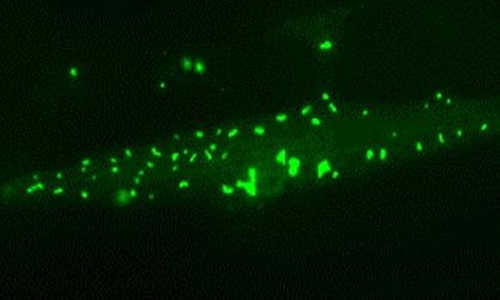
Kenong spent 10 years looking for this gene. Then one day, he said, “Come look at this experiment. You’ve got to see it.” I went to the greenhouse and I saw that the conventional variety that was flooded for 18 days had died, but the rice variety that we had genetically engineered with a new gene we had discovered, called Sub1, was alive. Kenong and I were amazed and excited that a single gene could have this dramatic effect. But this is just a greenhouse experiment. Would this work in the field?

Now, I’m going to show you a four-month time lapse video taken at the International Rice Research Institute. Breeders there developed a rice variety carrying the Sub1 gene using another genetic technique called precision breeding. On the left, you can see the Sub1 variety, and on the right is the conventional variety. Both varieties do very well at first, but then the field is flooded for 17 days. You can see the Sub1 variety does great. In fact, it produces three and a half times more grain than the conventional variety. I love this video because it shows the power of plant genetics to help farmers. Last year, with the help of the Bill and Melinda Gates Foundation, three and a half million farmers grew Sub1 rice.

(Applause)

Thank you.

Now, many people don’t mind genetic modification when it comes to moving rice genes around, rice genes in rice plants, or even when it comes to mixing species together through grafting or random mutagenesis. But when it comes to taking genes from viruses and bacteria and putting them into plants, a lot of people say, “Yuck.” Why would you do that? The reason is that sometimes it’s the cheapest, safest, and most effective technology for enhancing food security and advancing sustainable agriculture. I’m going to give you three examples.

First, take a look at papaya. It’s delicious, right? But now, look at this papaya. This papaya is infected with papaya ringspot virus. In the 1950s, this virus nearly wiped out the entire production of papaya on the island of Oahu in Hawaii. Many people thought that the Hawaiian papaya was doomed, but then, a local Hawaiian, a plant pathologist named Dennis Gonsalves, decided to try to fight this disease using genetic engineering. He took a snippet of viral DNA and he inserted it into the papaya genome. This is kind of like a human getting a vaccination. Now, take a look at his field trial. You can see the genetically engineered papaya in the center. It’s immune to infection. The conventional papaya around the outside is severely infected with the virus. Dennis’ pioneering work is credited with rescuing the papaya industry. Today, 20 years later, there’s still no other method to control this disease. There’s no organic method. There’s no conventional method. Eighty percent of Hawaiian papaya is genetically engineered.

Now, some of you may still feel a little queasy about viral genes in your food, but consider this: The genetically engineered papaya carries just a trace amount of the virus. If you bite into an organic or conventional papaya that is infected with the virus, you will be chewing on tenfold more viral protein.

Now, take a look at this pest feasting on an eggplant. The brown you see is frass, what comes out the back end of the insect. To control this serious pest, which can devastate the entire eggplant crop in Bangladesh, Bangladeshi farmers spray insecticides two to three times a week, sometimes twice a day, when pest pressure is high. But we know that some insecticides are very harmful to human health, especially when farmers and their families cannot afford proper protection, like these children. In less developed countries, it’s estimated that 300,000 people die every year because of insecticide misuse and exposure. Cornell and Bangladeshi scientists decided to fight this disease using a genetic technique that builds on an organic farming approach. Organic farmers like my husband Raoul spray an insecticide called B.T., which is based on a bacteria. This pesticide is very specific to caterpillar pests, and in fact, it’s nontoxic to humans, fish and birds. It’s less toxic than table salt. But this approach does not work well in Bangladesh. That’s because these insecticide sprays are difficult to find, they’re expensive, and they don’t prevent the insect from getting inside the plants. In the genetic approach, scientists cut the gene out of the bacteria and insert it directly into the eggplant genome. Will this work to reduce insecticide sprays in Bangladesh? Definitely. Last season, farmers reported they were able to reduce their insecticide use by a huge amount, almost down to zero. They’re able to harvest and replant for the next season.

Now, I’ve given you a couple examples of how genetic engineering can be used to fight pests and disease and to reduce the amount of insecticides. My final example is an example where genetic engineering can be used to reduce malnutrition. In less developed countries, 500,000 children go blind every year because of lack of Vitamin A. More than half will die. For this reason, scientists supported by the Rockefeller Foundation genetically engineered a golden rice to produce beta-carotene, which is the precursor of Vitamin A. This is the same pigment that we find in carrots. Researchers estimate that just one cup of golden rice per day will save the lives of thousands of children. But golden rice is virulently opposed by activists who are against genetic modification. Just last year, activists invaded and destroyed a field trial in the Philippines. When I heard about the destruction, I wondered if they knew that they were destroying much more than a scientific research project, that they were destroying medicines that children desperately needed to save their sight and their lives.

Some of my friends and family still worry: How do you know genes in the food are safe to eat? I explained the genetic engineering, the process of moving genes between species, has been used for more than 40 years in wines, in medicine, in plants, in cheeses. In all that time, there hasn’t been a single case of harm to human health or the environment. But I say, look, I’m not asking you to believe me. Science is not a belief system. My opinion doesn’t matter. Let’s look at the evidence. After 20 years of careful study and rigorous peer review by thousands of independent scientists, every major scientific organization in the world has concluded that the crops currently on the market are safe to eat and that the process of genetic engineering is no more risky than older methods of genetic modification. These are precisely the same organizations that most of us trust when it comes to other important scientific issues such as global climate change or the safety of vaccines.

Raoul and I believe that, instead of worrying about the genes in our food, we must focus on how we can help children grow up healthy. We must ask if farmers in rural communities can thrive, and if everyone can afford the food. We must try to minimize environmental degradation. What scares me most about the loud arguments and misinformation about plant genetics is that the poorest people who most need the technology may be denied access because of the vague fears and prejudices of those who have enough to eat.

We have a huge challenge in front of us. Let’s celebrate scientific innovation and use it. It’s our responsibility to do everything we can to help alleviate human suffering and safeguard the environment.

Thank you.

(Applause)

Thank you.

Chris Anderson: Powerfully argued. The people who argue against GMOs, as I understand it, the core piece comes from two things. One, complexity and unintended consequence. Nature is this incredibly complex machine. If we put out these brand new genes that we’ve created, that haven’t been challenged by years of evolution, and they started mixing up with the rest of what’s going on, couldn’t that trigger some kind of cataclysm or problem, especially when you add in the commercial incentive that some companies have to put them out there? The fear is that those incentives mean that the decision is not made on purely scientific grounds, and even if it was, that there would be unintended consequences. How do we know that there isn’t a big risk of some unintended consequence? Often our tinkerings with nature do lead to big, unintended consequences and chain reactions.

Pamela Ronald: Okay, so on the commercial aspects, one thing that’s really important to understand is that, in the developed world, farmers in the United States, almost all farmers, whether they’re organic or conventional, they buy seed produced by seed companies. So there’s definitely a commercial interest to sell a lot of seed, but hopefully they’re selling seed that the farmers want to buy. It’s different in the less developed world. Farmers there cannot afford the seed. These seeds are not being sold. These seeds are being distributed freely through traditional kinds of certification groups, so it is very important in less developed countries that the seed be freely available.

CA: Wouldn’t some activists say that this is actually part of the conspiracy? This is the heroin strategy. You seed the stuff, and people have no choice but to be hooked on these seeds forever?

PR: There are a lot of conspiracy theories for sure, but it doesn’t work that way. For example, the seed that’s being distributed, the flood-tolerant rice, this is distributed freely through Indian and Bangladeshi seed certification agencies, so there’s no commercial interest at all. The golden rice was developed through support of the Rockefeller Foundation. Again, it’s being freely distributed. There are no commercial profits in this situation. And now to address your other question about, well, mixing genes, aren’t there some unintended consequences? Absolutely — every time we do something different, there’s an unintended consequence, but one of the points I was trying to make is that we’ve been doing kind of crazy things to our plants, mutagenesis using radiation or chemical mutagenesis. This induces thousands of uncharacterized mutations, and this is even a higher risk of unintended consequence than many of the modern methods. And so it’s really important not to use the term GMO because it’s scientifically meaningless. I feel it’s very important to talk about a specific crop and a specific product, and think about the needs of the consumer.

CA: So part of what’s happening here is that there’s a mental model in a lot of people that nature is nature, and it’s pure and pristine, and to tinker with it is Frankensteinian. It’s making something that’s pure dangerous in some way, and I think you’re saying that that whole model just misunderstands how nature is. Nature is a much more chaotic interplay of genetic changes that have been happening all the time anyway.

PR: That’s absolutely true, and there’s no such thing as pure food. I mean, you could not spray eggplant with insecticides or not genetically engineer it, but then you’d be stuck eating frass. So there’s no purity there.

CA: Pam Ronald, thank you. That was powerfully argued. PR: Thank you very much. I appreciate it. (Applause)

Protein aggregates save cells during aging

from
BIOENGINEER.ORG http://bioengineer.org/protein-aggregates-save-cells-during-aging/

Aging is a complex biological process which is accompanied by an increasing number of toxic protein aggregates in the cells. Scientists consider them the cause of various neurodegenerative disorders, such as Alzheimer’s, Huntington’s and Parkinson’s disease.

proteomics

Muscle cell of a long-lived nematode worm: chaperone-rich protein aggregates (green) accumulate and save the cell during aging. Photo Credit: Prasad Kasturi / © MPI of Biochemistry

However, their exact role remains poorly understood. A collaborative team headed by F.-Ulrich Hartl at the MPIB now used the tiny nematode worm Caenorhabditis elegans (short: C. elegans) as a model organism to analyze the changes that occur in the proteome (the entirety of all proteins) during a lifespan. “The study is the most extensive of its kind in a whole organism quantifying more than 5,000 different proteins at multiple time points during aging, explains Prasad Kasturi, equally contributing first author together with Dirk Walther.

The researchers were able to show that the proteome undergoes extensive changes as the worms age. About one third of the quantified proteins significantly change in abundance. The normal relation between different proteins, which is critical for proper cell function, is lost. This shift overwhelms the machinery of protein quality control and impairs the functionality of the proteins. This is reflected in the widespread aggregation of surplus proteins ultimately contributing to the death of the animals.

Based on these findings, the researchers also analyzed how genetically changed worms with a substantially longer or shorter lifespan manage these changes. “We found that proteome imbalance sets in earlier and is increased in short-lived worms. In contrast, long-lived worms coped much better and their proteome composition deviated less dramatically from that of young animals”, as Kasturi says. Surprisingly, the long-lived worms increasingly deposited surplus and harmful proteins in insoluble aggregates, thus relieving pressure on the soluble, functional proteome. However, in contrast to the aggregates found in short-lived animals, these deposits were enriched with helper proteins — the so-called molecular chaperones — which apparently prevented the toxic effects normally exerted by aggregates.

“These findings demonstrate that the cells specifically accumulate chaperone-rich protein aggregates as a safety mechanism. Therefore, the aggregates seem to be an important part of healthy aging”, Kasturi explains. Indeed, it is known that insoluble protein aggregates also accumulate in the brains of healthy elderly people. So far, researchers assumed that neurodegeneration and dementia appear to be mainly caused by aberrant protein species accumulating in aggregates. This assumption may now being tested again: “Clearly, aggregates are not always harmful. Finding ways to concentrate harmful proteins in insoluble deposits might be a useful strategy to avoid or postpone neurodegenerative diseases as we age”, F.-Ulrich Hartl classifies the results.

Story Source:

The above story is based on materials provided by Max Planck Institute of Biochemistry .

Master orchestrator of the genome is discovered,

from
BIOENGINEER.ORG http://bioengineer.org/master-orchestrator-of-the-genome-is-discovered/

One of developmental biology’s most perplexing questions concerns what signals transform masses of undifferentiated cells into tremendously complex organisms, a process called ontogeny.

genom

UB research suggests a new paradigm, visualized in this diagram, for developmental global genome programming by the nuclear FGFR1 protein.

New research by University at Buffalo scientists, published last week in PLOS ONE, provides evidence that it all begins with a single “master” growth factor receptor that regulates the entire genome.

“The finding provides a new level of understanding of the fundamental aspects of how organisms develop,” says senior author Michal K. Stachowiak, PhD, professor in the Department of Pathology and Anatomical Sciences in the UB School of Medicine and Biomedical Sciences and senior author. He also directs the Stem Cell Engraftment and In Vivo Analysis Facility and the Stem Cell Culture and Training Facility at the Western New York Stem Cell Culture and Analysis Center at UB.

“Our research shows how a single growth factor receptor protein moves directly to the nucleus in order to program the entire genome,” he said.

The research challenges a long-held supposition in biology that specific types of growth factors only functioned at a cell’s surface. For two decades, Stachowiak’s team has been intrigued by the possibility that growth factors function from within the nucleus, a point, he says, this current paper finally proves.

A more advanced understanding of how organisms form, based on this work, has the potential to significantly enhance the understanding and treatment of cancers, which result from uncontrolled development as well as congenital diseases, the researchers say. The new research also will contribute to the understanding of how stem cells work.

This work was conducted on mouse embryonic stem cells, not human cells.

Organizing ‘this cacophony of genes’

“We’ve known that the human body has almost 30,000 genes that must be controlled by thousands of transcription factors that bind to those genes,” Stachowiak said, “yet we didn’t understand how the activities of genes were coordinated so that they properly develop into an organism.

“Now we think we have discovered what may be the most important player, which organizes this cacophony of genes into a symphony of biological development with logical pathways and circuits,” he said.

At the center of the discovery is a single protein called nuclear Fibroblast Growth Factor Receptor 1 (nFGFR1). “FGFR1 occupies a position at the top of the gene hierarchy that directs the development of multicellular animals,” said Stachowiak.

The FGFR1 gene is known to govern gastrulation, occurring in early development, where the three-layered embryonic structure forms. It also plays a major role in the development of the central and peripheral nervous systems and the development of the body’s major systems, including muscles and bones.

To study how nuclear FGFR1 worked, the UB team used genome-wide sequencing of mouse embryonic stem cells programmed to develop cells of the nervous system, with additional experiments in which nuclear FGFR1 was either introduced or blocked. The researchers found that the protein was responsible, either alone or with so-called partner nuclear receptors, for ensuring that embryonic stem cells develop into differentiated cells. By targeting thousands of genes, it controls the development of the major points of growth in the body (known as axes) as well as neuronal and muscle development.

The research shows that nuclear FGFR1 binds to promoters of genes that encode transcription factors, the proteins that control which genes are turned on or off in the genome.

“We found that this protein works as a kind of ‘orchestration factor,’ preferably targeting certain gene promoters and enhancers. The idea that a single protein could bind thousands of genes and then organize them into a hierarchy, that was unknown,” Stachowiak said. “Nobody predicted it.”

Sequencing advances

The discovery that a single protein can exert such a global genomic function stems from recent advances in DNA sequencing technologies, which allow for the sequencing of a complex genome in just hours.

“NextGen DNA sequencing allows us to analyze millions of DNA sequences selected by the interacting protein,” Stachowiak said.

In the UB research, the DNA sequencing data were processed by the supercomputer at the university’s Center for Computational Research (CCR). Stachowiak and his colleagues then spent weeks aligning these data to the genome and conducting further analyses.

“We imposed nuclear FGFR1 on every little corner of genome,” he said. “The computer spit out which genes are affected by nuclear FGFR1: it was an enormously complex network of genome activity.”
They found that the protein binds to genes that make neurons and muscles as well as to an important oncogene, TP53, which is involved in a number of common cancers.

Other studies in Stachowiak’s laboratory demonstrate that these interactions also take place in the human genome, controlling function and possibly underlying diseases like schizophrenia. Targeting of the nuclear FGFR1 allows for the reactivation of neural development in the adult brain in preclinical studies and thus, Stachowiak says, may offer unprecedented opportunity for regenerative medicine. Nuclear accumulation of nuclear FGFR1 may be altered in some cancer cells, and thus could become a focus in cancer therapy, he added.

Stachowiak concluded: “This seminal discovery lends new perspectives to the origin, nature and treatment of a variety of human diseases.”

Story Source:

The above story is based on materials provided by University at Buffalo.

Researchers develop custom artificial membranes to study the molecular basis of disease

from
BIOENGINEER.ORG http://bioengineer.org/researchers-develop-custom-artificial-membranes-to-study-the-molecular-basis-of-disease/

Decorating the outside of cells like tiny antenna, a diverse community of sugar molecules acts like a telecommunications system, sending and receiving information, recognizing and responding to foreign molecules and neighboring cells.

artificial membrane

“The sugar part of our biomembranes are as crucial to our health as our DNA, and yet we know almost nothing about it,” said Virgil Percec, a professor of chemistry in the University of Pennsylvania School of Arts and Sciences.

Part of the reason cell membrane sugars, called glycans, are so poorly understood is that scientists were unable to accurately model them until last year, when Percec’s lab devised a way of programming artificial membranes with a precise number and spatial arrangement of sugars.

Now, as a proof-of-concept for their new model, the team has tested its interactions with galectin-8, a cell signaling protein that, when mutated, may contribute to rheumatoid arthritis. Gal-8 is one of a large family of growth-regulatory proteins the team is testing their model against. By modifying a single building block in Gal-8’s structure, exactly as nature does in a portion of the population, the researchers dramatically impaired its ability to communicate with the artificial membrane, suggesting a possible molecular basis for the disease.

Percec’s new study demonstrates how researchers can use this membrane model to examine the interactions of cell surfaces with other biological molecules, with far ranging applications in medicine, biochemistry and biophysics.

“There are lots of membrane sugar-protein interactions that are important for disease,” Percec said. “Now, we have the critical tool we need to develop these disease models.”

Other team members from Penn include postdoctoral chemistry researchers Shaodong Zhang and Ralph-Oliver Moussodia. They collaborated with Temple University’s Michael Klein, as well as Sabine Vértesy and Sabine André and Hans-Joachim Gabius of Ludwig-Maximillians University in Munich.

The study was published in The Proceedings of the National Academy of Sciences.

Cell membranes are composed of two layers of fatty molecules known as phospholipids, each of which has a water-loving head and a water-repellant tail. The simplest form of a membrane, called a liposome or vesicle, will self-assemble when its phospholipid building blocks are placed in water. But vesicles are difficult to produce in the lab and don’t remain stable for long. For decades, these challenges hindered scientific efforts to create artificial membranes for research.

But in 2010 Percec and his lab discovered they could produce stable, self-assembling vesicles by replacing phospholipids with a class of molecules called amphiphilic Janus dendrimers, which have water-loving and water-hating branches, instead of heads and tails. Not only are dendrimer-based vesicles much easier to produce, their size, number of functional ends-groups and the number of concentric layers they contain can be precisely tuned.

“This was a big advance. It provided us the tool we were looking for while saving a huge amount of work,” Percec said. “The next step was to ask ‘can we add surface sugars to it?’”

Early efforts to mimic membrane surfaces in the lab were crude and simplistic, with no control over the number or distribution of sugars. That posed a major limitation to researchers, who need an accurate representation of these surfaces to study how other cells, proteins or viruses, will interact with them.

Building off their dendrimer-based vesicles, Percec’s lab constructed a library of amphiphilic glycodendrimer molecules: dendrimers with chemically bonded glycan sugars. By diluting these glycodendrimers to a series of different concentrations in an organic solvent and injecting them in water, the team found they could program vesicles, called glycodendrimersomes, with different surface sugar topologies. Details of this work were published in 2013 in the Journal of the American Chemical Society.

“As our molecules self-assemble, the vesicles formed have a precise number and spatial arrangement of the sugars, something never possible before,” Percec said.

One of the most important roles membrane sugars play is receiving messages from signaling proteins and communicating those messages to the cell. Many diseases are thought to be the result of communication errors that arise when a signaling protein incurs a mutation or the membrane’s glycan structure is altered. To demonstrate the utility of their new model, the researchers studied how mutant varieties of Gal-8 interacted with a custom artificial membrane containing Gal-8’s specific binding sugars. By modifying a single amino acid in the protein’s structure, as occurs naturally in human populations, they could significantly impair Gal-8’s ability to bind to the membrane.

“By testing this model with a sugar binding protein of human origin, we show that single mutation of an amino acid from a giant protein structure can induce a dramatic change in its interactions with the cell,” Percec said. “This demonstrates just how efficient and sensitive a model this is for biological membranes.”

In the future, the team will continue to develop and refine their glycodendrimersomes models, building membranes of increasing complexity and studying how membrane functions are affected. Besides Gal-8, there are many other biologically interesting signaling proteins, which researchers can now study using a robust and customizable membrane model.

Percec’s glycodendrimersome research is housed at Penn’s interdisciplinary Laboratory for Research on the Structure of Matter and in his own laboratory. Through the LRSM, Percec is collaborating with researchers in bioengineering, computational biology, biology, biophysics and biomedicine who are interested in using his programmable membranes for a variety of purposes, from visualizing the interactions between viruses and cells to developing biological capsules for vaccine and drug delivery.

“A biomembrane with a programmable surface topology is a tool to answer almost any question in cell biology,” Percec said.

Story Source:

The above story is based on materials provided by Penn News

7 Mayıs 2015 Perşembe

How the brain tells good from bad

from
BIOENGINEER.ORG http://bioengineer.org/how-the-brain-tells-good-from-bad/

Eating a slice of chocolate cake or spending time with a friend usually stimulates positive feelings, while getting in a car accident or anticipating a difficult exam is more likely to generate a fearful or anxious response.

happy neuron

Neuroscientists from MIT’s Picower Institute for Learning and Memory have identified two populations of neurons in the amygdala that process positive and negative emotions. Photo Credit: iStock (edited by Jose-Luis Olivares/MIT)

An almond-shaped brain structure called the amygdala is believed to be responsible for assigning these emotional reactions. Neuroscientists from MIT’s Picower Institute for Learning and Memory have now identified two populations of neurons in the amygdala that process positive and negative emotions. These neurons then relay the information to other brain regions that initiate the appropriate behavioral response.

The study, which appears in the April 29 issue of Nature, represents a significant step in understanding how the brain assigns emotions to different experiences, says senior study author Kay Tye, the Whitehead Career Development Assistant Professor in the Department of Brain and Cognitive Sciences.

“How do we tell if something is good or bad? Even though that seems like a very simple question, we really don’t know how that process works,” Tye says. “This study tells us that streams of information are hard-wired and are separated into good and bad at the level of the amygdala.”

The findings could also help scientists to better understand how mental illnesses such as depression arise, she says. Many psychiatric symptoms may reflect impairments in emotional processing. For example, people who are depressed do not find positive experiences rewarding, and people who suffer from addiction are not deterred by the negative outcomes of their behavior.

Graduate student Praneeth Namburi and postdoc Anna Beyeler are the paper’s lead authors.

The good, the bad, and the amygdala

For many years neuroscientists viewed the amygdala — and in particular, a subregion known as the basolateral amygdala — as a processing center for fear. However, more recent studies, including work that Tye did as a graduate student at the University of California at San Francisco, have highlighted the importance of the amygdala in processing reward.

Those findings raised the question of how the same structure could respond to both positive and negative inputs and initiate the appropriate behavioral response. The neurons of the basolateral amygdala are intermingled, making it difficult to distinguish which populations might be involved in different functions.
Tye and colleagues suspected they might be able to distinguish populations of neurons that respond to different emotions based on their targets elsewhere in the brain. Previous studies had suggested that some of these neurons project to the nucleus accumbens, which plays a role in reward learning, while others send information to another part of the amygdala known as the centromedial amygdala.

To identify these populations, the researchers delivered green and red fluorescent microspheres called retrobeads to the target cells in the nucleus accumbens and centromedial amygdala, respectively. These spheres traveled backwards until they reached the neurons of the basolateral amygdala, clearly marking two distinct populations.

After labeling these neurons, the researchers analyzed amygdala activity as the mice learned either a fear-conditioning task or a reward task. In the fear-conditioning task, the mice learned to associate a tone with a foot shock, and in the reward task the tone was paired with a drink of sugary water.
The next day, the researchers measured the strength of the connections coming into the two populations, which carry sensory information to the amygdala. They found that basolateral amygdala neurons that connect to the nucleus accumbens receive stronger input after reward learning, but their inputs are weakened after fear learning. Neurons that connect to the centromedial amygdala show the opposite response.

The results suggest that these two populations essentially function as a gate for sensory information coming into the amygdala, Namburi says. “There are sensory inputs coming in to either of these populations, and once learning happens, you’re shifting the flood onto one population or the other,” he says.
The researchers then found that by shutting down the pathway to the fear circuit, they not only impaired fear learning, but also enhanced reward learning.
“This was exciting because it suggests that these populations engage in a push-pull interaction with each other, which makes sense as seeking rewards and avoiding threats are often behaviors that present opposing forces,” Tye says. “Just as you might expect someone to lose their appetite if gunshots were fired, the activation of the fear circuit could suppress reward-related behaviors.”

Sheena Josselyn, an associate professor of psychology and physiology at the University of Toronto, describes the paper as “a huge advance in our understanding of how the brain processes different emotions.”

“Everyone knows that we can learn about both positive and negative experiences, but it has never been shown how one structure can contribute to encoding two diametrically opposed emotional outcomes,” says Josselyn, who was not involved in the research. “This work showed that where each cell projects determines whether it encodes a positive or a negative memory. Just looking at the cell doesn’t reveal its identity, one must consider the cell in the context of a broader circuit.”

Distinguishing traits

Once the researchers defined the functions of each cell population, they set out to identify other distinguishing characteristics. They found only minor differences in shape and in the electrophysiological properties of the neurons, but they did detect some intriguing differences in gene expression. Some of the genes that were more active in one cell type than the other encode receptors that sit on cell surfaces and bind to incoming neurotransmitters, which help transmit sensory information to the amygdala.

The researchers are particularly interested in one of these receptors, which interacts with a small protein called neurotensin. This protein helps to regulate the cells’ response to glutamate, one of the major neurotransmitters required to strengthen connections between neurons. In follow-up studies, they are now investigating the role neurotensin may play in reward- and fear-learning in the amydgala.

“This represents a new paradigm for therapeutic development,” Tye says. “‘Circuit-based drug discovery’ relies on first identifying how different components of the circuit work and then identifying what targets might control them.”

Story Source:

The above story is based on materials provided by MIT News Office, Anne Trafton.

‘Leg Bank’ hope for changing amputees’ lives

from
BIOENGINEER.ORG http://bioengineer.org/leg-bank-hope-for-changing-amputees-lives/

A ‘leg bank’ — providing life-changing prostheses to low-income people who have lost limbs — is being developed by a team including University of Strathclyde researchers.

Academics from Strathclyde, and members of Dutch-based social enterprise organisation ProPortion, plan to establish a service in Colombia offering high-quality artificial legs to people who have lost limbs, often through injuries from landmines. The team aims to help people who struggle to find work, meaning they cannot afford to buy prosthetic limbs, or to support themselves and their families.

A team led by Dr Arjan Buis, from Strathclyde’s Department of Biomedical Engineering, has developed innovative technology, known as Majicast, to manufacture lower limb prosthetic sockets which fit prostheses securely to patients’ residual limbs.

ProPortion has devised a potential business model which would encompass training for people who are currently operating at prosthetic technician level, enhancing the quality of their product and enabling the service to become self-sustaining. If successful, the venture could be adapted for use in other countries.

While an agreement has been reached in Colombia for a nationwide programme of landmine removal, the devices have killed or injured more than 10,000 people in the past 25 years.

Dr Buis said: “Colombia has a high incidence rate for amputations, not only owing to landmines but also to other causes such as diabetes and traffic accidents. However, many people there are unable to get access to prostheses.

“The Majicast is a straightforward, fully automated, easy-to-use device that will produce high quality prosthetic sockets. The Majicast’s relative ease of use is particularly important in low-income countries, where human resources are often scarce and the demand is high.

“The device has been scientifically tested and clinically validated; this method has also been shown to be more repeatable and consistent than traditional methods.

“The Majicast is currently being optimised into a market-ready product, with the help of a design agency. We expect that this social enterprise venture can be successfully piloted in Colombia, then developed for other parts of the world.

“We have fantastic inventions but the capacity and organisational structure to bring them to markets is lacking; only then does an invention become an innovation. That’s why we have chosen to team up with ProPortion — their purpose is to serve people with low incomes in developing economies. That brings value to us; it helps our work to be relevant and impactful for society.”

Merel Rumping, Project Incubator with ProPortion, developed the idea for the leg bank after hearing of a surgeon in Thailand who, frustrated by a lack of access to quality prostheses, created his own from plastic bottle caps. She was also inspired by the many amputees without prostheses she saw during her time working with street children and former child soldiers in Colombia.

ProPortion produced the plan for a social enterprise following a feasibility study conducted with designers from Delft University of Technology.

Ms Rumping said: “We did research and found out Colombia has the highest landmine victim rate in the world. We also learned that many of them do not have access to high quality and affordable prostheses.

“It became clear that currently available prostheses are made from expensive imported components. We also understood that, in theory, the majority of Colombians have access to a prosthesis via healthcare insurance companies but that, in reality, these companies often delay payment.

“Therefore, some people do not get a prosthesis at all and if they manage to get approval, they have to travel for hours to the main cities, where the professional prosthesis workplaces are located. Many amputees from rural areas do not have the financial means to travel at all, and won’t get a prosthesis.

“Those who can pay for their travel expenses have to wait up to two or three months to have their prosthesis made; rehabilitation takes even longer. In this period they cannot work, and they cannot take care of their families.

“We want low-income amputees in Colombia to become socially included and financially self-sustainable by having easy access to high quality, affordable prosthesis. A potential business model is to use Majicast in combination with appropriate training aimed at clinically active people who currently perform at prosthetic technician level. We believe this will decentralise prosthetic care, meaning amputees don’t have to travel and also ensuring they are able to have their rehabilitation in their own communities.”

The partners in the leg bank project are currently seeking funding for the venture. They are in talks with potential investors and intend to begin a crowdfunding campaign in the near future.

Story Source:

The above story is based on materials provided by University of Strathclyde.

Thermometer-like device could help diagnose heart attacks

from
BIOENGINEER.ORG http://bioengineer.org/thermometer-like-device-could-help-diagnose-heart-attacks/

Diagnosing a heart attack can require multiple tests using expensive equipment. But not everyone has access to such techniques, especially in remote or low-income areas. Now scientists have developed a simple, thermometer-like device that could help doctors diagnose heart attacks with minimal materials and cost. The report on their approach appears in the ACS journal Analytical Chemistry.

heart 2

A simple, thermometer-like device could make diagnosing heart attacks easier in remote or low-income locations. Photo Credit: American Chemical Society

Sangmin Jeon and colleagues note that one way to tell whether someone has had a heart attack involves measuring the level of a protein called troponin in the person’s blood. The protein’s concentration rises when blood is cut off from the heart, and the muscle is damaged. Today, detecting troponin requires bulky, expensive instruments and is often not practical for point-of-care use or in low-income areas. Yet three-quarters of the deaths related to cardiovascular disease occur in low- and middle-income countries. Early diagnosis could help curb these numbers, so Jeon’s team set out to make a sensitive, more accessible test.

Inspired by the simplicity of alcohol and mercury thermometers, the researchers created a similarly straightforward way to detect troponin. It involves a few easy steps, a glass vial, specialized nanoparticles, a drop of ink and a skinny tube. When human serum with troponin — even at a minute concentration — is mixed with the nanoparticles and put in the vial, the ink climbs up a protruding tube and can be read with the naked eye, just like a thermometer.

Story Source:

The above story is based on materials provided by American Chemical Society.

“Cognitive” control to underwater robots

from
BIOENGINEER.ORG http://bioengineer.org/cognitive-control-to-underwater-robots/

For the last decade, scientists have deployed increasingly capable underwater robots to map and monitor pockets of the ocean to track the health of fisheries, and survey marine habitats and species. In general, such robots are effective at carrying out low-level tasks, specifically assigned to them by human engineers — a tedious and time-consuming process for the engineers.

underwater

Researchers watch underwater footage taken by various AUVs exploring Australia’s Scott Reef. Courtesy of the researchers

When deploying autonomous underwater vehicles (AUVs), much of an engineer’s time is spent writing scripts, or low-level commands, in order to direct a robot to carry out a mission plan. Now a new programming approach developed by MIT engineers gives robots more “cognitive” capabilities, enabling humans to specify high-level goals, while a robot performs high-level decision-making to figure out how to achieve these goals.

For example, an engineer may give a robot a list of goal locations to explore, along with any time constraints, as well as physical directions, such as staying a certain distance above the seafloor. Using the system devised by the MIT team, the robot can then plan out a mission, choosing which locations to explore, in what order, within a given timeframe. If an unforeseen event prevents the robot from completing a task, it can choose to drop that task, or reconfigure the hardware to recover from a failure, on the fly.

In March, the team tested the autonomous mission-planning system during a research cruise off the western coast of Australia. Over three weeks, the MIT engineers, along with groups from Woods Hole Oceanographic Institution, the Australian Center for Field Robotics, the University of Rhode Island, and elsewhere, tested several classes of AUVs, and their ability to work cooperatively to map the ocean environment.

The MIT researchers tested their system on an autonomous underwater glider, and demonstrated that the robot was able to operate safely among a number of other autonomous vehicles, while receiving higher-level commands. The glider, using the system, was able to adapt its mission plan to avoid getting in the way of other vehicles, while still achieving its most important scientific objectives. If another vehicle was taking longer than expected to explore a particular area, the glider, using the MIT system, would reshuffle its priorities, and choose to stay in its current location longer, in order to avoid potential collisions.
“We wanted to show that these vehicles could plan their own missions, and execute, adapt, and re-plan them alone, without human support,” says Brian Williams, a professor of aeronautics and astronautics at MIT, and principal developer of the mission-planning system. “With this system, we were showing we could safely zigzag all the way around the reef, like an obstacle course.”

Williams and his colleagues will present the mission-planning system in June at the International Conference on Automated Planning and Scheduling, in Israel.

All systems go

When developing the autonomous mission-planning system, Williams’ group took inspiration from the “Star Trek” franchise and the top-down command center of the fictional starship Enterprise, after which Williams modeled and named the system.

Just as a hierarchical crew runs the fictional starship, Williams’ Enterprise system incorporates levels of decision-makers. For instance, one component of the system acts as a “captain,” making higher-level decisions to plan out the overall mission, deciding where and when to explore. Another component functions as a “navigator,” planning out a route to meet mission goals. The last component works as a “doctor,” or “engineer,” diagnosing and repairing problems autonomously.
“We can give the system choices, like, ‘Go to either this or that science location and map it out,’ or ‘Communicate via an acoustic modem, or a satellite link,’” Williams explains. “What the system does is, it makes those choices, but makes sure it satisfies all the timing constraints and doesn’t collide with anything along the way. So it has the ability to adapt to its environment.”

Autonomy in the sea

The system is similar to one that Williams developed for NASA following the loss of the Mars Observer, a spacecraft that, days before its scheduled insertion into Mars’ orbit in 1993, lost contact with NASA.

“There were human operators on Earth who were experts in diagnosis and repair, and were ready to save the spacecraft, but couldn’t communicate with it,” Williams recalls. “Subsequently, NASA realized they needed systems that could reason at the cognitive level like engineers, but that were onboard the spacecraft.”
Williams, who at the time was working at NASA’s Ames Research Center, was tasked with developing an autonomous system that would enable spacecraft to diagnose and repair problems without human assistance. The system was successfully tested on NASA’s Deep Space 1 probe, which performed an asteroid flyby in 1999.

“That was the first chance to demonstrate goal-directed autonomy in deep space,” Williams says. “This was a chance to do the same thing under the sea.”
By giving robots control of higher-level decision-making, Williams says such a system would free engineers to think about overall strategy, while AUVs determine for themselves a specific mission plan. Such a system could also reduce the size of the operational team needed on research cruises. And, most significantly from a scientific standpoint, an autonomous planning system could enable robots to explore places that otherwise would not be traversable. For instance, with an autonomous system, robots may not have to be in continuous contact with engineers, freeing the vehicles to explore more remote recesses of the sea.

“If you look at the ocean right now, we can use Earth-orbiting satellites, but they don’t penetrate much below the surface,” Williams says. “You could send sea vessels which send one autonomous vehicle, but that doesn’t show you a lot. This technology can offer a whole new way to observe the ocean, which is exciting.”

Story Source:

The above story is based on materials provided by MIT News office, Jennifer Chu.

Researchers Reverse Bacterial Resistance to Antibiotics

from
BIOENGINEER.ORG http://bioengineer.org/researchers-reverse-bacterial-resistance-to-antibiotics/

The rise of antibiotic-resistant bacteria is a growing problem in the United States and the world. New findings by researchers in evolutionary biology and mathematics could help doctors better address the problem in a clinical setting.

Miriam Barlow

Biologist Miriam Barlow of the University of California, Merced, and mathematician Kristina CronaOpens a new window of American University tested and found a way to return bacteria to a pre-resistant state. In research published in the open-access journal PLOS ONEOpens a new window, they show how to rewind the evolution of bacteria and verify treatment options for a family of 15 antibiotics used to fight common infections, including penicillin.

Their work could have major implications for doctors attempting to keep patient infections at bay using “antibiotic cycling,” in which a handful of different antibiotics are used on a rotating basis.

“Doctors don’t take an ordered approach when they rotate antibiotics,” Barlow said. “The doctors would benefit from a system of rotation that is proven. Our goal was to find a precise, ordered schedule of antibiotics that doctors could rely on and know that in the end, resistance will be reversed, and an antibiotic will work.”

Dangers of Antibiotic Resistance

When bacteria grow powerful enough that antibiotics no longer work, it can be a matter of life and death. Recently, at the Ronald Reagan UCLA Medical Center, two people died and seven were injured when a medical scope used in patient procedures harbored drug-resistant bacteria. In the U.S. annually, more than 2 million people get infections that are resistant to antibiotics and at least 23,000 people die as a result, according to the Centers for Disease Control and Prevention.

Resistance to antibiotics is a natural part of the evolution of bacteria, and unavoidable given the many types of bacteria and the susceptibility of the human host. To compensate for bacterial evolution, a doctor fighting infections in an intensive care unit may reduce, rotate or discontinue different antibiotics to get them to be effective in the short term.

The researchers — from UC Merced, AU and UC Berkeley — have been leading the way to uncover how to reverse resistance in the drug environment. They’ve done so by combining lab work with mathematics and computer technology.

“We have learned so much about the human genome as well as the sequencing of bacteria,” Crona said “Scientists now have lots and lots of data, but they need to make sense of it. Mathematics helps one to draw interpretations, find patterns and give insight into medical applications.”

Challenging Work Yields Important Results

After creating bacteria in a lab, the researchers exposed them to 15 different antibiotics and measured their growth rates. From there, they computed the probability of mutations to return the bacteria to its harmless state using the aptly named “Time Machine” software.

Managing resistance in any drug environment is extremely difficult, because bacteria evolve so quickly, becoming highly resistant after many mutations. To find optimal cycling strategies, the researchers tested up to six drugs in rotation at a time and found optimal plans for reversing the evolution of drug-resistant bacteria.

“This shows antibiotics cycling works. As a medical application, physicians can take a more strategic approach,” Crona said. “Uncovering optimal plans in antibiotics cycling presents a mathematical challenge. Mathematicians will need to create algorithms that can decipher optimal plans for a greater amount of antibiotics and bacteria.”

The researchers hope to next test the treatment paths in a clinical setting, working with doctors to rotate antibiotics to maximize their efficacy.

“This work shows that there is still hope for antibiotics if we use them intelligently,” Barlow said. “More research in this area and more research funding would make it possible to explore the options more comprehensively.”

Story Source:

The above story is based on materials provided by University of California, Merced.