from
BIOENGINEER.ORG http://bioengineer.org/humans-built-in-gps-is-our-3-d-sense-of-smell/
Like homing pigeons, humans have a nose for navigation because our brains are wired to convert smells into spatial information, new research from the University of California, Berkeley, shows.
While humans may lack the scent-tracking sophistication of, say, a search-and-rescue dog, we can sniff our way, blindfolded, toward a location whose scent we’ve smelled only once before, according to the UC Berkeley study published today (June 17) in the journal PLOS ONE.
Similar investigations have been conducted on birds and rodents, but this is the first time smell-based navigation has been field-tested on humans. The results evoke a GPS-like superpower one could call an “olfactory positioning system.”
“What we’ve found is that we humans have the capability to orient ourselves along highways of odors and crisscross landscapes using only our sense of smell,” said study lead author Lucia Jacobs, a UC Berkeley psychology professor who studies evolution and cognition in animals and humans.
Smell is a primitive sense that our early ancestors used for foraging, hunting and mating, among other skills necessary for survival. Early sailors and aviators gave anecdotal reports of using odors to navigate, but there have been no experiential scientific studies on this until now.
The process of smelling, or olfaction, is triggered by odor molecules traveling up the nasal passage, where they are identified by receptors that send signals to the olfactory bulb – which sits between the nasal cavity and the brain’s frontal lobe – and processes the information. A key to the connection between smell, memory and navigation is that olfactory bulbs have a strong neural link to the brain’s hippocampus, which creates spatial maps of our environment.
“Olfaction is like this background fabric to our world that we might not be conscious of, but we are using it to stay oriented,” Jacobs said. “We may not see a eucalyptus grove as we pass it at night, but our brain is encoding the smells and creating a map.”
Pigeons and rats, for example, are known to orient themselves using odor maps, or “smellscapes,” but sighted humans rely more heavily on visual landmarks, and so the study turned up some surprising results.
Two dozen young adults were tested on orientation and navigation tasks under various scenarios in which their hearing, sight or smell was blocked. The test location was a 25-by-20-foot room where 32 containers with sponges were placed at points around the edge of the room. Two of the sponges were infused with essential oils such as sweet birch, anise or clove.
In the smell-only experiment, study participants were led, one at a time, into the room wearing blindfolds, earplugs and headphones and walked in circles for disorientation purposes. They spent a minute at a specific point on the grid, where they inhaled a combination of two fragrances. After being walked in circles again for disorientation purposes, they were tasked with sniffing their way back to the starting point where they had smelled the two fragrances.
Overall, study participants navigated relatively closely to the targeted location when using only their sense of smell, compared to when other sensory inputs were blocked. Moreover, they were not just following one scent, but using information from both scents to orient themselves toward a point on an odor grid.
“We never thought humans could have a good enough sense of smell for this,” said Jacobs. But in retrospect, she noted, the results are “as obvious as the nose on my face.” Jacobs will be exploring this mechanism further as a scientist selected to be on the team of the National Science Foundation’s “Cracking the Olfactory Code” Ideas Lab, which takes place this summer.
Story Source:
The above story is based on materials provided by the University of California, Berkeley.
from
BIOENGINEER.ORG http://bioengineer.org/scientists-shows-aids-vaccine-candidate-successfully-primes-immune-system/
New research led by scientists at The Scripps Research Institute (TSRI), International AIDS Vaccine Initiative (IAVI) and The Rockefeller University shows in mice that an experimental vaccine candidate designed at TSRI can stimulate the immune system activity necessary to stop HIV infection. The findings could provide key information for the development of an effective AIDS vaccine.
Key authors of the new HIV/AIDS vaccine research from The Scripps Research Institute, International AIDS Vaccine Initiative and other institutions include (left to right) Dennis Burton, David Nemazee and William Schief. Photo Credit: The Scripps Research Institute.
The research, published June 18, 2015 in concurrent studies in the journals Cell and Science, represents a leap forward in the effort to develop a vaccine against HIV, which has so far struggled to elicit antibodies (immune system molecules) that can effectively fight off different strains of the virus.
“The results are pretty spectacular,” said Dennis Burton, chair of the TSRI Department of Immunology and Microbial Science and scientific director of two centers at TSRI, the IAVI Neutralizing Antibody Consortium (NAC) and the National Institutes of Health (NIH) Center for HIV/AIDS Vaccine Immunology and Immunogen Discovery (CHAVI-ID).
The Science study was co-led by Burton, TSRI Professor and IAVI NAC Associate Director of Vaccine Design William Schief, and TSRI Professor David Nemazee. The Cell study was co-led by Schief and Michel Nussenzweig, who is Zanvil A. Cohn and Ralph M. Steinman Professor at The Rockefeller University and a Howard Hughes Medical Institute (HHMI) investigator.
A Huge Challenge
The researchers’ long-term goal is to design a vaccine that prompts the body to produce antibodies that bind to HIV and prevent infection.
While many vaccines for other diseases use a dead or inactive version of the disease-causing microbe itself to trigger antibody production, immunizations with “native” HIV proteins are ineffective in triggering an effective immune response, due to HIV’s ability to evade detection from the immune system and mutate rapidly into new strains.
This challenge has led many researchers to believe that a successful AIDS vaccine will need to consist of a series of related, but slightly different proteins (immunogens) to train the body to produce broadly neutralizing antibodies against HIV–a twist on the traditional “booster” shot, where a person is exposed to the same immunogen multiple times.
In the new studies, the scientists tested one of these potential proteins, an immunogen called eOD-GT8 60mer, a protein nanoparticle designed to bind and activate B cells needed to fight HIV. The eOD-GT8 60mer was developed in the Schief lab and tested in mouse models engineered by the Nemazee lab to produce antibodies that resemble human antibodies.
Using a technique called B cell sorting, the researchers showed that immunization with eOD-GT8 60mer produced antibody “precursors”–with some of the traits necessary to recognize and block HIV infection. This suggested that eOD-GT8 60mer could be a good candidate to serve as the first in a series of immunizations against HIV.
“The vaccine appears to work well in our mouse model to ‘prime’ the antibody response,” added Nemazee.
In the Cell paper, researchers used the same eOD-GT8 60mer immunogen but used a slightly different mouse model. “The immunogen again launched the immune system in the right direction,” said Schief.
A concurrent study also in Science (led by Professor John Moore of Weill Cornell Medical College and including contributions from Schief, Burton, TSRI Associate Professor Andrew Ward, Ian Wilson, who is Hansen Professor of Structural Biology and chair of the Department of Integrative Structural and Computational Biology at TSRI, and other researchers) showed engineered immunogens also triggered immune responses in rabbit models and non-human primate models.
Next Steps
With eOD-GT8 60mer in the running as a potential contributor to an HIV vaccine, the researchers are now investigating other immunogens that could work in conjunction with it.
Schief said the Nemazee lab’s mouse models will be crucial resources for testing other engineered immunogens. He emphasized the importance of bringing different disciplines together to engineer mouse models, design antibodies and analyze results. “This was a beautiful collaboration of three labs at TSRI,” said Schief.
Story Source:
The above story is based on materials provided by The Scripps Research Institute.
from
BIOENGINEER.ORG http://bioengineer.org/tough-biogel-structures-produced-by-3-d-printing/
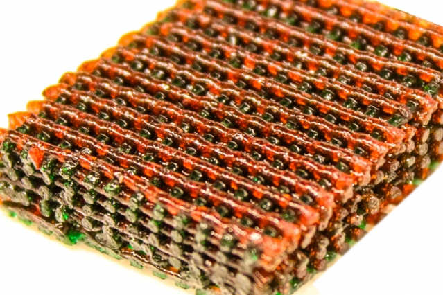
Researchers have developed a new way of making tough — but soft and wet — biocompatible materials, called “hydrogels,” into complex and intricately patterned shapes. The process might lead to injectable materials for delivering drugs or cells into the body; scaffolds for regenerating load-bearing tissues; or tough but flexible actuators for future robots, the researchers say.
Photo shows the open lattice of 3-D printed material, with materials having different characteristics of strength and flexibility indicated by different colors. Photo Credits: Researchers
The new process is described in a paper in the journal Advanced Materials, co-authored by MIT associate professor of mechanical engineering Xuanhe Zhao and colleagues at MIT, Duke University, and Columbia University.
Zhao says the new process can produce complex hydrogel structures that are “extremely tough and robust,” and compatible with the encapsulation of cells in the structures. That could make it possible to 3D-print complex hydrogel structures — for example, implants to be infused with cells and drugs and then placed in the body.
Hydrogels, defined by water molecules encased in rubbery polymer networks that provide shape and structure, are similar to natural tissues such as cartilage, which is used by the body as a natural shock absorber. The new 3-D printing process could eventually make it possible to produce tough hydrogel structures artificially for repair or replacement of load-bearing tissues, such as cartilage.
While synthetic hydrogels are commonly weak or brittle, a number of them that are tough and stretchable have been developed over the last decade. However, previous ways of making tough hydrogels have usually involved “harsh chemical environments” that would kill living cells encapsulated in them, Zhao says.
The new materials are benign enough to synthesize together with living cells — such as stem cells — which could then allow high viability of the cells, says Zhao, who holds a joint appointment in MIT’s Department of Civil and Environmental Engineering.
In addition, the previous work was not able to produce complex 3-D structures with tough hydrogels, Zhao says. The new biocompatible tough hydrogel can be printed into diverse 3-D structures such as a hollow cube, hemisphere, pyramid, twisted bundle, multilayer mesh, or physiologically relevant shapes, such as a human nose or ear.
The new method uses a commercially available 3D-printing mechanism, Zhao explains. “The innovation is really about the material — a new ink for 3-D printing of biocompatible tough hydrogel,” he says — specifically, a composite of two different biopolymers. “Each [material] individually is very weak and brittle, but once you put them together, it becomes very tough and strong. It’s like steel-reinforced concrete.”
One of the two polymers provides elasticity to the printed material, while the other allows it to dissipate energy under deformation without breaking. A third ingredient, a biocompatible “nanoclay,” makes it possible to fine-tune the viscosity of the material, improving the ability to control its flow through the 3D-printing nozzle.
The material can be made so flexible that a printed shape, such as a pyramid, can be compressed by 99 percent, and then spring back to its original shape, Sungmin Hong, a lead author of the paper and a former postdoc in Zhao’s group, says; it can also be stretched to five times its original size. Such resilience is a key feature of natural bodily tissues that need to withstand a variety of forces and impacts.
Such materials might eventually be used to custom-print shapes for the replacement of cartilaginous tissues in ears, noses, or load-bearing joints, Zhao says. Lab tests have already shown that the material is even tougher than natural cartilage.
The next step in the research will be to improve the resolution of the printer, which is currently limited to details about 500 micrometers in size, and to test the printed hydrogel structures in animal models. “We are enhancing the resolution,” Zhao says, “to be able to print more accurate structures for applications.”
In addition to biomedical applications, the same technique could be applied to printing a variety of soft but tough structural materials, he says, such as actuators for soft robotic systems.
“This is really beautiful work that demonstrates major advances in the utilization of tough hydrogels,” says David Mooney, a professor of bioengineering at Harvard University who was not involved in this work. “This builds off earlier work using other polymer systems, with some of this earlier work done by Dr. Zhao, but the demonstration that one can achieve similar mechanical performance with a common biomedical polymer is a substantial advance.”
Mooney adds, “It is also quite exciting that these new tough gels can be used for 3-D printing, as this is new for these gels, to my knowledge.”
Story Source:
The above post is reprinted from materials provided by Massachusetts Institute of Technology.
from
BIOENGINEER.ORG http://bioengineer.org/researchers-isolate-smallest-unit-of-sleep/
Scientists have grown a tiny group of brain cells that can be induced to fall asleep, wake up and even show rebound sleep after “staying up late.”
The study – the first to document that sleep originates in small neural networks – opens the door to deeper understanding of the genetic, molecular and electrical aspects underlying sleep disorders.
WSU Regents professor James Krueger and doctoral student Kathryn Jewett cultured neurons and glial cells that matured over two weeks into active neural networks exhibiting some of the same EEG sleep patterns seen in the brains of animals. The networks are the simplest unit of sleep identified to date.
Krueger predicted 20 years ago that sleep originates in small networks of neurons and glia. He said this study supports the hypothesis and will allow sleep to be independently isolated for research without the intrusion of physiological factors like changing body temperature.
The findings were recently published in the European Journal of Neuroscience. The work was funded by the National Institutes of Health/National Institute of Neurological Disease and Stroke and the National Science Foundation.
Sleep in a dish
Sleep clinics typically use EEG (electroencephalogram) measurements such as slow wave voltage (SW) and synchronization (SYN) to determine if a patient has fallen asleep. For the study, Krueger and Jewett used a number of readings including SW and SYN to determine sleep states of the mature cell cultures.
Krueger said the normal state of cultured neurons is sleep-like. To drive them into deeper sleep, the researchers added tumor necrosis factor (TNF) to the petri dish, which also contained an array of EEG electrodes. TNF is an immune protein that helps regulate sleep and is found in all animals from fruit flies to humans.
“TNF is also known to stabilize synapses – the junction between two neurons – so you don’t lose old memories and can simultaneously retain new memories,” he said.
Krueger and Jewett reversed the effects of TNF and awakened the neurons by applying mild electrical stimulation. During this period, the cells displayed EEG signs equivalent to wakeful activity.
When the researchers prolonged electrical stimulation, the cells responded with a miniature version of sleep homeostasis seen in animals; that is, after their extra activity, the nerve cells “slept in” the next day.
“Everyone has experienced this type of homeostasis,” said Krueger. “If you stay up late one night, you sleep more the next night to catch up.”
The researchers said the pathway for this sleep-wake cycle is consistent with the way sleep occurs in life. When the neuron cultures are activated they signal that activity by releasing ATP, which in turn releases TNF and another immune hormone called interleukin-1. Together they trigger the sleep phase.
Running on three cylinders
The study builds on the work of former WSU neuroscientist David Rector who, in 2005, showed that individual cortical columns in the brain go to sleep and wake up at different times. His research confirmed that after a day of unusual exertion certain parts of the brain will continue sleeping the next day. In effect, you really can be “half asleep.”
Rector and Krueger also showed that a cortical column could be driven into a sleep-like state by the application of TNF.
Krueger said that although it is late in his 40-year career, his discovery of the neural networks is opening up exciting new fields in sleep research and will help address the elusive mystery of sleep function.
“It is forcing scientists to see sleep as a small network property,” he said. “Before, people viewed sleep as a whole-brain phenomenon – using theories that often invoke “a miracle occurs and then you go to sleep.”
“In our theory, there is no miracle at all,” said Krueger. “Bits and pieces of the brain oscillate between sleep and wake-like states depending on how much activity they had the day before.
“And the reason they do that is because synapses are activity dependent – the more you use them, the better they get,” he said. “While our bodies rest, sleep stabilizes the neural network and provides an alternate pattern of stimulation to help preserve our memories.”
Story Source:
The above post is reprinted from materials provided by Washington State University.
from
BIOENGINEER.ORG http://bioengineer.org/pneumonia-found-to-harm-dna-in-lung-cells/
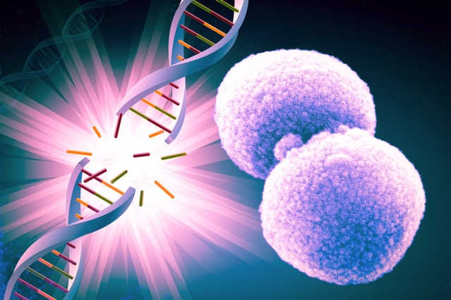
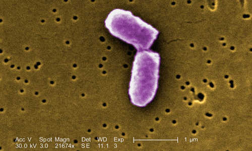
A bacterium that is the most common cause of pneumonia — a leading cause of death worldwide — can damage DNA in lung cells, a new study has shown.
The bacterium Streptococcus pneumoniae, a common cause of pneumonia, can damage DNA in lung cells, a new study shows. Photo Credit: Janice Carr/CDC (edited by MIT News)
Researchers from the Singapore-MIT Alliance for Research and Technology (SMART) demonstrated that hydrogen peroxide secreted by some strains of Streptococcus pneumoniae causes DNA in human lung cells to suffer double-strand breaks. Such breaks sever the DNA, creating broken ends that are highly toxic to cells, leading to cell suicide, or apoptosis.
“Secretion of hydrogen peroxide damages the DNA of lung cells, crippling the lungs’ defenses against invasion and making it easier for the bacteria to get into the bloodstream,” says Bevin Engelward, the paper’s senior author and a professor of biological engineering at MIT.
The discovery, she says, could lead to improved treatment for pneumonia patients by providing a means of measuring a person’s susceptibility to the disease. The study was led by Engelward, an expert in DNA damage and repair, and Vincent Chow, a professor and microbiologist at the National University of Singapore.
The study is published today in the Proceedings of the National Academy of Sciences. The lead author was Prashant Rai, a researcher in the infectious disease group at SMART.
“Hydrogen peroxide is commonly used as a disinfectant because of its ability to kill a wide range of microorganisms,” says Eric Rubin, a professor of immunology and infectious diseases at Harvard University who was not involved in the research. “[This study] turn[s] this concept around: It is the microbe, S. pneumoniae, that is using hydrogen peroxide, this time to damage the host.”
In addition to Rai and Engelward, the research team included MIT collaborators Marcus Parrish, Ian J.J. Tay, and Shelley Ackerman; Na Li and Chow at SMART’s infectious disease group; and Fang He and Jimmy Kwang from Temasek Life Sciences Laboratory.
Toxic breaks
Physicians and researchers alike have long known that bacteria can infect people weakened by influenza — and that those infections can be fatal.
“In the 1918 flu pandemic, most deaths were caused by secondary bacterial pneumonia, especially infection with S. pneumoniae,” Chow says. “There is autopsy evidence to show that it wasn’t influenza that directly killed patients, but mortality was mainly due to the secondary bacterial infection that followed. That was what really motivated our lab to study S. pneumoniae: It’s clearly tied to flu pathogenicity. We wanted to investigate if S. pneumoniae and its products, such as hydrogen peroxide, could inflict damage on DNA of the infected host.”
Until now, however, DNA double-strand breaks were not among the harms thought to be caused by S. pneumoniae.
“There have been previous reports [of DNA damage] for certain bacteria, but nobody expects DNA damage to play a role in an acute infection like pneumonia,” Rai says.
The team used fluorescent dyes to locate DNA-repair foci, phosphorylated histone proteins that form at the site of DNA double-strand breaks and are thought to play a role in repairing DNA damage. After testing three strains of S. pneumoniae, the team found that the presence of the bacteria resulted in a significant increase in the frequency of double-strand breaks, and that this DNA damage caused apoptosis.
In their experiments, the amount of hydrogen peroxide and the amount of DNA damage produced by the bacteria both depended on the bacterial strain. One strain in particular caused a relatively high number of DNA double-strand breaks; this same strain also produced the most hydrogen peroxide.
The researchers then aimed to determine how significant DNA damage from hydrogen peroxide was in the actual disease. To do this, the researchers tested two strains of S. pneumoniae in mice, some of which were infected with the strain the team had found to produce the most hydrogen peroxide; the other was genetically engineered so that it could not produce hydrogen peroxide. These experiments demonstrated that bacteria that produced hydrogen peroxide more easily invaded the lungs and bloodstream.
“It is an important piece of work that extends our knowledge of how bacterial infection can cause lung injury,” says David Dockrell, a professor of infectious diseases at Sheffield University who was not involved in the research.
Too much hydrogen peroxide
The human body produces hydrogen peroxide at sites of inflammation: If the body senses an infection, it activates an innate inflammatory response that includes immune cells, such as macrophages and neutrophils. “What they produce are reactive oxygen and nitrogen species, which includes an increase in hydrogen peroxide — really toxic chemicals meant to beat down the infective agent,” Engelward says.
“If the bacteria itself produces hydrogen peroxide, maybe it pushes past the threshold that the host can tolerate,” Rai adds.
Moreover, the inflammatory response might not work against hydrogen peroxide-producing bacteria. “S. pneumoniae strains that secrete hydrogen peroxide are predicted to be resistant to our body’s reactive oxygen species, used in an attempt to clear the infection, since the bacteria already tolerates its own reactive oxygen species,” Engelward says.
“The immune system’s ability to create reactive oxygen species may not be very helpful against certain strains of S. pneumoniae. It is possible that it might be hurting rather than helping,” Rai says.
Screening people and bacteria
The discovery that strains of S. pneumoniae cause DNA damage could open pathways to improved triage and treatment, based on the specific combination of patient and bacterial strain.
“It’s known that there is variability in DNA-repair capacity among people: Some people repair their DNA better than others,” Engelward says.
Combined with these latest research results, that suggests that a person with lower capacity for DNA repair might be more susceptible to certain types of pneumonia.
Story Source:
The above story is based on materials provided by MIT News.
from
BIOENGINEER.ORG http://bioengineer.org/dna-breakage-underlies-both-learning-age-related-damage/
The process that allows our brains to learn and generate new memories also leads to degeneration as we age, according to a new study by researchers at MIT.
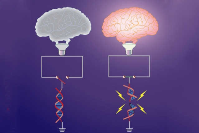
Early-response genes, which are important for synaptic plasticity, are “switched off” under basal conditions by topological constraints. Neuronal activity triggers DNA breaks in a subset of early-response genes, which overrides these topological constraints, and “switches on” gene expression. Shown here is the topological constraint to early-response genes represented as an open switch (left) that is tethered by intact DNA. Formation of the break severs the constraint, and promotes the circuit to be closed (right). The “brain bulb” represents the manifestation of neuronal activity. Photo Credits: Researchers
The finding, reported in a paper published in the journal Cell, could ultimately help researchers develop new approaches to preventing cognitive decline in disorders such as Alzheimer’s disease.
Each time we learn something new, our brain cells break their DNA, creating damage that the neurons must immediately repair, according to Li-Huei Tsai, the Picower Professor of Neuroscience and director of the Picower Institute for Learning and Memory at MIT.
This process is essential to learning and memory. “Cells physiologically break their DNA to allow certain important genes to be expressed,” Tsai says. “In the case of neurons, they need to break their DNA to enable the expression of early response genes, which ultimately pave the way for the transcriptional program that supports learning and memory, and many other behaviors.”
Slower DNA repair
However, as we age, our cells’ ability to repair this DNA damage weakens, leading to degeneration, Tsai says. “When we are young, our brains create DNA breaks as we learn new things, but our cells are absolutely on top of this and can quickly repair the damage to maintain the functionality of the system,” Tsai says. “But during aging, and particularly with some genetic conditions, the efficiency of the DNA repair system is compromised, leading to the accumulation of damage, and in our view this could be very detrimental.”
In previous research into Alzheimer’s disease in mice, the researchers found that even in the presymptomatic phase of the disorder, neurons in the hippocampal region of the brain contain a large number of DNA lesions, known as double strand breaks.
To determine how and why these double strand breaks are generated, and what genes are affected by them, the researchers began to investigate what would happen if they created such damage in neurons. They applied a toxic agent to the neurons known to induce double strand breaks, and then harvested the RNA from the cells for sequencing.
They discovered that of the 700 genes that showed changes as a result of this damage, the vast majority had reduced expression levels, as expected. Surprisingly though, 12 genes — known to be those that respond rapidly to neuronal stimulation, such as a new sensory experience — showed increased expression levels following the double strand breaks.
To determine whether these breaks occur naturally during neuronal stimulation, the researchers then treated the neurons with a substance that causes synapses to strengthen in a similar way to exposure to a new experience.
“Sure enough, we found that the treatment very rapidly increased the expression of those early response genes, but it also caused DNA double strand breaks,” Tsai says.
The good with the bad
In further studies the researchers were able to confirm that an enzyme known as topoisomerase IIβ is responsible for the DNA breaks in response to stimulation, according to the paper’s lead author Ram Madabhushi, a postdoc in Tsai’s laboratory.
“When we knocked down this enzyme, we found that both double strand break formation and the expression of early response genes was reduced,” Madabhushi says.
Finally, the researchers attempted to determine why the genes need such a drastic mechanism to allow them to be expressed. Using computational analysis, they studied the DNA sequences near these genes and discovered that they were enriched with a motif, or sequence pattern, for binding to a protein called CTCF. This “architectural” protein is known to create loops or bends in DNA.
In the early-response genes, the bends created by this protein act as a barrier that prevents different elements of DNA from interacting with each other — a crucial step in the genes’ expression.
The double strand breaks created by the cells allow them to collapse this barrier, and enable the early response genes to be expressed, Tsai says.
“Surprisingly then, even though conventional wisdom dictates that DNA lesions are very bad — as this ‘damage’ can be mutagenic and sometimes lead to cancer — it turns out that these breaks are part of the physiological function of the cell,” Tsai says.
Previous research has shown that the expression of genes involved in learning and memory is reduced as people age. So the researchers now plan to carry out further studies to determine how the DNA repair system is altered with age, and how this compromises the ability of cells to cope with the continued production and repair of double strand breaks.
They also plan to investigate whether certain chemicals could enhance this DNA repair capacity.
Story Source:
The above post is reprinted from materials provided by Massachusetts Institute of Technology.
from
BIOENGINEER.ORG http://bioengineer.org/how-your-brain-is-telling-you-to-vote/
A new joint study by researchers at the Montreal Neurological Institute and the Centre for the Study of Democratic Citizenship, both at McGill University, has cast some light on the brain mechanisms that support people’s voting decisions.
Evidence in the study shows that a part of the brain called the lateral orbitofrontal cortex (LOFC) must function properly if voters are to make choices that combine different sources of information about the candidates. The study found that damage to the LOFC leads people to base their vote on simpler information, namely the candidate’s good looks. Healthy individuals and those with brain damage affecting other parts of the frontal lobes spontaneously weighed both attractiveness and an assessment of the candidate’s competence when making their choices.
The new study provides the first evidence that the LOFC is critical for integrating different kinds of information to allow people to arrive at a preference.
Recent studies of political behaviour suggest that voting decisions can be influenced by “first-impression” social attributions based on physical appearance. Separate lines of research have implicated the orbitofrontal cortex in the judgement of social traits on one hand and economic decision-making on the other, implicating the orbitofrontal cortex region as a candidate for linking social attributes to voting decisions.
“How multiple attributes are combined in decision-making and how values are constructed is an important field that is just starting to be considered,” says Dr. Lesley Fellows, a neurologist and researcher at the Montreal Neurological Institute and senior author of the paper in the June 3 issue of the Journal of Neuroscience. “Recent research suggests that several areas in the brain carry information about the value of decision options, but it is not yet clear how these areas work together when we make a choice. The LOFC appears to be important when decisions are hard, helping to select the best from among options of similar value.”
The study tested subjects with and without damage affecting the LOFC. Participants took part in a simulated election task, where they were asked to vote for real-life but, unknown politicians based only on their photographs. Imagining themselves in an electoral period, participants were also asked to rate the perceived attractiveness and perceived competence of the candidates.
Participants without lesions in the LOFC appeared to make voting decisions based on both perceived attractiveness and perceived competence. Although subjects with LOFC damage could rate the competence of the candidates, they did not use this information when voting, instead relying only on the attractiveness factor
“This study provides a strong test of the function of this part of the brain,” says Dr. Fellows. “It shows that damage disrupts a specific aspect of how a decision is made. It provides evidence that LOFC is necessary for this function. This is the first time the brain basis of political behaviour has been studied with these methods.”
Story Source:
The above post is reprinted from materials provided by McGill University.
from
BIOENGINEER.ORG http://bioengineer.org/vitamin-d-status-related-to-immune-response-to-hiv-1/
Vitamin D plays an important part in the human immune response and deficiency can leave individuals less able to fight infections like HIV-1. Now an international team of researchers has found that high-dose vitamin D supplementation can reverse the deficiency and also improve immune response.
“Vitamin D may be a simple, cost-effective intervention, particularly in resource-poor settings, to reduce HIV-1 risk and disease progression,” the researchers report in today’s (June 15) online issue of Proceedings of the National Academy of Science.
The researchers looked at two ethnic groups in Cape Town, South Africa, to see how seasonal differences in exposure to ultraviolet B radiation, dietary vitamin D, genetics, and pigmentation affected vitamin D levels, and whether high-dose supplementation improved deficiencies and the cell’s ability to repel HIV-1.
“Cape Town, South Africa, has a seasonal ultraviolet B regime and one of the world’s highest rates of HIV-1 infection, peaking in young adults, making it an appropriate location for a longitudinal study like this one,” said Nina Jablonski, Evan Pugh Professor of Anthropology, Penn State, who led the research.
One hundred healthy young individuals divided between those of Xhosa ancestry — whose ancestors migrated from closer to the equator into the Cape area — and those self-identified as having Cape Mixed ancestry — a complex admixture of Xhosa, Khoisan, European, South Asian and Indonesian populations — were recruited for this study. The groups were matched for age and smoking. The Xhosa, whose ancestors came from a place with more ultraviolet B radiation, have the darkest skin pigmentation, while the Khoisan — the original inhabitants of the Cape — have adapted to the seasonally changing ultraviolet radiation in the area and are lighter skinned. The Cape Mixed population falls between the Xhosa and Khoisan in skin pigmentation levels.
Cape Town is situated in the southern hemisphere at about the same distance from the equator as the Florida panhandle, slightly more than 30 degrees latitude. Ultraviolet B levels show a winter decline anywhere above 30 degrees latitude, so Cape Town has a definite winter with low levels of the ultraviolet B wavelengths needed to produce precursor vitamin D3. Add to this the fact that people now spend more time indoors during winter and wear more clothing, and exposure to ultraviolet B in winter may be insufficient to prevent vitamin D deficiency.
The researchers note that sunscreen use is not a factor in these populations. However, the darker the skin’s pigment, the more ultraviolet B radiation necessary to trigger the precursor chemicals in the body to produce vitamin D.
“The skin of the indigenous people of the Cape, the Khoisan, is considerably lighter than that of either study group and may represent a long-established adaptation to seasonal UVB,” according to the researchers. “The darker skin of both study populations — Xhosia and Cape mixed — denotes a degree of mismatch between skin pigmentation and environmental UVB resulting from their recent migration into the region.”
The researchers found that both groups exhibited vitamin D deficiency during the winter, with women in both groups being more deficient, on average, than the men. Because of vitamin D’s impact on the immune system, the researchers provided six weeks of supplemental vitamin D3 to 30 of the Xhosa participants, which brought 77 percent of the participants to optimal vitamin D status.
Jablonski and her team determined that diet, genetics and other variables played very small roles in vitamin D status, although some genetic variations did influence the success of supplementation.
To test how vitamin D status affected the immune system and HIV-1 in particular, the researchers exposed blood samples from Xhosa and Cape mixed participants taken during the summer and winter when the subjects were vitamin D sufficient or deficient. They found that after nine days, the winter blood samples had greater infection than those taken in summer. After six weeks of vitamin D supplementation, the Xhosa blood sample levels of HIV-1 infection were the same as those during the summer.
“High-dosage oral vitamin D3 supplementation attenuated HIV-1 replication, increased circulating white blood cells and reversed winter-associated anemia,” the researchers reported. “Vitamin D3 presents a low-cost supplementation to improve HIV-associated immunity.”
Story Source:
The above post is reprinted from materials provided by Penn State.
from
BIOENGINEER.ORG http://bioengineer.org/scientists-grow-multiple-brain-structures-and-make-connections-between-them/
Opens up new avenues of research into human neuronal systems and interconnections, according to Restorative Neurology and Neuroscience report.
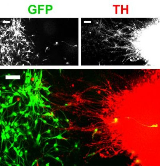
An example of TH-positive projections (red) from the inner chamber to the area of the outer chamber after 21 days. GFP (green) indicates neocortical neurons and TH (red) indicates mDA neurons. In the lower picture, each type has been tagged with fluorescent markers so that the locations and types can be identified. The fine tendrils indicate the growth of synapses between the neocortical and mDA neurons, mimicking the structures found in vivo. Photo Credit: Restorative Neurology and Neuroscience
Human stem cells can be differentiated to produce other cell types, such as organ cells, skin cells, or brain cells. While organ cells, for example, can function in isolation, brain cells require synapses, or connectors, between cells and between regions of the brain. In a new study published in Restorative Neurology and Neuroscience, researchers report successfully growing multiple brain structures and forming connections between them in vitro, in a single culture vessel, for the first time.
“We have developed a human pluripotent stem cell (hPSC)-based system for producing connections between neurons from two brain regions, the neocortex and midbrain,” explained lead investigator Chun-Ting Lee, PhD, working in the laboratory of William J. Freed, PhD, of the Intramural Research Program, National Institute on Drug Abuse, National Institutes of Health, Baltimore, MD.
Mesencephalic dopaminergic (mDA) neurons and their connections to other neurons in the brain are believed to be related to disorders including drug abuse, schizophrenia, Parkinson’s disease, and perhaps eating disorders, attention deficit-hyperactivity disorder, Tourette’s syndrome, and Lesch-Nyhan syndrome. However, studying mDA neurons and neocortical neurons in isolation does not reveal much data about how these cells actually interact in these conditions. This new capability to grow and interconnect two types of neurons in vitro now provides researchers with an excellent model for further study.
“This method, therefore, has the potential to expand the potential of hPSC-derived neurons to allow for studies of human neural systems and interconnections that have previously not been possible to model in vitro,” commented Dr. Lee.
Using a special container called an “ibidi wound healing dish,” which contains two chambers separated by a removable barrier, the researchers used hPSC to grow mDA neurons and neocortical neurons in the two individual chambers. The barrier was removed after colonies of both types of cells had formed and further growth resulted in the formation of synapses between neurons from each colony.
Future experiments could employ modifications of this method to examine connections between any two brain regions or neuronal subtypes that can be produced from hPSCs in vitro.
Story Source:
The above post is reprinted from materials provided by IOS Press BV.
from
BIOENGINEER.ORG http://bioengineer.org/speech-recognition-from-brain-activity/
Spoken Sentences Can Be Reconstructed from Activity Patterns of Human Brain Surface / ”Brain-to-Text“ Combines Knowledge from Neuroscience, Medicine, and Informatics
Speech is produced in the human cerebral cortex. Brain waves associated with speech processes can be directly recorded with electrodes located on the surface of the cortex. It has now been shown for the first time that is possible to reconstruct basic units, words, and complete sentences of continuous speech from these brain waves and to generate the corresponding text. Researchers at KIT and Wadsworth Center, USA present their ”Brain-to-Text“ system in the scientific journal Frontiers in Neuroscience (doi: 10.3389/fnins.2015.00217).
”It has long been speculated whether humans may communicate with machines via brain activity alone,” says Tanja Schultz, who conducted the present study with her team at the Cognitive Systems Lab of KIT. ”As a major step in this direction, our recent results indicate that both single units in terms of speech sounds as well as continuously spoken sentences can be recognized from brain activity.“
These results were obtained by an interdisciplinary collaboration of researchers of informatics, neuroscience, and medicine. In Karlsruhe, the methods for signal processing and automatic speech recognition have been developed and applied. ”In addition to the decoding of speech from brain activity, our models allow for a detailed analysis of the brain areas involved in speech processes and their interaction,” outline Christian Herff und Dominic Heger, who developed the Brain-to-Text system within their doctoral studies.
The present work is the first that decodes continuously spoken speech and transforms it into a textual representation. For this purpose, cortical information is combined with linguistic knowledge and machine learning algorithms to extract the most likely word sequence. Currently, Brain-to-Text is based on audible speech. However, the results are an important first step for recognizing speech from thought alone.
The brain activity was recorded in the USA from 7 epileptic patients, who participated voluntarily in the study during their clinical treatments. An electrode array was placed on the surface of the cerebral cortex (electrocorticography (ECoG)) for their neurological treatment. While patients read aloud sample texts, the ECoG signals were recorded with high resolution in time and space. Later on, the researchers in Karlsruhe analyzed the data to develop Brain-to-Text. In addition to basic science and a better understanding of the highly complex speech processes in the brain, Brain-to-Text might be a building block to develop a means of speech communication for locked-in patients in the future.
Story Source:
The above post is reprinted from materials provided by Karlsruhe Institute of Technology.
from
BIOENGINEER.ORG http://bioengineer.org/scientists-develop-new-technique-for-analyzing-the-epigenetics-of-bacteria/
Scientists from the Icahn School of Medicine at Mount Sinai have developed a new technique to more precisely analyze bacterial populations, to reveal epigenetic mechanisms that can drive virulence. The new methods hold the promise of a potent new tool to offset the growing challenge of antibiotic resistance by bacterial pathogens.
The research was published today in the journal Nature Communications, and conducted in collaboration with New York University Langone Medical Center and Brigham and Women’s Hospital of Harvard Medical School.
The information content of the genetic code in DNA is not limited to the primary nucleotide sequence of A’s, G’s, C’s and T’s. Individual DNA bases can be chemically modified, with significant functional consequences. In the bacterial kingdom, the most prevalent base modifications are in the form of DNA methylations, specifically to adenine and cytosine residuals. Beyond their participation in host defense, increasing evidence suggests that these modifications also play important roles in the regulation of gene expression, virulence and antibiotic resistance.
The research team employed the PacBio® RS II system from Pacific Biosciences, which can collect data on base modifications simultaneously as it collects DNA sequence data. PacBio’s single molecule, real-time sequencing enables the detection of N6-methyladenine and 4-methylcytosine, two major types of DNA modifications comprising the bacterial methylome. However, existing methods for studying bacterial methylomes rely on a population-level consensus that lack the single-cell resolution required to observe epigenetic heterogeneity.
‘We created a technique for the detection and phasing of DNA methylation at the single molecule level. We found that a typical clonal bacterial population that would otherwise be considered homogeneous using conventional techniques has epigenetically distinct subpopulations with different gene expression patterns’ said Gang Fang, Ph.D., assistant professor of genetics and genomics at the Icahn School of Medicine at Mount Sinai and senior author of the study. ‘Given that phenotypic heterogeneity within a bacterial population can increase its advantage of survival under stress conditions such as antibiotic treatment, this new technique is quite promising for future treatment of bacterial pathogens, as it enables de novo detection and characterization of epigenetic heterogeneity in a bacterial population.’
The researchers studied seven bacterial strains, demonstrating the new technique reveals distinct types of epigenetic heterogeneity. For Helicobacter pylori, a pathogenic bacterium that colonizes over 40 percent of the world population and is associated with gastric cancer, the team discovered that epigenetic heterogeneity can quickly emerge as a single cell divides, and different subpopulations with distinct methylation patterns have distinct gene expressions patterns. This may have contributed to the increasing rate of antibiotic resistance of Helicobacter pylori.
‘The application of this new technique will enable a more comprehensive characterization of the functions of DNA methylation and their impact on bacterial physiology. Resolving nucleotide modifications at the single molecule, single nucleotide level, especially when integrated with other single molecule- or single cell-level data, such as RNA and protein expression, will help resolve regulatory relationships that govern higher order phenotypes such as drug resistance’ said Eric Schadt, Ph.D., founding director of the Icahn Institute and professor of genomics at the Icahn School of Medicine at Mount Sinai. ‘The approach we developed can also be used to analyze DNA viruses and human mitochondrial DNA, both of which present significant epigenetic heterogeneity.’
Story Source:
The above story is based on materials provided by Icahn School of Medicine at Mount Sinai
from
BIOENGINEER.ORG http://bioengineer.org/the-first-synthetic-immune-organ-that-can-be-controlled-in-a-lab/
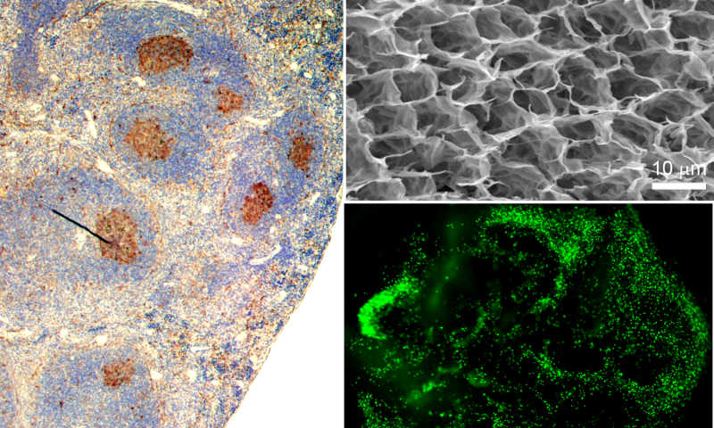
Cornell engineers have created a functional, synthetic immune organ that produces antibodies and can be controlled in the lab, completely separate from a living organism. The engineered organ has implications for everything from rapid production of immune therapies to new frontiers in cancer or infectious disease research.
When exposed to a foreign agent, such as an immunogenic protein, B cells in lymphoid organs undergo germinal center reactions. The image on the left is an immunized mouse spleen with activated B cells (brown) that produce antibodies. At right, top: a scanning electron micrograph of porous synthetic immune organs that enable rapid proliferation and activation of B cells into antibody-producing cells. At right, bottom: primary B cell viability and distribution is visible 24 hours following encapsulation procedure. Credit: Singh Lab
The immune organoid was created in the lab of Ankur Singh, assistant professor of mechanical and aerospace engineering, who applies engineering principles to the study and manipulation of the human immune system. The work was published online June 3 in Biomaterials and will appear later in print.
The synthetic organ is bio-inspired by secondary immune organs like the lymph node or spleen. It is made from gelatin-based biomaterials reinforced with nanoparticles and seeded with cells, and it mimics the anatomical microenvironment of lymphoid tissue. Like a real organ, the organoid converts B cells – which make antibodies that respond to infectious invaders – into germinal centers, which are clusters of B cells that activate, mature and mutate their antibody genes when the body is under attack. Germinal centers are a sign of infection and are not present in healthy immune organs.
The engineers have demonstrated how they can control this immune response in the organ and tune how quickly the B cells proliferate, get activated and change their antibody types. According to their paper, their 3-D organ outperforms existing 2-D cultures and can produce activated B cells up to 100 times faster.
The immune organ, made of a hydrogel, is a soft, nanocomposite biomaterial. The engineers reinforced the material with silicate nanoparticles to keep the structure from melting at the physiologically relevant temperature of 98.6 degrees.
The organ could lead to increased understanding of B cell functions, an area of study that typically relies on animal models to observe how the cells develop and mature.
What’s more, Singh said, the organ could be used to study specific infections and how the body produces antibodies to fight those infections – from Ebola to HIV.
“You can use our system to force the production of immunotherapeutics at much faster rates,” he said. Such a system also could be used to test toxic chemicals and environmental factors that contribute to infections or organ malfunctions.
The process of B cells becoming germinal centers is not well understood, and in fact, when the body makes mistakes in the genetic rearrangement related to this process, blood cancer can result.
“In the long run, we anticipate that the ability to drive immune reaction ex vivo at controllable rates grants us the ability to reproduce immunological events with tunable parameters for better mechanistic understanding of B cell development and generation of B cell tumors, as well as screening and translation of new classes of drugs,” Singh said.
Story Source:
The above story is based on materials provided by Cornell Chronicle.
from
BIOENGINEER.ORG http://bioengineer.org/injectable-device-delivers-nano-view-of-the-brain/
It’s a notion that might have come from the pages of a science-fiction novel — an electronic device that can be injected directly into the brain, or other body parts, and treat everything from neurodegenerative disorders to paralysis.
Sounds unlikely, until you visit Charles Lieber’s lab.
Led by Lieber, the Mark Hyman Jr. Professor of Chemistry, an international team of researchers has developed a method of fabricating nanoscale electronic scaffolds that can be injected via syringe. The scaffolds can then be connected to devices and used to monitor neural activity, stimulate tissues, or even promote regeneration of neurons. The research is described in a June 8 paper in Nature Nanotechnology.
Contributors to the work include Jia Liu, Tian-Ming Fu, Zengguang Cheng, Guosong Hong, Tao Zhou, Lihua Jin, Madhavi Duvvuri, Zhe Jiang, Peter Kruskal, Chong Xie, Zhigang Suo, and Ying Fang.
“I do feel that this has the potential to be revolutionary,” said Lieber, who holds a joint appointment in the Harvard Paulson School of Engineering and Applied Sciences. “This opens up a completely new frontier where we can explore the interface between electronic structures and biology. For the past 30 years, people have made incremental improvements in micro-fabrication techniques that have allowed us to make rigid probes smaller and smaller, but no one has addressed this issue — the electronics/cellular interface — at the level at which biology works.”
In an earlier study, scientists in Lieber’s lab demonstrated that cardiac or nerve cells grown with embedded scaffolds could be used to create “cyborg” tissue. Researchers were then able to record electrical signals generated by the tissue, and to measure changes in those signals as they administered cardio- or neuro-stimulating drugs.
“We were able to demonstrate that we could make this scaffold and culture cells within it, but we didn’t really have an idea how to insert that into pre-existing tissue,” Lieber said. “But if you want to study the brain or develop the tools to explore the brain-machine interface, you need to stick something into the body. When releasing the electronic scaffold completely from the fabrication substrate, we noticed that it was almost invisible and very flexible, like a polymer, and could literally be sucked into a glass needle or pipette. From there, we simply asked, ‘Would it be possible to deliver the mesh electronics by syringe needle injection?’”
Though not the first attempt at implanting electronics into the brain — deep brain stimulation has been used to treat a variety of disorders for decades — the nanofabricated scaffolds operate on a completely different scale.
“Existing techniques are crude relative to the way the brain is wired,” Lieber said. “Whether it’s a silicon probe or flexible polymers … they cause inflammation in the tissue that requires periodically changing the position or the stimulation.
“But with our injectable electronics, it’s as if it’s not there at all. They are one million times more flexible than any state-of-the-art flexible electronics and have subcellular feature sizes. They’re what I call ‘neuro-philic’ — they actually like to interact with neurons.”
The process for fabricating the scaffolds is similar to that used to etch microchips, and begins with a dissolvable layer deposited on a substrate. To create the scaffold, researchers lay out a mesh of nanowires sandwiched in layers of organic polymer. The first layer is then dissolved, leaving the flexible mesh, which can be drawn into a needle and administered like any other injection.
The input-output of the mesh can then be connected to standard measurement electronics so that the integrated devices can be addressed and used to stimulate or record neural activity.
“These type of things have never been done before, from both a fundamental neuroscience and medical perspective,” Lieber said. “It’s really exciting — there are a lot of potential applications.”
Going forward, researchers hope to better understand how the body reacts to the injectable electronics over longer periods.
Harvard’s Office of Technology Development has filed for a provisional patent on the technology and is actively seeking commercialization opportunities.
“The idea of being able to precisely position and record from very specific areas, or even from specific neurons over an extended period of time — this could, I think, make a huge impact on neuroscience,” Lieber said.
Story Source:
The above story is based on materials provided by harvard gazette
from
BIOENGINEER.ORG http://bioengineer.org/microchip-captures-clusters-of-circulating-tumor-cells/
Researchers have developed a microfluidic chip that can capture rare clusters of circulating tumor cells, which could yield important new insights into how cancer spreads. The work was funded by the National Institute of Biomedical Imaging and Bioengineering (NIBIB), part of the National Institutes of Health.
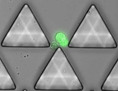
Fluorescently labelled cancer cell cluster balancing on the tip of a post within Cluster-Chip. Credit: Mehmet Toner, BioMicroElectroMechanical Systems Resource Center at MGH
Circulating tumor cells (CTCs) are cells that break away from a tumor and move through a cancer patient’s bloodstream. Single CTCs are extremely rare, typically fewer than 1 in 1 billion cells. These cells can take up residence in distant organs, and researchers believe this is one mode by which cancer spreads.
Even less common than single CTCs are small groups of CTCs, or clusters. While the existence of CTC clusters has been known for more than 50 years, their prevalence in the blood as well as their role in metastasis has not been thoroughly investigated, mostly because they are so elusive. However, recent advances in biomedical technologies that enable researchers to capture single CTCs have renewed interest in CTC clusters, which are occasionally captured along with single CTCs.
Now, researchers led by Mehmet Toner, Ph.D., professor of surgery (biomedical engineering) at the Massachusetts General Hospital (MGH) and the Harvard-MIT Division of Health & Sciences Technology, report the development of a novel microfluidic chip that is specifically designed for the efficient capture of CTC clusters from whole, unprocessed blood.
“Very little is known about CTC clusters and their role in the progression and metastasis of cancer. This unique technology presents an exciting opportunity to capture these exceptionally rare groups of cells for further analysis in a way that is minimally-invasive,” said NIBIB Director Roderic I. Pettigrew, Ph.D., M.D. “This is the kind of breakthrough technology that could have a very large impact on cancer research.”
The new technology — called Cluster-Chip — was developed with support from a Quantum Grant from NIBIB, which funds transformative technological innovation designed to solve major medical problems with a substantial disease burden, such as preventing cancer metastasis or precisely tailoring therapeutics to an individual’s cancer cell biology.
Toner and his collaborator Dr. Daniel Haber, M.D., Ph.D., also at MGH, recently used Cluster-Chip to capture and analyze CTC clusters in a group of 60 patients with metastatic breast, prostate, and melanoma cancers. The researchers found CTC clusters — ranging from two to 19 cells — in 30-40 percent of the patients.
“The presence of these clusters is far more common than we thought in the past,” said Toner. “The fact that we saw clusters in this many patients is really a remarkable finding.”
Further analysis of the patients’ CTC clusters yielded new insights into the biology of CTC clusters. The researchers published their results in the May 18, 2015 advance online issue of Nature Methods.
The chip is designed to slowly push blood through many rows of microscopic triangle-shaped posts. The posts are arranged in such a way that every two posts funnels cells towards the tip of a third post. At the tip, single cells — including blood cells and single CTCs — easily slide to either side of the post and continue through the chip until reaching the next tip; however CTC clusters are left at the tip, hanging in the balance due to forces pulling them down the post in opposite directions.
To determine the efficiency of Cluster-Chip, the researchers introduced fluorescently tagged cell clusters (ranging from 2-30 cells) into the chip and counted the number of clusters that were captured and the number that flowed through undetected. At a blood flow rate of 2.5ml/hr, the chip captured 99 percent of clusters containing four or more cells, 70 percent of three-cell clusters, and 41 percent of two-cell clusters. Comparison of the clusters under a microscope before and after capture found that the chip had no negative effects on the integrity of the clusters as a whole.
The researchers next compared the efficiency of their novel chip to two currently-used methods that have had some success capturing CTC clusters. They found that at similar blood flow rates, the Cluster-Chip was significantly more efficient than a filter-based method, which pushes blood through a membrane with pores only large enough to let single cells pass through. The chip was also more efficient than a different microfluidic chip — previously developed by Toner — that isolates CTCs and occasionally clusters using antibodies that stick to special proteins found on the surface of some tumor cells.
The results highlight the importance of the unique Cluster-Chip capture technique, which is based on the structural properties of CTC clusters rather than their size or the presence of surface proteins. This latter property makes the Cluster-Chip well-suited for capturing CTC clusters from a range of cancer types, including those that lose surface proteins during metastasis and those that never express them, such as melanoma.
The researchers went on to test the Cluster-Chip in a small trial of 60 patients with metastatic cancer. In this study, the chip captured CTC clusters in 11 of 27 (40.7 percent) breast cancer patients, 6 of 20 (30 percent) melanoma patients, and 4 of 13 (31 percent) prostate patients. The large number of clusters found in the patient samples suggests a possibly greater role for clusters in the metastatic cascade. While the significance of CTC clusters has not been fully established, a previous study published by Toner and the Haber team in Cell (2014) found an association between increased number of CTC clusters in patients with metastatic breast cancer and reduced survival, and an association between the presence of clusters and reduced survival in prostate cancer patients.
To characterize the biology of the clusters, the researchers measured a marker of tumor cell proliferation — an indicator of increased invasiveness and poor outcomes — in one breast cancer patient with high numbers of both single CTCs and clusters. Approximately half of the cells in the patient’s clusters were positive for the proliferative marker, demonstrating that clusters can contain both actively proliferating and quiescent cells.
The researchers also noted the rare presence of non-tumor cells within clusters in less than 5 percent of patients. “The fact that some CTC clusters contain immune cells is of particular interest,” said Pettigrew. “Given the increasing number of cancer therapies that engage the immune system, the ability to monitor tumor-immune cell interactions via the blood could be of great value.”
Toner anticipates that the Cluster-Chip will play an important role in stimulating new research on CTC cluster biology: “It’s like poking a sleeping bear. It could really awaken the field to go after clusters and to develop even better technologies to understand their biology in cancer metastasis.”
Story Source:
The above story is based on materials provided by National Institutes of Health (NIH).
from
BIOENGINEER.ORG http://bioengineer.org/bioengineers-develop-a-computer-that-operates-on-water-droplets/
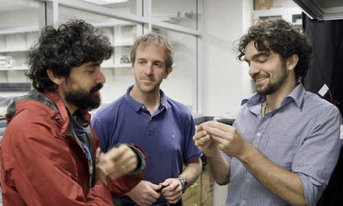
Manu Prakash, an assistant professor of bioengineering at Stanford, and his students have developed a synchronous computer that operates using the unique physics of moving water droplets. Their goal is to design a new class of computers that can precisely control and manipulate physical matter.
Computers and water typically don’t mix, but in Manu Prakash’s lab, the two are one and the same. Prakash, an assistant professor of bioengineering at Stanford, and his students have built a synchronous computer that operates using the unique physics of moving water droplets.
The computer is nearly a decade in the making, incubated from an idea that struck Prakash when he was a graduate student. The work combines his expertise in manipulating droplet fluid dynamics with a fundamental element of computer science – an operating clock.
“In this work, we finally demonstrate a synchronous, universal droplet logic and control,” Prakash said.
Because of its universal nature, the droplet computer can theoretically perform any operation that a conventional electronic computer can crunch, although at significantly slower rates. Prakash and his colleagues, however, have a more ambitious application in mind.
“We already have digital computers to process information. Our goal is not to compete with electronic computers or to operate word processors on this,” Prakash said. “Our goal is to build a completely new class of computers that can precisely control and manipulate physical matter. Imagine if when you run a set of computations that not only information is processed but physical matter is algorithmically manipulated as well. We have just made this possible at the mesoscale.”
Stanford Assistant Professor Manu Prakash, left, and graduate students Jim Cybulski and Georgios Katsikis developed the water drop computer.
The ability to precisely control droplets using fluidic computation could have a number of applications in high-throughput biology and chemistry, and possibly new applications in scalable digital manufacturing.
The results are published in the current edition of Nature Physics.
The crucial clock
For nearly a decade since he was in graduate school, an idea has been nagging at Prakash: What if he could use little droplets as bits of information and utilize the precise movement of those drops to process both information and physical materials simultaneously. Eventually, Prakash decided to build a rotating magnetic field that could act as clock to synchronize all the droplets. The idea showed promise, and in the early stages of the project, Prakash recruited a graduate student, Georgios “Yorgos” Katsikis, who is the first author on the paper.
Computer clocks are responsible for nearly every modern convenience. Smartphones, DVRs, airplanes, the Internet – without a clock, none of these could operate without frequent and serious complications. Nearly every computer program requires several simultaneous operations, each conducted in a perfect step-by-step manner. A clock makes sure that these operations start and stop at the same times, thus ensuring that the information synchronizes.
The results are dire if a clock isn’t present. It’s like soldiers marching in formation: If one person falls dramatically out of time, it won’t be long before the whole group falls apart. The same is true if multiple simultaneous computer operations run without a clock to synchronize them, Prakash explained.
“The reason computers work so precisely is that every operation happens synchronously; it’s what made digital logic so powerful in the first place,” Prakash said.
A magnetic clock
Developing a clock for a fluid-based computer required some creative thinking. It needed to be easy to manipulate, and also able to influence multiple droplets at a time. The system needed to be scalable so that in the future, a large number of droplets could communicate amongst each other without skipping a beat. Prakash realized that a rotating magnetic field might do the trick.
Katsikis and Prakash built arrays of tiny iron bars on glass slides that look something like a Pac-Man maze. They laid a blank glass slide on top and sandwiched a layer of oil in between. Then they carefully injected into the mix individual water droplets that had been infused with tiny magnetic nanoparticles.
Next, they turned on the magnetic field. Every time the field flips, the polarity of the bars reverses, drawing the magnetized droplets in a new, predetermined direction, like slot cars on a track. Every rotation of the field counts as one clock cycle, like a second hand making a full circle on a clock face, and every drop marches exactly one step forward with each cycle.
A camera records the interactions between individual droplets, allowing observation of computation as it occurs in real time. The presence or absence of a droplet represents the 1s and 0s of binary code, and the clock ensures that all the droplets move in perfect synchrony, and thus the system can run virtually forever without any errors.
“Following these rules, we’ve demonstrated that we can make all the universal logic gates used in electronics, simply by changing the layout of the bars on the chip,” said Katsikis. “The actual design space in our platform is incredibly rich. Give us any Boolean logic circuit in the world, and we can build it with these little magnetic droplets moving around.”
The current paper describes the fundamental operating regime of the system and demonstrates building blocks for synchronous logic gates, feedback and cascadability – hallmarks of scalable computation. A simple-state machine including 1-bit memory storage (known as “flip-flop”) is also demonstrated using the above basic building blocks.
A new way to manipulate matter
The current chips are about half the size of a postage stamp, and the droplets are smaller than poppy seeds, but Katsikis said that the physics of the system suggests it can be made even smaller. Combined with the fact that the magnetic field can control millions of droplets simultaneously, this makes the system exceptionally scalable.
“We can keep making it smaller and smaller so that it can do more operations per time, so that it can work with smaller droplet sizes and do more number of operations on a chip,” said graduate student and co-author Jim Cybulski. “That lends itself very well to a variety of applications.”
Prakash said the most immediate application might involve turning the computer into a high-throughput chemistry and biology laboratory. Instead of running reactions in bulk test tubes, each droplet can carry some chemicals and become its own test tube, and the droplet computer offers unprecedented control over these interactions.
From the perspective of basic science, part of why the work is so exciting, Prakash said, is that it opens up a new way of thinking of computation in the physical world. Although the physics of computation has been previously applied to understand the limits of computation, the physical aspects of bits of information has never been exploited as a new way to manipulate matter at the mesoscale (10 microns to 1 millimeter).
Because the system is extremely robust and the team has uncovered universal design rules, Prakash plans to make a design tool for these droplet circuits available to the public. Any group of people can now cobble together the basic logic blocks and make any complex droplet circuit they desire.
“We’re very interested in engaging anybody and everybody who wants to play, to enable everyone to design new circuits based on building blocks we describe in this paper or discover new blocks. Right now, anyone can put these circuits together to form a complex droplet processor with no external control – something that was a very difficult challenge previously,” Prakash said.
“If you look back at big advances in society, computation takes a special place. We are trying to bring the same kind of exponential scale up because of computation we saw in the digital world into the physical world.”
Story Source:
The above story is based on materials provided by Standford University.
from
BIOENGINEER.ORG http://bioengineer.org/first-circadian-clock-transplant-allows-bacteria-to-keep-time/
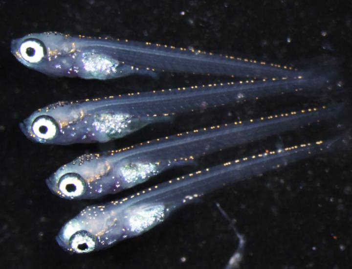
The first successful transplant of a circadian rhythm into a naturally non–circadian species could lead to precisely timed release of drugs and other innovative therapeutic applications
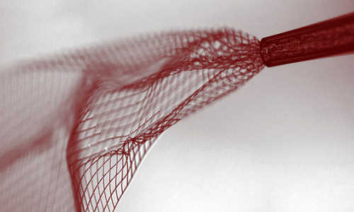
The E. coli say it is 10.30 Photo Credit: Cultura/Rex_Shutterstock
Often referred to as the “body clock”, circadian rhythm controls what time of day people are most alert, hungry, tired or physically primed due to a complex biological process that is not unique to humans. Circadian rhythms, which oscillate over a roughly 24–hour cycle in adaptation to the Earth’s rotation, have been observed in most of the planet’s plants, animals, fungi and cyanobacteria, and are responsible for regulating many aspects of organisms’ physiological, behavioral and metabolic functions.
Now, scientists led by the pioneering Harvard synthetic biologist Pamela Silver, Ph.D., have harnessed the circadian mechanism found in cyanobacteria to transplant the circadian wiring into a common species of bacteria that is naturally non–circadian. The novel work, which for the first time demonstrates the transplant of a circadian rhythm, is reported in a new study in Science Advances.
“By looking at systems in nature as modular, we think like engineers to manipulate and use biological circuits in a predictable, programmable way,” said Silver, who is a Core Faculty member at the Wyss Institute for Biologically Inspired Engineering at Harvard University and a Professor in the Department of Systems Biology at Harvard Medical School.
Silver’s team used this methodology to successfully transplant a circadian rhythm into the bacterial species E. coli, which is widely used as a “workhorse” cell species by biologists due to how well it is understood and the ease in which E. coli can be genetically altered. The genetically engineered circadian E. coli designed by Silver could one day be used in probiotic pills as a way to monitor the gut microbiota, which is the collective and diverse set of bacterial species that flourish in the human gastrointestinal tract and contribute to many different health factors.
To create a circadian rhythm in E. coli, the protein circuit responsible for regulating circadian oscillations was modularly removed from cyanobacteria, a photosynthetic bacteria species that is the only bacteria known to naturally contain a circadian rhythm. The protein circuit was then transplanted into E. coli, where it can be connected to additional gene expression components to potentially influence metabolic and behavioral functions in programmable relation to the day–night cycle. In the experimental E. coli, the circuit was linked to fluorescent proteins that lit up each time the circadian oscillations were triggered, causing the E. coli to glow rhythmically in a stunning visual confirmation of the transplant’s success.
“The ultimate dream application would be to deliver these circadian E. coli to an individual in pill form, which could allow the circadian rhythm to be linked to additional biological circuits in order to perform a precisely–timed release of drugs, or to be able to sense and influence the host’s circadian rhythm,” said the study’s first author, Anna Chen, a systems biology graduate student at the Wyss Institute and Harvard Medical School.
The human circadian rhythm has been shown to impact metabolism, which when disturbed can contribute to obesity and glucose intolerance. What’s more, many drugs including those commonly used to treat various cancers have been shown to fluctuate in their efficacy based on the time of day and the point in a patient’s circadian cycle in which they are administered. Using circadian E. coli, therapeutic strategies to influence a patient’s circadian rhythm or to program drugs to release internally at a specific point in the patient’s circadian rhythm could therefore energize treatments for cancer and metabolic diseases, among other clinical applications.
And further down the road, even the condition known as “jet lag”, which is caused when high–speed air travel disrupts a person’s body clock, might someday be combated using circadian E. coli that could potentially be leveraged to re–program and adjust the body’s circadian rhythm to match the travel destination’s day–night cycle.
“What’s really amazing is that we’ve demonstrated the modularity of biological systems – this finding goes beyond the transplantability of a circadian rhythm to open new doors to understanding how other modular biological circuits could be transplanted from one species to another,” said Silver.
“Circadian rhythm has an enormous impact on our health and how we respond to our environment – so the opportunity to tap into those controls, using genome engineering to rewire genetic circuits, opens an exciting new path for treating disease and regulating a wide range of physiological behaviors,” said Wyss Institute Founding Director Donald Ingber, M.D., Ph.D., who is also the Judah Folkman Professor of Vascular Biology at Harvard Medical School and Boston Children’s Hospital and Professor of Bioengineering at the Harvard John A. Paulson School of Engineering and Applied Sciences.
Story Source:
The above story is based on materials provided by Wyss Institute for Biologically Inspired Engineering at Harvard.
from
BIOENGINEER.ORG http://bioengineer.org/cell-density-remains-constant-as-brain-shrinks-with-age/
New, ultra-high-field magnetic resonance images (MRI) of the brain by researchers at the University of Illinois at Chicago provide the most detailed images to date to show that while the brain shrinks with age, brain cell density remains constant.
Brain cell density remains constant with age among cognitively normal adults. Photo Credit: Dr. Keith Thulborn
The study, of cognitively normal young and old adults, was published in the journal NMR in Biomedicine.
The images provide the first evidence that in normal aging, cell density is preserved throughout the brain, not just in specific regions, as previous studies on human brain tissue have shown. The findings also suggest that the maintenance of brain cell density may protect against cognitive impairment as the brain gradually shrinks in normal aging.
The images were made at UIC by a powerful 9.4-Tesla MRI, the first of its kind for human imaging. The 9.4 T magnetic field is more than three times stronger than that of a typical MRI machine in a doctor’s office and is currently approved only for research. The strongest MRI units approved for clinical use are 3 T.
‘The information provided by these 9.4-Tesla scans may be very useful in helping us to detect tiny losses of brain cells and the reduction in cell density that characterizes the early stages of neurodegenerative diseases that can take decades to develop before symptoms appear, like Alzheimer’s disease,’ said Dr. Keith Thulborn, director of MRI research in the UIC College of Medicine and lead author of the report.
‘If we can identify when Alzheimer’s pathology starts, the efficacy of new drugs or other interventions to slow or prevent Alzheimer’s disease can be tested and monitored when the disease starts, instead of after it’s developed for 20 or 30 years and becomes clinically apparent,’ said Thulborn, who is professor of radiology, physiology and biophysics.
Neuroscientists have long known that the brain shrinks with age, but for a long time they thought the loss in volume was associated with a loss of brain cells. That was disproven by studies that showed it is the neurons themselves that shrink while the number of cells remains the same in normal older adults.
Thulborn and his colleagues scanned the brains of 49 cognitively normal adults ranging in age from 21 to 80. All had at least a college education and were employed or, if retired, were active in their communities. All were without major medical, neurological or psychiatric disease and scored within the normal range for mental status.
The 9.4 Tesla MRI measures sodium ions, which are less concentrated by several orders of magnitude than the fat and water molecules detected with standard MRI. Sodium ions are present throughout the body and are pumped in and out of neurons to generate the electric potentials needed to spark nerve impulses. Sodium concentrations in the brain reflect neuron density. Areas of low sodium concentration indicate lots of neurons packed tightly together, while higher sodium concentration can indicate more space between cells — or the loss of cells, as in the case of neurodegenerative diseases.
Thulborn thinks the ultra-high-field scanners eventually will be approved for clinical use.
‘We can use the 9.4 T to look at brain cell loss in real time in patients experiencing stroke, or to see whether chemotherapy for brain tumors is working in higher resolution that is just not available using the current 3 T clinical scanners,’ he said.
Story Source:
The above story is based on materials provided by the University of Illinois.
from
BIOENGINEER.ORG http://bioengineer.org/to-be-sperm-or-not-to-be-sperm/
Researchers in Japan have found, for the first time in vertebrates, a genetic switch that determines whether germ cells become sperm or eggs.
The gene is named foxl3, and has been identified using a small fish called medaka (Oryzias latipes). In medaka without this gene’s functionality, surprisingly, sperm are produced in the ovaries of females. The sperm that are produced function normally, and have been confirmed to produce normal offspring. These results will be announced in the journal Science through Science Express the online pre-print version on June 11th.
This image shows young medaka born using normal eggs and sperm from female fish lacking functional foxl3 genes. Photo Credit: National Institute for Basic Biology
Dr. Toshiya Nishimura, Associate Professor Minoru Tanaka and their colleagues of the National Institute for Basic Biology, National Institutes of Natural Sciences, in collaboration with Dr. Satoru Kobayashi of the Okazaki Institute for Integrative Bioscience, and through joint research with Kyushu University’s Dr. Mikita Suyama and Dr. Yasuyuki Ohkawa, revealed that the foxl3 gene works in the germ cells of females “to suppress differentiation into sperm”.
In females lacking functional foxl3 genes, the small fish’s body appearance is still totally female, however a large number of sperm are formed in the ovaries, and a small number of eggs are formed at the same time.
It has also become clear that in these females lacking the function of foxl3 functional sperm can be obtained in a shorter period of time than in normal wild-type males. Applied research into uses for aquaculture is already underway.
“In spite of the environment surrounding the germ cells being female, the fact that functional sperm has been made surprised me greatly. That this sexual switch present in the Germ Cells is independent of the body’s sex is an entirely new finding,” Dr. Nishimura said.
Dr. Tanaka commented: “While germ cells can become either sperm or eggs, nobody knew that in vertebrates the germ cells have a switch mechanism to decide their own sperm or egg fate. Our result indicates that once the decision is made the germ cells have the ability to go all the way to the end. I believe it is of very large significance that this mechanism has been found.”
Story Source:
The above story is based on materials provided by NIBB’s Office of Public Relations.
from
BIOENGINEER.ORG http://bioengineer.org/reprogramming-of-dna-observed-in-human-germ-cells-for-first-time/
A team of researchers led by the University of Cambridge has described for the first time in humans how the epigenome – the suite of molecules attached to our DNA that switch our genes on and off – is comprehensively erased in early primordial germ cells prior to the generation of egg and sperm.
However, the study, published in the journal Cell, shows some regions of our DNA – including those associated with conditions such as obesity and schizophrenia – resist complete reprogramming.
Although our genetic information – the ‘code of life’ – is written in our DNA, our genes are turned on and off by epigenetic ‘switches’. For example, small methyl molecules attach to our DNA in a process known as methylation and contribute to the regulation of gene activity, which is important for normal development. Methylation may also occur spontaneously or through our interaction with the environment – for example, periods of famine can lead to methylation of certain genes – and some methylation patterns can be potentially damaging to our health. Almost all of this epigenetic information is, however, erased in germ cells prior to transmission to the next generation.
Professor Azim Surani from the Wellcome Trust/Cancer Research UK Gurdon Institute at the University of Cambridge, explains: “Epigenetic information is important for regulating our genes, but any abnormal methylation, if passed down from generation to generation, may accumulate and be detrimental to offspring. For this reason, the information needs to be reset in every generation before further information is added to regulate development of a newly fertilised egg. It’s like erasing a computer disk before you add new data.”
When an egg cell is fertilised by a sperm, it begins to divide into a cluster of cells known as a blastocyst, the early stage of the embryo. Within the blastocyst, some cells are reset to their master state, becoming stem cells, which have the potential to develop into any type of cell within the body. A small number of these cells become primordial germ cells with the potential to become sperm or egg cells.
In a study funded primarily by the Wellcome Trust, Professor Surani and colleagues showed that a process of reprogramming the epigenetic information contained in these primordial germ cells is initiated around two weeks into the embryo’s development and continues through to around week nine. During this period, a genetic network acts to inhibit the enzymes that maintain or programme the epigenome until the DNA is almost clear of its methylation patterns.
Crucially, however, the researchers found that this process does not clear the entire epigenome: around 5% of our DNA appears resistant to reprogramming. These ‘escapee’ regions of the genome contain some genes that are particularly active in neuronal cells, which may serve important functions during development. However, data analysis of human diseases suggests that such genes are associated with conditions such as schizophrenia, metabolic disorders and obesity.
Walfred Tang, a PhD student who is the first author on the study, adds: “Our study has given us a good resource of potential candidates of regions of the genome where epigenetic information is passed down not just to the next generation but potentially to future generations, too. We know that some of these regions are the same in mice, too, which may provide us with the opportunity to study their function in greater detail.”
Epigenetic reprogramming also has potential consequences for the so-called ‘dark matter’ within our genome. As much as half of human DNA is estimated to be comprised of ‘retroelements’, regions of DNA that have entered our genome from foreign invaders including bacteria and plant DNA. Some of these regions can be beneficial and even drive evolution – for example, some of the genes important to the development of the human placenta started life as invaders. However, others can have a potentially detrimental effect – particularly if they jump about within our DNA, potentially interfering with our genes. For this reason, our bodies employ methylation as a defence mechanism to suppress the activity of these retroelements.
“Methylation is effective at controlling potentially harmful retroelements that might harm us, but if, as we’ve seen, methylation patterns are erased in our germ cells, we could potentially lose the first line of our defence,” says Professor Surani.
In fact, the researchers found that a notable fraction of the retroelements in our genome are ‘escapees’ and retain their methylation patterns – particularly those retroelements that have entered our genome in our more recent evolutionary history. This suggests that our body’s defence mechanism may be keeping some epigenetic information intact to protect us from potentially detrimental effects.
Story Source:
The above story is based on materials provided by University of Cambridge.
from
BIOENGINEER.ORG http://bioengineer.org/scientists-find-genetic-basis-of-brain-networks-seen-in-imaging-studies/
A new study by researchers at the Stanford University School of Medicine found that synchronized physiological interactions between remote brain regions have genetic underpinnings.
The research was performed at Stanford but was made possible by collaborations with the Seattle-based Allen Institute for Brain Science and the IMAGEN Consortium, a multicenter European project, said the study’s senior author, Michael Greicius, M.D., an associate professor of neurology and neurological sciences.
Past neuroimaging studies have defined several “functional networks” in which remote regions of the brain appear to operate in synchrony. A new study from Stanford provides the molecular underpinnings for this theory. Photo Credit: Jonas Richiardi
An emerging consensus among neuroscientists is that cognitive operations are performed not by individual brain regions working in isolation, but by networks consisting of several discrete brain regions—anatomically connected either directly via white-matter tracts or indirectly through intermediary nodes—that share ‘functional connectivity,’ meaning that activity in these regions is tightly coupled.
Any given functional network is normally most active during the performance of the task associated with that network, as in the case of autobiographical memory (‘What did I eat for dinner last night?’). But the synchronous activity of component regions persists when networks are idling. Well over a dozen functional networks have been identified via a technique called resting-state functional magnetic resonance imaging, said Greicius, who is the medical director of the Stanford Center for Memory Disorders.
In resting-state fMRI scans, the individual is asked to simply lie still and relax for several minutes. The results of these scans indicate that even at rest, the brain’s functional networks continue to hum along at their own distinguishable frequencies and phases, like different radio stations playing simultaneously, but quietly, on the same radio.
Digging into fMRI images
However, whether resting-state fMRI-derived images, which measure local blood flows in different places throughout the brain, actually reflect neuronal activity has been controversial.
‘There’s been some skepticism regarding the validity of resting-state network activity,’ said Greicius. ‘We wanted to dig deeper and get to the molecular underpinnings of these imaging results, which indicated that the brain maintains its exquisite functional-network architecture even at rest.’
To start, Greicius and his colleagues computationally blended resting-state fMRI data they’d obtained from eight-minute scans of 15 healthy adults whose sole instructions were to lie still and relax. This enabled them to pinpoint numerous well-delineated functional networks.
Hoping to find genes that might promote or at least be involved in functional connectivity, the investigators next sought gene-expression profiles—measurements of activity levels of each of the human genome’s approximately 20,000 known genes—of regions within corresponding functional networks.
There’s no noninvasive way to obtain gene-profile expressions of brain tissue in living humans. But Jonas Richiardi, Ph.D., a postdoctoral scholar in Greicius’ lab now at the University of Geneva in Switzerland, made use of massive amounts of carefully annotated and meticulously archived data derived from the Allen Institute’s collection of six post-mortem human brain samples. The institute’s scientists have obtained gene-expression profiles of several hundred tissue samples excised from specific locations throughout the brain. Richiardi shares lead authorship of the study with neurology instructor Andre Altmann, Ph.D., who was also a postdoctoral scholar during the study’s duration.
Greicius and his colleagues narrowed their focus to cortical areas associated with four functional networks that are all well characterized in the imaging literature, consist of discrete, noncontiguous regions in both hemispheres, and are well represented in the Allen Institute’s human-brain database. Along with the default-mode network associated with autobiographical memory, they looked at gene-expression profiles in component regions of the brain’s sensorimotor, visuospatial and salience (emotion) networks.
Zeroing in on gene activity
The researchers were hunting specifically for a set of genes whose expression rose or fell in a more synchronized
fashion from region to region within a given network than between networks or outside any network. Using sophisticated statistical methods, they identified a set of 136 genes that showed a correlated pattern of gene expression in regions within each network.
These 136 genes weren’t specific to any single network, Greicius noted. Rather, ‘any one of these genes that was being expressed at a high, intermediate or low level in one region of any network, regardless of which network you’d picked, was also being expressed at corresponding levels in the other regions of that network,’ he said.
Importantly, a number of these genes encode proteins that aid in nerve cells’ signature activity: propagating electrical impulses. Some are ion channels, which maintain and modulate voltage differences across nerve cells’ outer membranes. Others are found at the junctions where one nerve cell in a circuit contacts another.
The Stanford team validated their findings by turning to another database. The IMAGEN Consortium has conducted widespread imaging, cognitive and genetic tests on 14-year-olds in an effort to predict who’s at high risk of encountering problems such as substance abuse by age 16. Among other things, the IMAGEN database contains detailed information on tiny variations from the norm in subjects’ genomic sequences. Altmann spearheaded an analysis of the variants present in the 136 genes of interest in 259 healthy adolescents. These subjects’ network-connectivity strength was determined, in part, by the genetic-variant profiles of these 136 genes.
Additional experiments using tissue samples obtained from two additional data sets, the Allen Institute’s mouse-brain and mouse-connectivity atlases, confirmed and amplified the findings from research on human brains. The reliance on large, shared data sets was another important feature of the study and, Altmann said, ‘highlights the value of making scientific data freely available. We had an idea and found collaborators willing to share their painstakingly collected data.’
The identification of functional-connectivity-associated genes sets the stage for targeted clinical applications, such as finding out how neurodegeneration propagates within a network.
‘Our work holds potential implications for a number of neuropsychiatric disorders,’ said Richiardi.
Evidence suggests, for instance, that Alzheimer’s disease spreads from one brain region to the next within the brain’s so-called default-mode network, which is activated when a person is recalling recent autobiographical events. Resting-state imaging holds exceptional potential in cases where task-based fMRI isn’t applicable. Alzheimer’s patients, for example, have difficulty focusing on memory-based tasks. Future work will focus on genes whose expression is correlated within one network, but not in other networks. Focusing on default-mode network-specific genes, for example, may lend novel insights into Alzheimer’s disease.
Story Source:
The above story is based on materials provided by Helmholtz Zentrum München – German Research Center for Environmental Health.