17 Temmuz 2015 Cuma

Researchers develop basic computing elements for bacteria

from
BIOENGINEER.ORG http://bioengineer.org/researchers-develop-basic-computing-elements-for-bacteria/

The “friendly” bacteria inside our digestive systems are being given an upgrade, which may one day allow them to be programmed to detect and ultimately treat diseases such as colon cancer and immune disorders.

bacteria

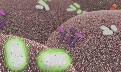
The illustration depicts Bacteroides thetaiotaomicron (white) living on mammalian cells in the gut (large pink cells coated in microvilli) and being activated by exogenously added chemical signals (small green dots) to express specific genes, such as those encoding light-generating luciferase proteins (glowing bacteria). Photo Credit: Janet Iwasa

In a paper published today in the journal Cell Systems, researchers at MIT unveil a series of sensors, memory switches, and circuits that can be encoded in the common human gut bacterium Bacteroides thetaiotaomicron.

These basic computing elements will allow the bacteria to sense, memorize, and respond to signals in the gut, with future applications that might include the early detection and treatment of inflammatory bowel disease or colon cancer.

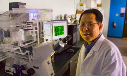
Researchers have previously built genetic circuits inside model organisms such as E. coli. However, such strains are only found at low levels within the human gut, according to Timothy Lu, an associate professor of biological engineering and of electrical engineering and computer science, who led the research alongside Christopher Voigt, a professor of biological engineering at MIT.

“We wanted to work with strains like B. thetaiotaomicron that are present in many people in abundant levels, and can stably colonize the gut for long periods of time,” Lu says.

The team developed a series of genetic parts that can be used to precisely program gene expression within the bacteria. “Using these parts, we built four sensors that can be encoded in the bacterium’s DNA that respond to a signal to switch genes on and off inside B. thetaiotaomicron,” Voigt says.

These can be food additives, including sugars, which allow the bacteria to be controlled by the food that is eaten by the host, Voigt adds.

Bacterial “memory”

To sense and report on pathologies in the gut, including signs of bleeding or inflammation, the bacteria will need to remember this information and report it externally. To enable them to do this, the researchers equipped B. thetaiotaomicron with a form of genetic memory. They used a class of proteins known as recombinases, which can record information into bacterial DNA by recognizing specific DNA addresses and inverting their direction.

The researchers also implemented a technology known as CRISPR interference, which can be used to control which genes are turned on or off in the bacterium. The researchers used it to modulate the ability of B. thetaiotaomicron to consume a specific nutrient and to resist being killed by an antimicrobial molecule.

The researchers demonstrated that their set of genetic tools and switches functioned within B. thetaiotaomicron colonizing the gut of mice. When the mice were fed food containing the right ingredients, they showed that the bacteria could remember what the mice ate.

Expanded toolkit

The researchers now plan to expand the application of their tools to different species of Bacteroides. That is because the microbial makeup of the gut varies from person to person, meaning that a particular species might be the dominant bacteria in one patient, but not in others.

“We aim to expand our genetic toolkit to a wide range of bacteria that are important commensal organisms in the human gut,” Lu says.

The concept of using microbes to sense and respond to signs of disease could also be used elsewhere in the body, he adds.

In addition, more advanced genetic computing circuits could be built upon this genetic toolkit in Bacteroides to enhance their performance as noninvasive diagnostics and therapeutics.

“For example, we want to have high sensitivity and specificity when diagnosing disease with engineered bacteria,” Lu says. “To achieve this, we could engineer bacteria to detect multiple biomarkers, and only trigger a response when they are all present.”

Tom Ellis, group leader of the Centre for Synthetic Biology at Imperial College London, who was not involved in the research, says the paper takes many of the best tools that have been developed for synthetic biology applications with E. coli and moves them over to use with a common class of gut bacteria.

“Whereas others have developed tools and applications for engineering genetic circuits, or biosensors, in bacteria that are then placed in the gut, this paper stands out from the crowd by first engineering a member of the Bacteroides genus, the most common type of bacteria found in our guts,” Ellis says.

The study has so far shown the efficacy of the approach in mice, and there will be a long road ahead before it can be approved for use in humans, Ellis says.

However, the paper really opens up the possibility of one day having engineered cells resident in our guts for long periods of time, he says. “These could do tasks like sensing and recording, or even in-situ synthesis of therapeutic molecules as and when they are needed.”

Story Source:

The above post is reprinted from materials provided by MIT News.

Electrical signals could help repair injured spinal cords

from
BIOENGINEER.ORG http://bioengineer.org/electrical-signals-could-help-repair-injured-spinal-cords/

Wichita State University’s Li Yao is taking a special approach to the study of spinal cord injuries through research that uses an electrical signal to repair tissue damage.

spinal cord

Wichita State University researcher Li Yao is studying how an electric signal can help promote repairs to injured spinal cords. Photo Credit: Wichita State University

When a person suffers neurological damage to their spinal cord, the tissues surrounding the injury site can die. But one of the body’s defense mechanisms is the regeneration and migration of a type of support cell — called Schwann cells — to the injury.

Those cells, as has been discovered in recent years, help myelinate — or cover — nerve axons where the injury has occurred, which promotes the recovery of some of the spinal cord’s function.

Yao, a biological sciences assistant professor, is studying how electrical signals can aim those cells directly to the injury site. His research, he hopes, will open new doors for the medical field to use electrical fields in the treatment of neural injuries.

“Electrical signal is a kind of ignored approach that may generate significant biological function in neural regeneration,” Yao says.

Yao’s research studies the molecular mechanism of cell migration in electric fields using next-generation RNA sequencing to look at the signaling pathways that regulate cell migration.

So far, he has discovered that the precision of the cell migration toward the injury increased significantly as the strength of the electrical field increased. The electrical field did not, however, change the speed at which the cells moved.

Still, Yao’s early findings suggest that the use of electrical fields in cell migration could become a burgeoning area of study in regenerative medicine.

“Our work has implications for central nervous system repair, and the application of an electrical field may assist with that,” Yao says.

Story Source:

The above post is reprinted from materials provided by Wichita State University.

16 Temmuz 2015 Perşembe

A most singular nano-imaging technique

from
BIOENGINEER.ORG http://bioengineer.org/a-most-singular-nano-imaging-technique/

Just as proteins are one of the basic building blocks of biology, nanoparticles can serve as the basic building blocks for next generation materials. In keeping with this parallel between biology and nanotechnology, a proven technique for determining the three dimensional structures of individual proteins has been adapted to determine the 3D structures of individual nanoparticles in solution.

collaidal

SINGLE uses in situ TEM imaging of platinum nanocrystals freely rotating in a graphene liquid cell to determine the 3-D structures of individual colloidal nanoparticles. Photo Credit: Berkeley Lab

A multi-institutional team of researchers led by the U.S. Department of Energy (DOE)’s Lawrence Berkeley National Laboratory (Berkeley Lab), has developed a new technique called “SINGLE” that provides the first atomic-scale images of colloidal nanoparticles. SINGLE, which stands for 3D Structure Identification of Nanoparticles by Graphene Liquid Cell Electron Microscopy, has been used to separately reconstruct the 3D structures of two individual platinum nanoparticles in solution.

“Understanding structural details of colloidal nanoparticles is required to bridge our knowledge about their synthesis, growth mechanisms, and physical properties to facilitate their application to renewable energy, catalysis and a great many other fields,” says Berkeley Lab director and renowned nanoscience authority Paul Alivisatos, who led this research. “Whereas most structural studies of colloidal nanoparticles are performed in a vacuum after crystal growth is complete, our SINGLE method allows us to determine their 3D structure in a solution, an important step to improving the design of nanoparticles for catalysis and energy research applications.”

Alivisatos, who also holds the Samsung Distinguished Chair in Nanoscience and Nanotechnology at the University of California Berkeley, and directs the Kavli Energy NanoSciences Institute at Berkeley (Kavli ENSI), is the corresponding author of a paper detailing this research in the journal Science. The paper is titled “3D Structure of Individual Nanocrystals in Solution by Electron Microscopy.” The lead co-authors are Jungwon Park of Harvard University, Hans Elmlund of Australia’s Monash University, and and Peter Ercius of Berkeley Lab. Other co-authors are Jong Min Yuk, David Limmer, Qian Chen, Kwanpyo Kim, Sang Hoon Han, David Weitz and Alex Zettl.

Colloidal nanoparticles are clusters of hundreds to thousands of atoms suspended in a solution whose collective chemical and physical properties are determined by the size and shape of the individual nanoparticles. Imaging techniques that are routinely used to analyze the 3D structure of individual crystals in a material can’t be applied to suspended nanomaterials because individual particles in a solution are not static. The functionality of proteins are also determined by their size and shape, and scientists who wanted to image 3D protein structures faced a similar problem. The protein imaging problem was solved by a technique called “single-particle cryo-electron microscopy,” in which tens of thousands of 2D transmission electron microscope (TEM) images of identical copies of an individual protein or protein complex frozen in random orientations are recorded then computationally combined into high-resolution 3D reconstructions. Alivisatos and his colleagues utilized this concept to create their SINGLE technique.

“In materials science, we cannot assume the nanoparticles in a solution are all identical so we needed to develop a hybrid approach for reconstructing the 3D structures of individual nanoparticles,” says co-lead author of the Science paper Peter Ercius, a staff scientist with the National Center for Electron Microscopy (NCEM) at the Molecular Foundry, a DOE Office of Science User Facility.

“SINGLE represents a combination of three technological advancements from TEM imaging in biological and materials science,” Ercius says. “These three advancements are the development of a graphene liquid cell that allows TEM imaging of nanoparticles rotating freely in solution, direct electron detectors that can produce movies with millisecond frame-to-frame time resolution of the rotating nanocrystals, and a theory for ab initio single particle 3D reconstruction.”

The graphene liquid cell (GLC) that helped make this study possible was also developed at Berkeley Lab under the leadership of Alivisatos. TEM imaging uses a beam of electrons rather than light for illumination and magnification but can only be used in a high vacuum because molecules in the air disrupt the electron beam. Since liquids evaporate in high vacuum, samples in solutions must be hermetically sealed in special solid containers – called cells – with a very thin viewing window before being imaged with TEM. In the past, liquid cells featured silicon-based viewing windows whose thickness limited resolution and perturbed the natural state of the sample materials. The GLC developed at Berkeley lab features a viewing window made from a graphene sheet that is only a single atom thick.

“The GLC provides us with an ultra-thin covering of our nanoparticles while maintaining liquid conditions in the TEM vacuum,” Ercius says. “Since the graphene surface of the GLC is inert, it does not adsorb or otherwise perturb the natural state of our nanoparticles.”

Working at NCEM’s TEAM I, the world’s most powerful electron microscope, Ercius, Alivisatos and their colleagues were able to image in situ the translational and rotational motions of individual nanoparticles of platinum that were less than two nanometers in diameter. Platinum nanoparticles were chosen because of their high electron scattering strength and because their detailed atomic structure is important for catalysis.

“Our earlier GLC studies of platinum nanocrystals showed that they grow by aggregation, resulting in complex structures that are not possible to determine by any previously developed method,” Ercius says. “Since SINGLE derives its 3D structures from images of individual nanoparticles rotating freely in solution, it enables the analysis of heterogeneous populations of potentially unordered nanoparticles that are synthesized in solution, thereby providing a means to understand the structure and stability of defects at the nanoscale.”

The next step for SINGLE is to recover a full 3D atomic resolution density map of colloidal nanoparticles using a more advanced camera installed on TEAM I that can provide 400 frames-per-second and better image quality.

“We plan to image defects in nanoparticles made from different materials, core shell particles, and also alloys made of two different atomic species,” Ercius says.

Story Source:

The above story is based on materials provided by Berkely University.

Device delivers drugs to brain via remote control

from
BIOENGINEER.ORG http://bioengineer.org/device-delivers-drugs-to-brain-via-remote-control/

A team of researchers has developed a wireless device the width of a human hair that can be implanted in the brain and activated by remote control to deliver drugs.

drug delivery

Tiny, implantable devices are capable of delivering light or drugs to specific areas of the brain, potentially improving drug delivery to targeted regions of the brain and reducing side effects. Eventually, the devices may be used to treat pain, depression, epilepsy and other neurological disorders in people. Photo Credit: Alex David Jerez Roman

The technology, demonstrated for the first time in mice, one day may be used to treat pain, depression, epilepsy and other neurological disorders in people by targeting therapies to specific brain circuits, according to the researchers at Washington University School of Medicine in St. Louis and the University of Illinois at Urbana-Champaign.

The research is a major step forward in pharmacology and builds on earlier work in optogenetics, a technology that makes individual brain cells sensitive to light and then activates those targeted populations of cells with flashes of light. Because it’s not yet practical to re-engineer human neurons, the researchers made the tiny wireless devices capable of delivering drugs directly into the brain, with the remote push of a button.

“In the future, it should be possible to manufacture therapeutic drugs that could be activated with light,” said co-principal investigator Michael R. Bruchas, PhD, associate professor of anesthesiology and neurobiology at Washington University. “With one of these tiny devices implanted, we could theoretically deliver a drug to a specific brain region and activate that drug with light as needed. This approach potentially could deliver therapies that are much more targeted but have fewer side effects.”

The study will be published online July 16 in the journal Cell and appear in the July 30 print issue.

Previous attempts to deliver drugs or other agents, such as enzymes or other compounds, to experimental animals have required the animals to be tethered to pumps and tubes that restricted their movement. But the new devices were built with four chambers to carry drugs directly into the brain. By activating brain cells with drugs and with light, the scientists are getting an unprecedented look at the inner workings of the brain.

“This is the kind of revolutionary tool development that neuroscientists need to map out brain circuit activity,” said James Gnadt, PhD, program director at the National Institute of Neurological Disorders and Stroke at the National Institutes of Health (NIH). “It’s very much in line with the goals of the NIH’s BRAIN Initiative.”

The NIH BRAIN (Brain Research through Advancing Innovative Technologies) Initiative is a program designed to accelerate the development and application of new technologies to shed light on the complex links between brain function and behavior.

The new devices ultimately may help people with neurological disorders and other problems, according to co-first authors Jae-Woong Jeong, PhD, a former postdoctoral researcher at the University of Illinois and now assistant professor of electrical, computer and energy engineering at the University of Colorado, Boulder, and Jordan G. McCall, PhD, a graduate student in the Bruchas lab.

“Now, we literally can deliver drug therapy with the press of a button,” McCall said. “We’ve designed it to exploit infrared technology, similar to that used in a TV remote. If we want to influence an animal’s behavior with light or with a particular drug, we can simply point the remote at the animal and press a button.”

Jeong added: “The device embeds microfluid channels and microscale pumps, but it is soft like brain tissue and can remain in the brain and function for a long time without causing inflammation or neural damage.”

As part of the study, the researchers showed that by delivering a drug to one side of an animal’s brain, they could stimulate neurons involved in movement, which caused the mouse to move in a circle.

In other mice, shining a light directly onto brain cells expressing a light-sensitive protein prompted the release of dopamine, a neurotransmitter that rewarded the mice by making them feel good. The mice then returned to the same location in a maze to seek another reward. But the researchers were able to interfere with that light-activated pursuit by remotely controlling the release of a drug that blocks the action of dopamine on its receptors.

The researchers also believe that similar, more flexible devices could have applications in areas of the body other than the brain, including peripheral organs.

“We’ve successfully produced and demonstrated an implantable, cellular-scale microfluidic and micro-optical interface to biology, with application opportunities not only in the brain but in other parts of the nervous system and other organs as well,” said the study’s other co-principal investigator, John A. Rogers, PhD, professor of materials science and engineering at the University of Illinois.

For now, the devices contain only four chambers for drugs, but in the future, the researchers hope to incorporate a design much like a printer’s ink cartridge so that drugs can continue to be delivered to specific cells in the brain, or elsewhere in the body, for as long as required without the need to replace the entire device.

Story Source:

The above story is based on materials provided by Washington University School of Medicine.

Neuroscientists establish brain-to-brain networks in primates,

from
BIOENGINEER.ORG http://bioengineer.org/neuroscientists-establish-brain-to-brain-networks-in-primates/

Neuroscientists at Duke University have introduced a new paradigm for brain-machine interfaces that investigates the physiological properties and adaptability of brain circuits, and how the brains of two or more animals can work together to complete simple tasks.

brain network

Brain concept (stock image). In two separate experiments, the brains of monkeys and the brains of rats are linked, allowing the animals to exchange sensory and motor information in real time to control movement or complete computations. Photo Credit: © krishnacreations / Fotolia

These brain networks, or Brainets, are described in two articles to be published in the July 9, 2015, issue of Scientific Reports. In separate experiments reported in the journal, the brains of monkeys and the brains of rats are linked, allowing the animals to exchange sensory and motor information in real time to control movement or complete computations.

In one example, scientists linked the brains of rhesus macaque monkeys, who worked together to control the movements of the arm of a virtual avatar on a digital display in front of them. Each animal controlled two of three dimensions of movement for the same arm as they guided it together to touch a moving target.

In the rodent experiment, scientists networked the brains of four rats complete simple computational tasks involving pattern recognition, storage and retrieval of sensory information, and even weather forecasting.

Brain-machine interfaces (BMIs) are computational systems that allow subjects to use their brain signals to directly control the movements of artificial devices, such as robotic arms, exoskeletons or virtual avatars.

The Duke researchers, working at the Center for Neuroengineering, have previously built BMIs to capture and transmit the brain signals of individual rats, monkeys, and even human subjects to artificial devices.

“This is the first demonstration of a shared brain-machine interface, a paradigm that has been translated successfully over the past decades from studies in animals all the way to clinical applications,” said Miguel Nicolelis, M.D., Ph. D., co-director of the Center for Neuroengineering at the Duke University School of Medicine and principal investigator for the study. “We foresee that shared BMIs will follow the same track, and could soon be translated to clinical practice.”

To complete the experiments, Nicolelis and his team outfitted the animals with arrays implanted in their motor and somatosensory cortices to capture and transmit their brain activity.

For one experiment highlighted in the primate article, researchers recorded the electrical activity of more than 700 neurons from the brains of three monkeys as they moved a virtual arm toward a target. In this experiment, each monkey mentally controlled two out of three dimensions (i.e., x-axis and y-axis) of the virtual arm.

The monkeys could be successful only when at least two of them synchronized their brains to produce continuous 3-D signals that moved the virtual arm. As the animals gained more experience and training in the motor task, researchers found that they adapted to the challenge.

The study described in the second paper used groups of three or four rats whose brains were interconnected via microwire arrays in the somatosensory cortex of the brain and received and transmitted information via those wires.

In one experiment, rats received temperature and barometric pressure information and were able to combine information with the other rats to predict an increased or decreased chance of rain. Under some conditions, the authors observed that the rat Brainet could perform at the same level or better than one rat on its own.

These results support the original claim of the same group that Brainets may serve as test beds for the development of organic computers created by the interfacing of multiple animal brains with computers.

Nicolelis and colleagues of the Walk Again Project, based in the project’s laboratory in Brazil, are currently working on a non-invasive human Brainet to be used for neuro-rehabilitation training in paralyzed patients.

Story Source:

The above post is reprinted from materials provided by Duke University Medical Center.

Making ‘miniature brains’ from skin cells to better understand autism

from
BIOENGINEER.ORG http://bioengineer.org/making-miniature-brains-from-skin-cells-to-better-understand-autism/

A larger head size — or macrocephaly — is seen in many children with severe autism spectrum disorder (ASD). A new stem cell study of these children by Yale School of Medicine researchers could help predict ASD and may lead to new drug targets for autism treatment.

mini brain

The findings are published in the July 16 issue of the journal Cell.

ASD is known to appear during brain development, but most cases of the disorder lack a clear origin or genetic basis. Recent studies of genetic mutations in rare cases of ASD hint that development of the cerebral cortex in the fetal period is abnormal in autism. The Yale research team sought to pinpoint what goes wrong as the cerebral cortex develops.

The team simulated early cerebral cortex development using stem cells generated from skin biopsies of four patients with ASD. They grew the stem cells into three-dimensional simulated miniature human brains (brain organoids). They then compared gene expression and developing cell types between the patients and their family members — typically their fathers — without ASD. Patients in the study had enlarged heads, which indicates worse autism outcomes.

“Brain growth abnormalities such as accelerated cell cycles, overproduction of inhibitory neurons, and synaptic overgrowth may all be precursors of a trajectory of brain development found in children with severe ASD,” said the study’s lead author Flora Vaccarino, the Harris Professor of Child Psychiatry and Professor of Neurobiology at Yale School of Medicine. “Our data provides a framework for studying normal human brain development and its disorders, including autism.”

“We discovered that the patients’ cells divided at a faster pace, and that they produced more inhibitory neurons and more synapses,” Vaccarino added. She and her team also noted a 10-fold increase in a gene called FOXG1, which is important in the early growth and development of neurons in the embryonic brain.

“By regulating FOXG1 expression levels in patients’ neural cells, we were able to reverse some of the neurobiological alterations,” said Vaccarino. “Indeed, correcting the FOXG1 overexpression prevented the overproduction of inhibitory neurons in patient’s cells. Remarkably, we also found a link between the extent of change in gene expression and the degree of a patient’s macrocephaly and autism severity.”

Vaccarino added that FOXG1 could be used as potential biomarkers or molecular signature of severe ASD and a potential drug target.

Story Source:

The above post is reprinted from materials provided by Yale University.

Model for robots with bacteria-controlled brains

from
BIOENGINEER.ORG http://bioengineer.org/model-for-robots-with-bacteria-controlled-brains/

Forget the Vulcan mind-meld of the Star Trek generation — as far as mind control techniques go, bacteria is the next frontier.

brain

Waren Ruder used a mathematical model to demonstrate that bacteria can control the behavior of an inanimate device like a robot. Photo Credit: Virginia Tech

In a paper published July 16 in Scientific Reports, which is part of the Nature Publishing Group, a Virginia Tech scientist used a mathematical model to demonstrate that bacteria can control the behavior of an inanimate device like a robot.

“Basically we were trying to find out from the mathematical model if we could build a living microbiome on a nonliving host and control the host through the microbiome,” said Ruder, an assistant professor of biological systems engineering in both the College of Agriculture and Life sciences and the College of Engineering.

“We found that robots may indeed be able to have a working brain,” he said.

For future experiments, Ruder is building real-world robots that will have the ability to read bacterial gene expression levels in E. coli using miniature fluorescent microscopes. The robots will respond to bacteria he will engineer in his lab.

On a broad scale, understanding the biochemical sensing between organisms could have far reaching implications in ecology, biology, and robotics.

In agriculture, bacteria-robot model systems could enable robust studies that explore the interactions between soil bacteria and livestock. In healthcare, further understanding of bacteria’s role in controlling gut physiology could lead to bacteria-based prescriptions to treat mental and physical illnesses. Ruder also envisions droids that could execute tasks such as deploying bacteria to remediate oil spills.

The findings also add to the ever-growing body of research about bacteria in the human body that are thought to regulate health and mood, and especially the theory that bacteria also affect behavior.

The study was inspired by real-world experiments where the mating behavior of fruit flies was manipulated using bacteria, as well as mice that exhibited signs of lower stress when implanted with probiotics.

Ruder’s approach revealed unique decision-making behavior by a bacteria-robot system by coupling and computationally simulating widely accepted equations that describe three distinct elements: engineered gene circuits in E. coli, microfluid bioreactors, and robot movement.

The bacteria in the mathematical experiment exhibited their genetic circuitry by either turning green or red, according to what they ate. In the mathematical model, the theoretical robot was equipped with sensors and a miniature microscope to measure the color of bacteria telling it where and how fast to go depending upon the pigment and intensity of color.

The model also revealed higher order functions in a surprising way. In one instance, as the bacteria were directing the robot toward more food, the robot paused before quickly making its final approach — a classic predatory behavior of higher order animals that stalk prey.

Ruder’s modeling study also demonstrates that these sorts of biosynthetic experiments could be done in the future with a minimal amount of funds, opening up the field to a much larger pool of researchers.

The Air Force Office of Scientific Research funded the mathematical modeling of gene circuitry in E. coli, and the Virginia Tech Student Engineers’ Council has provided funding to move these models and resulting mobile robots into the classroom as teaching tools.

Ruder conducted his research in collaboration with biomedical engineering doctoral student Keith Heyde, who studies phyto-engineering for biofuel synthesis.

“We hope to help democratize the field of synthetic biology for students and researchers all over the world with this model,” said Ruder. “In the future, rudimentary robots and E. coli that are already commonly used separately in classrooms could be linked with this model to teach students from elementary school through Ph.D.-level about bacterial relationships with other organisms.”

Ruder spoke about his development in a recent video.

Story Source:

The above post is reprinted from materials provided by Virginia Tech.