28 Ocak 2016 Perşembe

A cancer’s surprise origins, caught in action

from
BIOENGINEER.ORG http://bioengineer.org/cancers-surprise-origins-caught-action/

Researchers at Harvard-affiliated Boston Children’s Hospital have, for the first time, visualized the origins of cancer from the first affected cell and watched its spread in a live animal. Their work, published in the Jan. 29 issue of Science, could change the way scientists understand melanoma and other cancers and lead to new, early treatments before the cancer has taken hold.

A zebrafish melanoma model revealed the emergence of neural crest identity during melanoma initiation. With further research, these cancer-originating cells may offer scientists the ability to stop cancer before it even begins. Credit: The Zon Laboratory/Boston Children’s Hospital

“An important mystery has been why some cells in the body already have mutations seen in cancer, but do not yet fully behave like the cancer,” says the paper’s first author, Charles Kaufman, a postdoctoral fellow in the Zon Laboratory at Boston Children’s Hospital. “We found that the beginning of cancer occurs after activation of an oncogene or loss of a tumor suppressor, and involves a change that takes a single cell back to a stem cell state.”

That change, Kaufman and colleagues found, involves a set of genes that could be targeted to stop cancer from ever starting.

The study imaged live zebrafish over time to track the development of melanoma. All the fish had the human cancer mutation BRAFV600E — found in most benign moles — and had also lost the tumor suppressor gene p53.

Cancer from the beginning

Kaufman and colleagues engineered the fish to light up in fluorescent green if a gene called crestin was turned on — a “beacon” indicating activation of a genetic program characteristic of stem cells. This program normally shuts off after embryonic development, but occasionally, in certain cells and for reasons not yet known, crestin and other genes in the program turn back on.

“Every so often we would see a green spot on a fish,” said Leonard Zon, director of the Stem Cell Research Program at Boston Children’s and senior investigator on the study. “When we followed them, they became tumors 100 percent of the time.”

The cell that caused melanoma
When Kaufman, Zon, and colleagues looked to see what was different about these early cancer cells, they found that crestin and the other activated genes were the same ones turned on during zebrafish embryonic development — specifically, in the stem cells that give rise to the pigment cells known as melanocytes, within a structure called the neural crest.

“What’s cool about this group of genes is that they also get turned on in human melanoma,” said Zon, who is also a member of the Harvard Stem Cell Institute and a Howard Hughes Medical Institute investigator. “It’s a change in cell fate, back to neural crest status.”

Finding these cancer-originating cells was tedious. Wearing goggles and using a microscope with a fluorescent filter, Kaufman examined the fish as they swam around, shooting video with his iPhone. Scanning 50 fish could take two to three hours. In 30 fish, Kaufman spotted a small cluster of green-glowing cells about the size of the head of a Sharpie marker — and in all 30 cases, these spots grew into melanomas. In two cases, he was able to see on a single green-glowing cell and watch it divide and ultimately become a tumor mass.

“It’s estimated that only one in tens or hundreds of millions of cells in a mole eventually becomes a melanoma,” says Kaufman, who is also an instructor at the Harvard-affiliated Dana-Farber Cancer Institute. “Because we can also efficiently breed many fish, we can look for these very rare events. The rarity is very similar in both humans and fish, which suggests that the underlying process of melanoma formation is probably much the same in humans.”

Zon, the Grousbeck Professor of Pediatric Medicine at Harvard Medical School, and Kaufman believe that their findings could lead to a new genetic test for suspicious moles to see whether the cells are behaving like neural crest cells, indicating that the stem-cell program has been turned on. They are also investigating the regulatory elements that turn on the genetic program (known as super-enhancers). These DNA elements have epigenetic functions that are similar in zebrafish and human melanoma, and could potentially be targeted with drugs to stop a mole from becoming cancerous.

“Every so often we would see a green spot on a fish,” said Leonard Zon, director of the Stem Cell Research Program at Boston Children’s Hospital. “When we followed them, they became tumors 100 percent of the time.” Zon (pictured) has used zebrafish in other related research. File photo by Justin Ide

A paradigm shift for cancer?
Zon and Kaufman posit a new model for cancer formation, going back to a decades-old concept of “field cancerization.” They propose that normal tissue becomes primed for cancer when oncogenes are activated and tumor suppressor genes are silenced or lost, but that cancer develops only when a cell in the tissue reverts to a more primitive, embryonic state and starts dividing. They believe this model may apply to most if not all cancers, not just melanoma.

The study was supported by the National Institutes of Health, the National Institute of Arthritis and Musculoskeletal and Skin Diseases, the Ellison Foundation, the Melanoma Research Alliance, the V Foundation, and the Howard Hughes Medical Institute. Zon is a founder and stockholder of Fate, Inc., and Scholar Rock.

Co-authors on the study were Christian Mosimann (University of Zürich), Zi Peng Fan (Whitehead Institute and MIT), Justin Tan (Genome Institute of Singapore), Richard White (Memorial Sloan Kettering Cancer Center), Dominick Matos (Massachusetts General Hospital), Ann-Christin Puller (University Medical Center Hamburg-Eppendorf, Germany), Eric Liao (Harvard Stem Cell Institute and MGH), Richard Young (Whitehead Institute and MIT), and, at Boston Children’s Hospital, Song Yang, Andrew Thomas, Julien Ablain, Rachel Fogley, Ellen van Rooijen, Elliott Hagedorn, Christie Ciarlo, and Cristina Santoriello.

Story Source:

The above post is reprinted from materials provided by Harvard News
19 Ocak 2016 Salı

Scientists demonstrate basics of nucleic acid computing inside cells

from
BIOENGINEER.ORG http://bioengineer.org/scientists-demonstrate-basics-nucleic-acid-computing-inside-cells/

Using strands of nucleic acid, scientists have demonstrated basic computing operations inside a living mammalian cell. The research could lead to an artificial sensing system that could control a cell’s behavior in response to such stimuli as the presence of toxins or the development of cancer.

nucleic acide Scientists demonstrate basics of nucleic acid computing inside cells nucleic acide

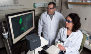
Using strands of nucleic acid, scientists have demonstrated basic computing operations inside a living mammalian cell. Shown examining a cellular “AND” gate are associate professor Philip Santangelo and research scientist Chiara Zurla. Photo Credit: Rob Felt, Georgia Tech

The research uses DNA strand displacement, a technology that has been widely used outside of cells for the design of molecular circuits, motors and sensors. Researchers modified the process to provide both “AND” and “OR” logic gates able to operate inside the living cells and interact with native messenger RNA (mRNA).

The tools they developed could provide a foundation for bio-computers able to sense, analyze and modulate molecular information at the cellular level. Supported by the Defense Advanced Research Projects Agency (DARPA) and the National Science Foundation (NSF), the research was reported December 21 in the journal Nature Nanotechnology.

“The whole idea is to be able to take the logic that is used in computers and port that logic into cells themselves,” said Philip Santangelo, an associate professor in the Wallace H. Coulter Department of Biomedical Engineering at Georgia Tech and Emory University. “These devices could sense an aberrant RNA, for instance, and then shut down cellular translation or induce cell death.”

Strand displacement reactions are the biological equivalent of the switches or gates that form the foundation for silicon-based computing. They can be programmed to turn on or off in response to an external stimuli such as a molecule. An “AND” gate, for example, would switch when both conditions were met, while an “OR” gate would switch when either condition was met.

In the switches the researchers used, a fluorophore reporter molecule and its complementary quenching molecule were placed side-by-side to create an “off” mode. Binding of RNA in one of the strands then displaced a portion of nucleic acid, separating the molecules and allowing generation of a signal that created an “on” mode. Two “on” modes on adjacent nucleic acid strands created an “AND” gate.

“Demonstrating individual logic gates is only a first step,” said Georg Seelig, assistant professor of computer science and engineering and electrical engineering at the University of Washington. “In the longer term, we want to expand this technology to create circuits with many inputs, such as those we have constructed in cell-free settings.”

The researchers used ligands designed to bind to specific portions of the nucleic acid strands, which can be created as desired and produced by commercial suppliers.

“We sensed molecules and showed that we could respond to them,” said Santangelo. “We showed that we could utilize native molecules in the cell as part of the circuit, though we haven’t been able to control a cell yet.”

Getting basic computing operations to function inside cells was no easy task, and the research required a number of years to accomplish. Among the challenges were getting the devices into the cells without triggering the switches, providing operation rapid enough to be useful, and not killing the human cell lines that researchers used in the lab.

“We had to chemically change the probes to get them to work inside the cell and to make them stable enough inside the cells,” said Santangelo. “We found that these strand displacement reactions can be slow within the cytosol, so to get them to work faster, we built scaffolding onto the messenger RNA that allowed us to amplify the effects.”

The nucleic acid computers ultimately operated as desired, and the next step is to use their switching to trigger the production of signaling chemicals that would prompt the desired reaction from the cells. Cellular activity is normally controlled by the production of proteins, so the nucleic acid switches will have to be given the ability to produce enough signaling molecules to induce a change.

“We need to generate enough of whatever final signal is needed to get the cell to react,” Santangelo explained. “There are amplification methods used in strand displacement technology, but none of them have been used so far in living cells.”

Even without that final step, the researchers feel they’ve built a foundation that can be used to attain the goal.

“We were able to design some of the basic logical constructs that could be used as building blocks for future work,” Santangelo said. “We know the concentrations of chemicals and the design requirements for individual components, so we can now start putting together a more complicated set of circuits and components.”

Cells, of course, already know how to sense toxic molecules and the development malignant tendencies, and to then take action. But those safeguards can be turned off by viruses or cancer cells that know how to circumvent natural cellular processes.

“Our mechanism would just give cells a hand at doing this,” Santangelo said. “The idea is to add to the existing machinery to give the cells enhanced capabilities.”

Applying an engineering approach to the biological world sets this example apart from other efforts to control cellular machinery.

“What makes DNA strand displacement circuits unique is that all components are fully rationally designed at the level of the DNA sequence,” said Seelig. “This really makes this technology ideal for an engineering approach. In contrast, many other approaches to controlling the cellular machinery rely on components that are borrowed from biology and are not fully understood.”

###

Beyond those already mentioned, the research team included Benjamin Groves, Yuan-Jyue Chen and Sergii Pochekailov from the University of Washington and Chiara Zurla and Jonathan Kirschman from Georgia Tech and Emory University.

This material is based on work supported by the Defense Advanced Research Projects Agency (DARPA) under contract W911NF-11-2-0068 and by National Science Foundation CAREER award 1253691. The content is solely the responsibility of the authors and does not necessarily represent the official views of DARPA or the NSF.

CITATION: Benjamin Groves, et al., “Computing in mammalian cells with nucleic acid strand exchange,” (Nature Nanotechnology, 2015). http://dx.doi.org/10.1038/nnano.2015.278

Media Contact

John Toon
jtoon@gatech.edu
404-894-6986
@GeorgiaTech

http://www.gatech.edu

18 Ocak 2016 Pazartesi

Biologists unravel drug-resistance mechanism in tumor cells

from
BIOENGINEER.ORG http://bioengineer.org/biologists-unravel-drug-resistance-mechanism-tumor-cells/

About half of all tumors are missing a gene called p53, which helps healthy cells prevent genetic mutations. Many of these tumors develop resistance to chemotherapy drugs that kill cells by damaging their DNA.

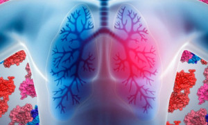
lung 1 Biologists unravel drug-resistance mechanism in tumor cells lung 1

P53, which helps healthy cells prevent genetic mutations, is missing from about half of all tumors. Researchers have found that a backup system takes over when p53 is disabled and encourages cancer cells to continue dividing. In the background of this illustration are crystal structures of p53 DNA-binding domains. Photo Credit: Jose-Luis Olivares/MIT (p53 illustration by Richard Wheeler/Wikimedia Commons)

MIT cancer biologists have now discovered how this happens: A backup system that takes over when p53 is disabled encourages cancer cells to continue dividing even when they have suffered extensive DNA damage. The researchers also discovered that an RNA-binding protein called hnRNPA0 is a key player in this pathway.

“I would argue that this particular RNA-binding protein is really what makes tumor cells resistant to being killed by chemotherapy when p53 is not around,” says Michael Yaffe, the David H. Koch Professor in Science, a member of the Koch Institute for Integrative Cancer Research, and the senior author of the study, which appears in the Oct. 22 issue of Cancer Cell.

The findings suggest that shutting off this backup system could make p53-deficient tumors much more susceptible to chemotherapy. It may also be possible to predict which patients are most likely to benefit from chemotherapy and which will not, by measuring how active this system is in patients’ tumors.

Rewired for resistance

In healthy cells, p53 oversees the cell division process, halting division if necessary to repair damaged DNA. If the damage is too great, p53 induces the cell to undergo programmed cell death.

In many cancer cells, if p53 is lost, cells undergo a rewiring process in which a backup system, known as the MK2 pathway, takes over part of p53’s function. The MK2 pathway allows cells to repair DNA damage and continue dividing, but does not force cells to undergo cell suicide if the damage is too great. This allows cancer cells to continue growing unchecked after chemotherapy treatment.

“It only rescues the bad parts of p53’s function, but it doesn’t rescue the part of p53’s function that you would want, which is killing the tumor cells,” says Yaffe, who first discovered this backup system in 2013.

In the new study, the researchers delved further into the pathway and found that the MK2 protein exerts control by activating the hnRNPA0 RNA-binding protein.

RNA-binding proteins are proteins that bind to RNA and help control many aspects of gene expression. For example, some RNA-binding proteins bind to messenger RNA (mRNA), which carries genetic information copied from DNA. This binding stabilizes the mRNA and helps it stick around longer so the protein it codes for will be produced in larger quantities.

“RNA-binding proteins, as a class, are becoming more appreciated as something that’s important for response to cancer therapy. But the mechanistic details of how those function at the molecular level are not known at all, apart from this one,” says Ian Cannell, a research scientist at the Koch Institute and the lead author of the Cancer Cell paper.

In this paper, Cannell found that hnRNPA0 takes charge at two different checkpoints in the cell division process. In healthy cells, these checkpoints allow the cell to pause to repair genetic abnormalities that may have been introduced during the copying of chromosomes.

One of these checkpoints, known as G2/M, is controlled by a protein called Gadd45, which is normally activated by p53. In lung cancer cells without p53, hnRNPA0 stabilizes mRNA coding for Gadd45. At another checkpoint called G1/S, p53 normally turns on a protein called p21. When p53 is missing, hnRNPA0 stabilizes mRNA for a protein called p27, a backup to p21. Together, Gadd45 and p27 help cancer cells to pause the cell cycle and repair DNA so they can continue dividing.

Personalized medicine

The researchers also found that measuring the levels of mRNA for Gadd45 and p27 could help predict patients’ response to chemotherapy. In a clinical trial of patients with stage 2 lung tumors, they found that patients who responded best had low levels of both of those mRNAs. Those with high levels did not benefit from chemotherapy.

“You could measure the RNAs that this pathway controls, in patient samples, and use that as a surrogate for the presence or absence of this pathway,” Yaffe says. “In this trial, it was very good at predicting which patients responded to chemotherapy and which patients didn’t.”

“The most exciting thing about this study is that it not only fills in gaps in our understanding of how p53-deficient lung cancer cells become resistant to chemotherapy, it also identifies actionable events to target and could help us to identify which patients will respond best to cisplatin, which is a very toxic and harsh drug,” says Daniel Durocher, a senior investigator at the Samuel Lunenfeld Research Institute of Mount Sinai Hospital in Toronto, who was not part of the research team.

The MK2 pathway could also be a good target for new drugs that could make tumors more susceptible to DNA-damaging chemotherapy drugs. Yaffe’s lab is now testing potential drugs in mice, including nanoparticle-based sponges that would soak up all of the RNA binding protein so it could no longer promote cell survival.

This work was supported in part by the Charles and Marjorie Holloway Foundation.

Story Source:

The above post is reprinted from materials provided by MIT NEWS

17 Ocak 2016 Pazar

Organ-on-a-chip

from
BIOENGINEER.ORG http://bioengineer.org/organ-on-a-chip/

This image demonstrates tissue patterns that emerge from genetically programmed human pluripotent stem cells, and was taken on day nine of the MIT study. It shows immunostainings of cell nuclei for CEBPA (red, endodermal marker), SOX10 (green, ectodermal marker), and DAPI (blue, binds to DNA in the nucleus). Organ-on-a-chip MIT programmable organoids 1 0 1 1

This image demonstrates tissue patterns that emerge from genetically programmed human pluripotent stem cells, and was taken on day nine of the MIT study. It shows immunostainings of cell nuclei for CEBPA (red, endodermal marker), SOX10 (green, ectodermal marker), and DAPI (blue, binds to DNA in the nucleus).

Courtesy of the researchers

A new technique for programming human stem cells to produce different types of tissue on demand may ultimately allow personalized organs to be grown for transplant patients.

The technique, which also has near-term implications for growing organ-like tissues on a chip, was developed by researchers at MIT and is unveiled in a study published today in the journal Nature Communications.

Growing organs on demand, using stem cells derived from patients themselves, could eliminate the lengthy wait that people in need of a transplant are often forced to endure before one becomes available.

It could also reduce the risk of a patient’s immune system rejecting the transplant, since the tissue would be grown from the patient’s own cells, according to Ron Weiss, professor of biological engineering at MIT, who led the research.

“Imagine that there is a patient with liver complications,” Weiss says. “We could take skin cells from that person and then [convert] them into stem cells, and then genetically program them to make the liver tissue, and transplant that into the patient.”

A rudimentary organ

The researchers developed the new technique while investigating whether they could use stem cells to produce pancreatic beta cells for treating patients with diabetes.

In order to do this, the researchers needed to devise a means to convert stem cells into pancreatic beta cells on demand.

As a first step in this process, they took human induced pluripotent stem (IPS) cells — stem cells generated from adult fibroblast, or skin cells — and converted them into “endoderm,” one of the three primary cell types in a developing organism. Endoderm, mesoderm, and ectoderm make up the three so-called germ layers that contribute to nearly all of the different cell types in the body. “They are the first real step of [cell] differentiation,” Weiss says.

The researchers developed a method to use a type of small molecule called dox to induce the IPS cells to express a protein known as GATA6. This protein can convert IPS cells into endoderm.

Rather than immediately attempting to convert these endoderm cells into pancreatic cells though, the paper’s lead author, Patrick Guye, a former postdoc in Weiss’ lab and currently laboratory head with Sanofi-Aventis in Frankfurt, Germany, then decided to allow the cells to continue growing, to monitor their progress.

After two weeks, the researchers found that the endoderm, and some mesoderm also present in the cell culture, had matured further, to form a liver “bud,” or small, rudimentary liver.

“We observed the development of many cells types found in the fetal liver, including the development of blood vessel-like networks, various mesenchymal precursors, and the formation of early red and white blood cells within our liver-like tissue,” Guye says. “This is especially exciting, as the process looks very similar if not identical to what is happening in the early liver bud in vivo, that is, in our own development.”

What’s more, the researchers discovered that only those IPS cells that had been exposed to more of the genetic programming, and had therefore gone on to produce more GATA6, became liver tissue. Alongside these were IPS cells that did not make much GATA6, which went on to form ectoderm instead, and then further matured to become early telencephalon, or forebrain.

By controlling how much GATA6 the cells expressed, the researchers were able to determine how much liver bud and how much forebrain tissue was generated, Weiss says.

This suggests that the technique could be used to produce not just individual tissue types, but different combinations of tissue, he says.

“The fact that we are able to produce endoderm, mesoderm, and ectoderm gives us great hope that we can take each of these germ layers and hopefully grow any kind of tissue we want,” he says.

Liver-on-a-chip

While it is likely to be some time before the technique can be used to generate transplant organs, it could be used almost immediately to grow different human tissue on which to test new drugs, Weiss says.

Using human stem cell-derived organ tissue to test new treatments could be far more reliable than testing on animals, since different species may react differently to a drug, he says.

The technique could also allow clinicians to carry out patient-specific drug testing. “If you are not sure whether you will have complications from taking a particular drug, then before you take it you could try it out on your own liver-on-a-chip,” Weiss says.

Similarly, the organ-on-a-chip could be used to monitor the interaction between different drugs that people may be taking.

“As people age, some are taking 10, 15, or 20 drugs together, and it’s impossible for the pharmaceutical companies to test all of these combinations for every individual. But we would be able to test that out,” he says. “That is something that can be done now.”

In addition to these therapeutic applications, the technique could allow researchers to gain a better understanding of the development of different types of tissue, such as the liver and neurons.

The paper reveals some intrinsic mechanisms underlying the interactions of stem cells during liver development, and provides a useful model that sheds light on the complex process of embryogenesis, says Bing Song, a professor of tissue engineering at Cardiff University in the UK, who was not involved in the research.

"In my field, which is combining genetically modified stem cells and physical stimulation (electrical and magnetic) to cure spinal cord injuries and degenerative disease, the paper has given me some very useful ideas," he says.

The researchers now hope to investigate whether they can use the technique to grow other organs on demand, such as a pancreas.

Story Source:

The above post is reprinted from materials provided by MIT NEWS

Related

New system for human genome editing has potential to increase power and precision of DNA engineering

from
BIOENGINEER.ORG http://bioengineer.org/new-system-for-human-genome-editing-has-potential-to-increase-power-and-precision-of-dna-engineering/

CRISPR systems are found in many different bacterial species, and have evolved to protect host cells against infection by viruses. New system for human genome editing has potential to increase power and precision of DNA engineering MIT Crispr cpf1 0 1 1

CRISPR systems are found in many different bacterial species, and have evolved to protect host cells against infection by viruses.

Image courtesy of Broad Institute/Science Photo Images

A team including the scientist who first harnessed the CRISPR-Cas9 system for mammalian genome editing has now identified a different CRISPR system with the potential for even simpler and more precise genome engineering.

In a study published today in Cell, Feng Zhang and his colleagues at the Broad Institute of MIT and Harvard and the McGovern Institute for Brain Research at MIT, with co-authors Eugene Koonin at the National Institutes of Health, Aviv Regev of the Broad Institute and the MIT Department of Biology, and John van der Oost at Wageningen University, describe the unexpected biological features of this new system and demonstrate that it can be engineered to edit the genomes of human cells.

“This has dramatic potential to advance genetic engineering,” says Eric Lander, director of the Broad Institute. “The paper not only reveals the function of a previously uncharacterized CRISPR system, but also shows that Cpf1 can be harnessed for human genome editing and has remarkable and powerful features. The Cpf1 system represents a new generation of genome editing technology.”

CRISPR sequences were first described in 1987, and their natural biological function was initially described in 2010 and 2011. The application of the CRISPR-Cas9 system for mammalian genome editing was first reported in 2013, by Zhang and separately by George Church at Harvard University.

In the new study, Zhang and his collaborators searched through hundreds of CRISPR systems in different types of bacteria, searching for enzymes with useful properties that could be engineered for use in human cells. Two promising candidates were the Cpf1 enzymes from bacterial species Acidaminococcus and Lachnospiraceae, which Zhang and his colleagues then showed can target genomic loci in human cells.

“We were thrilled to discover completely different CRISPR enzymes that can be harnessed for advancing research and human health,” says Zhang, the W.M. Keck Assistant Professor in Biomedical Engineering in MIT’s Department of Brain and Cognitive Sciences.

The newly described Cpf1 system differs in several important ways from the previously described Cas9, with significant implications for research and therapeutics, as well as for business and intellectual property:

  • First: In its natural form, the DNA-cutting enzyme Cas9 forms a complex with two small RNAs, both of which are required for the cutting activity. The Cpf1 system is simpler in that it requires only a single RNA. The Cpf1 enzyme is also smaller than the standard SpCas9, making it easier to deliver into cells and tissues.
  • Second, and perhaps most significantly: Cpf1 cuts DNA in a different manner than Cas9. When the Cas9 complex cuts DNA, it cuts both strands at the same place, leaving “blunt ends” that often undergo mutations as they are rejoined. With the Cpf1 complex the cuts in the two strands are offset, leaving short overhangs on the exposed ends. This is expected to help with precise insertion, allowing researchers to integrate a piece of DNA more efficiently and accurately.
  • Third: Cpf1 cuts far away from the recognition site, meaning that even if the targeted gene becomes mutated at the cut site, it can likely still be recut, allowing multiple opportunities for correct editing to occur.
  • Fourth: The Cpf1 system provides new flexibility in choosing target sites. Like Cas9, the Cpf1 complex must first attach to a short sequence known as a PAM, and targets must be chosen that are adjacent to naturally occurring PAM sequences. The Cpf1 complex recognizes very different PAM sequences from those of Cas9. This could be an advantage in targeting some genomes, such as in the malaria parasite as well as in humans.

“The unexpected properties of Cpf1 and more precise editing open the door to all sorts of applications, including in cancer research,” says Levi Garraway, an institute member of the Broad Institute, and the inaugural director of the Joint Center for Cancer Precision Medicine at the Dana-Farber Cancer Institute, Brigham and Women’s Hospital, and the Broad Institute. Garraway was not involved in the research.

An open approach to empower research

Zhang, along with the Broad Institute and MIT, plan to share the Cpf1 system widely. As with earlier Cas9 tools, these groups will make this technology freely available for academic research via the Zhang lab’s page on the plasmid-sharing website Addgene, through which the Zhang lab has already shared Cas9 reagents more than 23,000 times with researchers worldwide to accelerate research. The Zhang lab also offers free online tools and resources for researchers through its website.

The Broad Institute and MIT plan to offer nonexclusive licenses to enable commercial tool and service providers to add this enzyme to their CRISPR pipeline and services, further ensuring availability of this new enzyme to empower research. These groups plan to offer licenses that best support rapid and safe development for appropriate and important therapeutic uses.

“We are committed to making the CRISPR-Cpf1 technology widely accessible,” Zhang says. “Our goal is to develop tools that can accelerate research and eventually lead to new therapeutic applications. We see much more to come, even beyond Cpf1 and Cas9, with other enzymes that may be repurposed for further genome editing advances.”

Story Source:

The above post is reprinted from materials provided by MIT NEWS

Monitoring the rise and fall of the microbiome

from
BIOENGINEER.ORG http://bioengineer.org/monitoring-the-rise-and-fall-of-the-microbiome/

The bacterium, Enterococcus faecalis, which lives in the human gut, is just one microbe type that will be studied as part of NIH's Human Microbiome Project Monitoring the rise and fall of the microbiome MIT Gut Bacteria 1 1

The bacterium, Enterococcus faecalis, which lives in the human gut, is just one microbe type that will be studied as part of NIH's Human Microbiome Project

Courtesy of the Centers for Disease Control and Prevention

Trillions of bacteria live in each person’s digestive tract. Scientists believe that some of these bacteria help digest food and stave off harmful infections, but their role in human health is not well understood.

To help shed light on the role of these bacteria, a team of researchers led by MIT associate professor Eric Alm recently tracked fluctuations in the bacterial populations of two research subjects over a full year. The findings, described in the July 25 issue of the journal Genome Biology, suggest that while these populations are fairly stable, they undergo daily fluctuations in response to changes in diet and other factors.

“On any given day, the amount of one species could change manyfold, but after a year, that species would still be at the same median level,” says Alm, the Karl Van Tassel Career Development Associate Professor of Biological and Environmental Engineering and senior author of the paper. “To a large extent, the main factor we found that explained a lot of that variance was the diet.”

Alm and Lawrence David, an assistant professor at Duke University and the paper’s lead author, began the study in 2009, around the same time the National Institutes of Health launched the Human Microbiome Project — an effort to analyze the composition of bacterial communities found in humans, including the gut but also other organs such as the skin, nasal passages, and mouth.

There are a few thousand strains of bacteria that can inhabit the human gut, but only a few hundred of those are found in any given individual, Alm says. For one year, the two subjects in the study collected daily stool samples so bacterial populations could be measured. They also used an iPhone app to track lifestyle factors such as diet, sleep, mood, and exercise, generating a huge amount of data.

Analysis of this data revealed that dietary changes could produce daily variations in the populations of different strains of bacteria. For example, an increase in fiber correlated with a boost in the populations of Bifidobacteria, Roseburia, and Eubacterium rectale. Four strains — including Faecalibacterium prausnitzii, which has been implicated in protecting against inflammatory bowel disease — were correlated with eating citrus.

During the study, each of the two subjects experienced an event that dramatically altered the gut microbiome. Subject B experienced food poisoning caused by Salmonella, and Subject A traveled to a developing nation, where he experienced diarrheal illness for two weeks.

During Subject B’s infection, Salmonella leapt from 10 percent of the gut microbiome to nearly 30 percent. At the same time, populations of bacteria from the phylum Firmicutes, believed to be beneficial to human health, nearly disappeared. After the subject recovered, Firmicutes rebounded to about 40 percent of the total microbiome, but most of the strains were different from those originally present.

“There really wasn’t an overall change in the level of Firmicutes. The levels are very different from person to person, but whatever it is about a person that is maintaining a certain level is able to survive a huge insult like that. What’s keeping it at that level is a really interesting research question,” Alm says, adding that the answer may lie in dietary differences.

Subject A also exhibited severe disruptions to his microbiome during his trip, but once he returned to the United States, it returned to normal. Unlike Subject B’s recovery from food poisoning, Subject A’s populations returned to their original composition.

“This is a fundamental manuscript that defines a very important component of ‘microbiome law,’ namely that the microbial profile of individuals is both stable over very long timeframes, and unique to the individual over that timeframe,” says Jack Gilbert, an associate professor of ecology and evolution at the University of Chicago. “This is not the first time these trends have been shown, but the scale of detail in this paper makes these trends incontrovertible.”

The researchers now plan to investigate why microbiome populations tend to return to an average value, specific to the individual host, after going way up or down. They also plan to study how the immune system responds to changes in the microbiome by measuring daily changes in cytokine and hormone levels. To make such follow-up studies easier to do, they are also working on wearable sensors that would collect much of the needed data with less effort required from the subjects.

The ultimate goal, Alm says, is to generate data from individual patients that could be analyzed to produce a personalized monitoring system for people suffering from inflammatory bowel disease or other diseases prone to occasional flare-ups. Such a system could detect when someone is heading for a flare-up and recommend dietary changes that could help avoid it.

The research was funded by the National Science Foundation, the MIT Whitaker Health Sciences Fund, and the Crohn’s and Colitis Foundation of America.

Story Source:

The above post is reprinted from materials provided by MIT NEWS

Bone marrow lesions can help predict rapidly progressing joint disease

from
BIOENGINEER.ORG http://bioengineer.org/bone-marrow-lesions-can-help-predict-rapidly-progressing-joint-disease/

A new study from the Medical Research Council Lifecourse Epidemiology Unit, University of Southampton, shows lesions, which can best be seen on MRI scans, could help identify individuals who are more likely to suffer from more rapidly progressing osteoarthritis.

Osteoarthritis is the most common type of arthritis in the UK and can cause the joints to become painful and stiff. Almost any joint can be affected, but it most often causes problems in the knees, hips, and small joints of the hands. It can progress at varying speeds.

The SEKOIA study, a major international osteoarthritis disease-modifying trial, carried out MRI scanning on the knees of 176 men and women over 50 years old. They were then followed up for an average of three years with repeated knee x-rays. Individuals with abnormalities on their MRI scans at the first appointment were compared to those without to examine the effect on disease progression.

Individuals with bone marrow lesions (BMLs) on their MRI scan were found to have osteoarthritis that progressed more rapidly than those that did not. On average, the space within the joint is lost at a rate of 0.15mm per year however the Southampton study shows that, overall, individuals with BMLs had a loss rate that was 0.10mm per year faster than those without BMLs. This may lead to earlier need for joint replacement or other intervention.

BMLs show up on MRI as regions of bone beneath the cartilage with ill-defined high signal and represent areas of bone marrow oedema, fibrosis, and necrosis. The Southampton researchers believe that therapies to target these abnormalities may slow the progression of this disabling joint disease, but further work is required to examine this.

Dr Mark Edwards, Clinical Lecturer in Rheumatology at the MRC Lifecourse Epidemiology Unit, University of Southampton, led the study which has been published in The Journal of Rheumatology.

He comments: "Osteoarthritis causes a significant burden to individuals and the healthcare system as a whole. If we can identify those people who may experience a rapid progression of the disease, this may be of benefit to both physicians and patients. The next step would be to explore the mechanisms through which bone marrow lesions might influence the progression of osteoarthritis and whether this could lead to a novel treatment."

Professor Cyrus Cooper, Professor of Rheumatology and Director of the MRC Lifecourse Epidemiology Unit, University of Southampton adds: "This study points to the utility of data derived from large randomised controlled trials in deriving predictive models which will facilitate a stratified approach to therapy in knee osteoarthritis, the commonest cause of arthritis worldwide."

###

Media Contact

Becky Attwood
r.attwood@soton.ac.uk
44-023-805-92128
@unisouthampton

http://www.southampton.ac.uk/

Mentally challenging activities key to a healthy aging mind

from
BIOENGINEER.ORG http://bioengineer.org/mentally-challenging-activities-key-to-a-healthy-aging-mind/

One of the greatest challenges associated with the growing numbers of aged adults is how to maintain a healthy aging mind. Taking up a new mental challenge such as digital photography or quilting may help maintain cognitive vitality, say researchers reporting in Restorative Neurology and Neuroscience.

Recent evidence suggests that engaging in enjoyable and enriching lifestyle activities may be associated with maintaining cognitive vitality. However, the underlying mechanism accounting for cognitive enhancement effects have been poorly understood.

Investigators at the University of Texas at Dallas proposed that only tasks that involved sustained mental effort and challenge would facilitate cognitive function. Senior author Denise Park and lead author Ian McDonough compared changes in brain activity in 39 older adults that resulted from the performance of high-challenge activities that required new learning and sustained mental effort compared to low-challenge activities that did not require active learning. All of the participants underwent a battery of cognitive tests and brain scans using functional magnetic resonance imaging (fMRI), an MRI technology that measures brain activity by detecting changes associated with blood flow.

Participants were randomly assigned to the high-challenge, low-challenge, or placebo groups. The high-challenge group spent at least 15 hours per week for 14 weeks learning progressively more difficult skills in digital photography, quilting, or a combination of both. The low-challenge group met for 15 hours per week to socialize and engage in activities related to subjects such as travel and cooking with no active learning component. The placebo group engaged in low-demand cognitive tasks such as listening to music, playing simple games, or watching classic movies. All participants were tested before and after the 14-week period and a subset was retested a year later.

The high-challenge group demonstrated better memory performance after the intervention, and an increased ability to modulate brain activity more efficiently to challenging judgments of word meaning in the medial frontal, lateral temporal, and parietal cortex regions of the brain. These are brain areas associated with attention and semantic processing. Some of this enhanced brain activity was maintained a year later. This increased neural efficiency in judging words was demonstrated by participants showing lowered brain activity when word judgments were easy and increasing activity when they became hard. This is a pattern of response typical of young adults. Before participating in the high-challenge intervention, the older adults were processing every item, both easy and hard, with maximum brain activity. After participation, they were able to modulate their brain activity to the demands of the task, thus showing a more efficient use of neural resources. This change in modulation was not observed in the low-challenge group.

The findings show that mentally demanding activities may be neuroprotective and an important element for maintaining a healthy brain into late adulthood.

"The present findings provide some of the first experimental evidence that mentally-challenging leisure activities can actually change brain function and that it is possible that such interventions can restore levels of brain activity to a more youth-like state. However, we would like to conduct much larger studies to determine the universality of this effect and understand who will benefit the most from such an intervention," explained senior author Denise C. Park, PhD, of the Center for Vital Longevity, School of Behavioral and Brain Sciences, University of Texas at Dallas.

Ian McDonough, who is now an assistant professor of Psychology at the University of Alabama and was first author on the study, said: "The study clearly illustrates that the enhanced neural efficiency was a direct consequence of participation in a demanding learning environment. The findings superficially confirm the familiar adage regarding cognitive aging of 'Use it or lose it.'"

Denise Park added, "Although there is much more to be learned, we are cautiously optimistic that age-related cognitive declines can be slowed or even partially restored if individuals are exposed to sustained, mentally challenging experiences."

###

Media Contact

Daphne Watrin
d.watrin@iospress.nl
31-206-883-355
@IOSPress_STM

http://www.iospress.com

CU researchers study hospital readmissions from post-acute care facilities

from
BIOENGINEER.ORG http://bioengineer.org/cu-researchers-study-hospital-readmissions-from-post-acute-care-facilities/

AURORA, Colo. (Jan. 15, 2016) – Better coordination between hospitals and post-acute care facilities could reduce patient readmission to hospitals and mortality rates, according to a new study of risk factors by researchers from the University of Colorado School of Medicine.

In a review of more than 3,200 hospitalizations followed by stays in post-acute care facilities, the researchers found specific risk factors that may contribute to the need for readmission to the hospital. Nearly half of the readmissions occurred within 14 days of being released from the hospital.

The study, published online in JAMDA, the Journal of Post-Acute and Long Term Care Medicine, identified the patient's need for an invasive device, such as a feeding tube or urinary catheter, and the patient's need for advanced care, such as dialysis and oxygen therapy, as factors more common in readmitted patents.

The causes of hospital readmission from post-acute care facilities, which are also called skilled nursing facilities, are critical areas to study in order to improve the quality of patient care and to prepare for reimbursement models that penalize hospitals if patients are readmitted.

"Patients who experienced readmission during their stay in a post-acute care facility were less likely to return to the community," said lead author Robert Burke, MD, academic hospitalist and health services researcher at the Denver VA Medical Center and an assistant professor at the CU School of Medicine.

Readmitted patients had a higher mortality rate too.

"Readmitted patients were twice as likely as non-readmitted patients to die in the 30 days following hospital discharge and nearly four times as likely to die in the 100 days post-hospital discharge," the authors write.

The authors also found that payment systems matter and affect patient outcomes.

"Under a prospective payment system, hospitals are incentivized to discharge these patients as early as possible, and in contrast to discharges home, hospitals are not currently penalized for readmissions from PAC (post-acute care) facilities," the authors write. "PAC facilities may be substituting for prolonged hospital care in some cases."

Hospitals and post-acute care facilities need to focus on patient selection and on processes for transitioning care from the hospital to the post-acute care facility.

###

Burke's fellow authors on the article are Emily Whitfield, PhD, of the Denver-Seattle Center of Innovation at the Denver VA Medical Center; and David Hittle, PhD, Sung-Joon Min, PhD, Cari Levy, MD, PhD, Allan V. Prochazka, MD, MSc, Eric A. Coleman, MD, MPH, Robert Schwartz, MD, and Adit A. Ginde, MD, MPH, all of whom are faculty members of the University of Colorado School of Medicine.

The research was funded by the Hartford Foundation/Jahnigen Center of Excellence at the University of Colorado.

About the University of Colorado School of Medicine

Faculty at the University of Colorado School of Medicine work to advance science and improve care. These faculty members include physicians, educators and scientists at University of Colorado Health, Children's Hospital Colorado, Denver Health, National Jewish Health, and the Denver Veterans Affairs Medical Center. The school is located on the Anschutz Medical Campus, one of four campuses in the University of Colorado system. To learn more about the medical school's care, education, research and community engagement, visit its web site.

Media Contact

Mark Couch
mark.couch@ucdenver.edu
303-724-5377
@CUAnschutz

http://www.ucdenver.edu

UT Southwestern researchers identify process that causes chronic neonatal lung disease

Engineers design ‘living materials’

from
BIOENGINEER.ORG http://bioengineer.org/engineers-design-living-materials/

Inspired by natural materials such as bone — a matrix of minerals and other substances, including living cells — MIT engineers have coaxed bacterial cells to produce biofilms that can incorporate nonliving materials, such as gold nanoparticles and quantum dots.

engineers Engineers design ‘living materials’ engineers

An artist’s rendering of a bacterial cell engineered to produce amyloid nanofibers that incorporate particles such as quantum dots (red and green spheres) or gold nanoparticles. Photo Credit: Yan Liang

These “living materials” combine the advantages of live cells, which respond to their environment, produce complex biological molecules, and span multiple length scales, with the benefits of nonliving materials, which add functions such as conducting electricity or emitting light.

The new materials represent a simple demonstration of the power of this approach, which could one day be used to design more complex devices such as solar cells, self-healing materials, or diagnostic sensors, says Timothy Lu, an assistant professor of electrical engineering and biological engineering. Lu is the senior author of a paper describing the living functional materials in the March 23 issue of Nature Materials.

“Our idea is to put the living and the nonliving worlds together to make hybrid materials that have living cells in them and are functional,” Lu says. “It’s an interesting way of thinking about materials synthesis, which is very different from what people do now, which is usually a top-down approach.”

The paper’s lead author is Allen Chen, an MIT-Harvard MD-PhD student. Other authors are postdocs Zhengtao Deng, Amanda Billings, Urartu Seker, and Bijan Zakeri; recent MIT graduate Michelle Lu; and graduate student Robert Citorik.

Self-assembling materials

Lu and his colleagues chose to work with the bacterium E. coli because it naturally produces biofilms that contain so-called “curli fibers” — amyloid proteins that help E. coli attach to surfaces. Each curli fiber is made from a repeating chain of identical protein subunits called CsgA, which can be modified by adding protein fragments called peptides. These peptides can capture nonliving materials such as gold nanoparticles, incorporating them into the biofilms.

By programming cells to produce different types of curli fibers under certain conditions, the researchers were able to control the biofilms’ properties and create gold nanowires, conducting biofilms, and films studded with quantum dots, or tiny crystals that exhibit quantum mechanical properties. They also engineered the cells so they could communicate with each other and change the composition of the biofilm over time.

First, the MIT team disabled the bacterial cells’ natural ability to produce CsgA, then replaced it with an engineered genetic circuit that produces CsgA but only under certain conditions — specifically, when a molecule called AHL is present. This puts control of curli fiber production in the hands of the researchers, who can adjust the amount of AHL in the cells’ environment. When AHL is present, the cells secrete CsgA, which forms curli fibers that coalesce into a biofilm, coating the surface where the bacteria are growing.

The researchers then engineered E. coli cells to produce CsgA tagged with peptides composed of clusters of the amino acid histidine, but only when a molecule called aTc is present. The two types of engineered cells can be grown together in a colony, allowing researchers to control the material composition of the biofilm by varying the amounts of AHL and aTc in the environment. If both are present, the film will contain a mix of tagged and untagged fibers. If gold nanoparticles are added to the environment, the histidine tags will grab onto them, creating rows of gold nanowires, and a network that conducts electricity.

‘Cells that talk to each other’

The researchers also demonstrated that the cells can coordinate with each other to control the composition of the biofilm. They designed cells that produce untagged CsgA and also AHL, which then stimulates other cells to start producing histidine-tagged CsgA.

“It’s a really simple system but what happens over time is you get curli that’s increasingly labeled by gold particles. It shows that indeed you can make cells that talk to each other and they can change the composition of the material over time,” Lu says. “Ultimately, we hope to emulate how natural systems, like bone, form. No one tells bone what to do, but it generates a material in response to environmental signals.”

To add quantum dots to the curli fibers, the researchers engineered cells that produce curli fibers along with a different peptide tag, called SpyTag, which binds to quantum dots that are coated with SpyCatcher, a protein that is SpyTag’s partner. These cells can be grown along with the bacteria that produce histidine-tagged fibers, resulting in a material that contains both quantum dots and gold nanoparticles.

These hybrid materials could be worth exploring for use in energy applications such as batteries and solar cells, Lu says. The researchers are also interested in coating the biofilms with enzymes that catalyze the breakdown of cellulose, which could be useful for converting agricultural waste to biofuels. Other potential applications include diagnostic devices and scaffolds for tissue engineering.

“I think this is really fantastic work that represents a great integration of synthetic biology and materials engineering,” says Lingchong You, an associate professor of biomedical engineering at Duke University who was not part of the research team.

The research was funded by the Office of Naval Research, the Army Research Office, the National Science Foundation, the Hertz Foundation, the Department of Defense, the National Institutes of Health, and the Presidential Early Career Award for Scientists and Engineers.

Story Source:

The above post is reprinted from materials provided by MIT NEWS

4 Ocak 2016 Pazartesi

Researchers develop a new means of killing harmful bacteria

from
BIOENGINEER.ORG http://bioengineer.org/researchers-develop-new-means-killing-harmful-bacteria/

The global rise in antibiotic resistance is a growing threat to public health, damaging our ability to fight deadly infections such as tuberculosis.

bacteria Researchers develop a new means of killing harmful bacteria bacteria

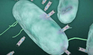
In this illustration, phagemid plasmids infect a targeted bacteria. Image: Christine Daniloff and Jose-Luis Olivares/MIT (plasmid illustration courtesy of the researchers)

What’s more, efforts to develop new antibiotics are not keeping pace with this growth in microbial resistance, resulting in a pressing need for new approaches to tackle bacterial infection.

In a paper published online in the journal Nano Letters, researchers at MIT, the Broad Institute of MIT and Harvard, and Harvard University reveal that they have developed a new means of killing harmful bacteria.

The researchers have engineered particles, known as “phagemids,” capable of producing toxins that are deadly to targeted bacteria.

Bacteriophages — viruses that infect and kill bacteria — have been used for many years to treat infection in countries such as those in the former Soviet Union. Unlike traditional broad-spectrum antibiotics, these viruses target specific bacteria without harming the body’s normal microflora.

But bacteriophages can also cause potentially harmful side effects, according to James Collins, the Termeer Professor of Medical Engineering and Science in MIT’s Department of Biological Engineering and Institute of Medical Engineering and Science, who led the research.

“Bacteriophages kill bacteria by lysing the cell, or causing it to burst,” Collins says. “But this is problematic, as it can lead to the release of nasty toxins from the cell.”

These toxins can lead to sepsis and even death in some cases, he says.

A gentler burst

In previous research, Collins and his colleagues engineered bacteriophages to express proteins that did not actually burst the cells, but instead increased the effectiveness of antibiotics when delivered at the same time.

To build on this earlier work, the researchers set out to develop a related technology that would target and kill specific bacteria, without bursting the cells and releasing their contents.

The researchers used synthetic biology techniques to develop a platform of particles called phagemids. These particles infect bacteria with small DNA molecules known as plasmids, which are able to replicate independently inside a host cell.

Once inside the cell, the plasmids are engineered to express different proteins or peptides — molecules made up of short chains of amino acids — that are toxic to the bacteria, Collins says.

“We systematically tested different antimicrobial peptides and bacterial toxins, and demonstrated that when you combine a number of these within the phagemids, you can kill the great majority of cells within a culture,” he says.

The expressed toxins are designed to disrupt different cellular processes, such as bacterial replication, causing the cell to die without bursting open.

Precise targeting

The phagemids will also only infect a specific species of bacteria, resulting in a highly targeted system, Collins says.

“You can use this to kill off very specific species of bacteria as part of an infection therapy, while sparing the rest of the microbiome,” he says.

When the researchers monitored the response of the bacteria to repeated reinfection with the phagemids, they did not witness signs of significant resistance to the particles. “This means you can do multiple rounds of delivery of the phagemids, in order to get a more effective therapy,” he says.

This is in contrast to repeated infection with bacteriophages, where the researchers found that the bacteria did develop resistance over time.

Although Collins acknowledges that bacteria will ultimately develop resistance to any stress that is placed upon them, the research suggests that it is likely to take them far longer to develop resistance to phagemids than to conventional bacteriophage therapy, he says.

A “cocktail” of different phagemids could be given to patients to treat an unclassified infection, in a similar way to the broad-spectrum antibiotics used today.

But they are more likely to be used in conjunction with rapid diagnostic tools, currently in development, which would allow physicians to treat specific infections, Collins says. “You would first run a fast diagnostic test to identify the bacteria your patient has, and then give the appropriate phagemid to kill off the pathogen,” he says.

The researchers are planning to expand their platform by developing a broader range of phagemids. They have so far experimented with a set of phagemids specific to E. coli, but now hope to create particles capable of killing off pathogens such as Clostridium difficile and the cholera-causing bacterium Vibrio cholerea.

The paper demonstrates that using synthetic biology to modify a gene in a phage to make it more toxic to a pathogen can lead to more effective antimicrobial particles than classical approaches, says Alfonso Jaramillo, a professor of synthetic biology at the University of Warwick in the U.K., who was not involved in the research.

“Combining synthetic genetic devices with phages as delivery vehicles allows a systematic approach to reprogram pathogenic bacteria for death,” Jaramillo explains. “The [researchers’] focus on nonreplicative phages is also very appropriate because those particles are more feasible for use in people, as they are not considered genetically modified organisms,” he says.

The researchers have created an improved form of phage therapy that may become the antibiotics of the future, he adds.

Story Source:

The above post is reprinted from materials provided by MIT NEWS

Scientists bioengineered behavior in ants by turning on genes in the brain

from
BIOENGINEER.ORG http://bioengineer.org/scientists-bioengineered-behavior-ants-turning-genes-brain/

Certain threats — such as starvation or an attack by enemies — turn on genes in carpenter ants that change their behavior in ways that help their colony survive, according to a study co-authored by NYU Langone researchers and published in the Jan. 1, 2016, edition of the journal Science. With related molecular pathways present in humans, the study may provide insights into mechanisms behind behavioral disorders.

Specifically, the research team found that compounds known to block the action of a group of enzymes, histone deacetylases (HDACs), activated genes that made one kind of carpenter ant worker behave like another, and without changing the instructions encoded in their genes.

“The study results are exciting because they show that behavior can be manipulated in social animals using compounds that bring about quick changes in the action of genes,” says Danny Reinberg, PhD, the Terry and Mel Karmazin Professor in the Department of Biochemistry and Molecular Pharmacology at NYU Langone, and a corresponding study author. “Changing the code in genes inherited from parents is slow and fraught with difficulties, but chemical, epigenetic changes that turn existing genes on or off may someday provide an agile way to reverse diseases with behavioral components.”

The newly published study is the result of an eight year collaboration among research teams led by Reinberg, Shelley Berger from the University of Pennsylvania, and Juergen Liebig from Arizona State University.

“Over millions of years of evolution, the same enzymes, including HDACs, have continued to play critical roles in the regulation of learning, memory and behavior,” says Reinberg, also an investigator for the Howard Hughes Medical Institute, which funded the work. “While no one is saying that ant behavior extends to humans, we believe, nonetheless, that this work promises to help guide the future use of HDAC inhibitors, which are already being studied as potential treatments for schizophrenia, depression, and neurodegenerative diseases.” Further investigation of HDAC inhibitors may also help to explain how the brain lays down nerve networks as memories form.

Epigenetic Changes Make Guards Act like Scouts

The current study results revolve around the organization of genes and how they are regulated. The blueprint for both ant and human bodies is encoded in genes, many of which hold the information needed to build proteins, the molecules that make up the body’s structures and signals.

Research in recent years has shown that the instructions coded in genes are just one part of a larger genetic machine. As a result, many mechanisms enable cells to put the same genes to several uses by switching them on or off in different situations. These epigenetic switches also enable an animal to adjust to environmental changes during its lifetime, and instead of waiting for evolutionary changes to occur in genes over many generations.

In the species Camponotus floridanus, more commonly known as the Florida carpenter ant, the research team found that epigenetic regulation is key to determining whether any young female carpenter ant matures into a muscular ‘major’ charged with protecting the colony or a smaller ‘minor’ that scouts for food.

These two ‘castes’ have the same genes, but develop early in adulthood to assume different roles.

Specifically, the study found that foraging behavior as a caste-specific trait in the ant C. floridanus is controlled by the interplay between well-known families of enzymes: histone acetyltransferases (HATs), and histone deacetylases (HDACs). As their names suggest, HAT enzymes attach acetyl groups to histones, protein spools that DNA is wrapped around, to turn on genes. HDACs remove the groups from histones to turn off gene expression.

Using HDAC inhibitors, researchers were able to program ants to make those in the major caste start acting like minors, increasing their likelihood to go searching for food in a HAT-dependent manner. In ants that scouted more thanks to a diet of HDAC inhibitors, changes were detected in the action of hundreds of genes in the central ant brain linked to hormone signaling, the sending of signals along nerve pathways, and the building of connections between nerve cells.

To examine caste-based behavioral change, the researchers examined scouting behaviors after feeding workers a small-molecule HDAC inhibitor, valproic acid (VPA). As expected, VPA blocked HDAC activity to increase levels of acetylation at key histones, which caused VPA-treated minors and majors to do much more foraging than those not treated. The team concluded that HDAC normally inhibits foraging behavior in major workers, until blocked by naturally occurring regulatory mechanisms or related drugs.

Story Source:

The above post is reprinted from materials provided by NYU Langone.

3 Ocak 2016 Pazar

Biologists find an early sign of cancer

from
BIOENGINEER.ORG http://bioengineer.org/biologists-early-sign-cancer/

Years before they show any other signs of disease, pancreatic cancer patients have very high levels of certain amino acids in their bloodstream, according to a new study from MIT, Dana-Farber Cancer Institute, and the Broad Institute.

cancer sign Biologists find an early sign of cancer cancer sign

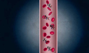
Years before they show any other signs of disease, pancreatic cancer patients have very high levels of certain amino acids in their bloodstream, according to a new study. Photo Credit: Christine Daniloff/MIT

This finding, which suggests that muscle tissue is broken down in the disease’s earliest stages, could offer new insights into developing early diagnostics for pancreatic cancer, which kills about 40,000 Americans every year and is usually not caught until it is too late to treat.

The study, which appears today in the journal Nature Medicine, is based on an analysis of blood samples from 1,500 people participating in long-term health studies. The researchers compared samples from people who were eventually diagnosed with pancreatic cancer and samples from those who were not. The findings were dramatic: People with a surge in amino acids known as branched chain amino acids were far more likely to be diagnosed with pancreatic cancer within one to 10 years.

“Pancreatic cancer, even at its very earliest stages, causes breakdown of body protein and deregulated metabolism. What that means for the tumor, and what that means for the health of the patient — those are long-term questions still to be answered,” says Matthew Vander Heiden, an associate professor of biology, a member of MIT’s Koch Institute for Integrative Cancer Research, and one of the paper’s senior authors.

The paper’s other senior author is Brian Wolpin, an assistant professor of medical oncology at Dana-Farber. Wolpin, a clinical epidemiologist, assembled the patient sample from several large public-health studies. All patients had their blood drawn when they began participating in the studies and subsequently filled out annual health questionnaires.

Working with researchers at the Broad Institute, the team analyzed blood samples for more than 100 different metabolites — molecules, such as proteins and sugars, produced as the byproducts of metabolic processes.

“What we found was that this really interesting signature fell out as predicting pancreatic cancer diagnosis, which was elevation in these three branched chain amino acids: leucine, isoleucine, and valine,” Vander Heiden says. These are among the 20 amino acids — the building blocks for proteins — normally found in the human body.

Some of the patients in the study were diagnosed with pancreatic cancer just one year after their blood samples were taken, while others were diagnosed two, five, or even 10 years later.

“We found that higher levels of branched chain amino acids were present in people who went on to develop pancreatic cancer compared to those who did not develop the disease,” Wolpin says. “These findings led us to hypothesize that the increase in branched chain amino acids is due to the presence of an early pancreatic tumor.”

Early protein breakdown

Vander Heiden’s lab tested this hypothesis by studying mice that are genetically programmed to develop pancreatic cancer. “Using those mouse models, we found that we could perfectly recapitulate these exact metabolic changes during the earliest stages of cancer,” Vander Heiden says. “What happens is, as people or mice develop pancreatic cancer, at the very earliest stages, it causes the body to enter this altered metabolic state where it starts breaking down protein in distant tissues.”

“This is a finding of fundamental importance in the biology of pancreatic cancer,” says David Tuveson, a professor at the Cancer Center at Cold Spring Harbor Laboratory who was not involved in the work. “It really opens a window of possibility for labs to try to determine the mechanism of this metabolic breakdown.”

The researchers are now investigating why this protein breakdown, which has not been seen in other types of cancer, occurs in the early stages of pancreatic cancer. They suspect that pancreatic tumors may be trying to feed their own appetite for amino acids that they need to build cancerous cells. The researchers are also exploring possible links between this early protein breakdown and the wasting disease known as cachexia, which often occurs in the late stages of pancreatic cancer.

Also to be answered is the question of whether this signature could be used for early detection. The findings need to be validated with more data, and it may be difficult to develop a reliable diagnostic based on this signature alone, Vander Heiden says. However, he believes that studying this metabolic dysfunction further may reveal additional markers, such as misregulated hormones, that could be combined to generate a more accurate test.

The findings may also allow scientists to pursue new treatments that would work by targeting tumor metabolism and cutting off a tumor’s nutrient supply, Vander Heiden says.

MIT’s contribution to this research was funded by the Lustgarten Foundation, the National Institutes of Health, the Burroughs Wellcome Fund, and the Damon Runyon Cancer Research Foundation.

Story Source:

The above post is reprinted from materials provided by MIT NEWS