15 Temmuz 2014 Salı

Scientists criticize Europe’s $1.6B brain project

from

Bioengineer.org http://bioengineer.org/scientists-criticize-europes-1-6b-brain-project/


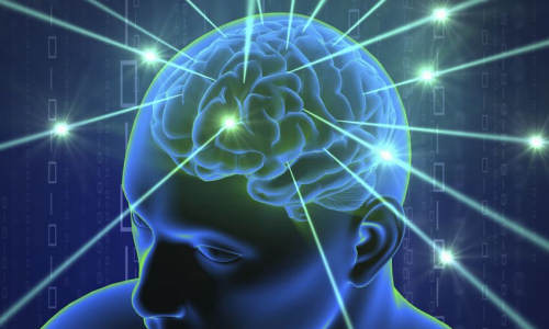
Dozens of neuroscientists are protesting Europe’s $1.6 billion attempt to recreate the functioning of the human brain on supercomputers, fearing it will waste vast amounts of money and harm neuroscience in general.


Scientists criticize Europe's $1


The 10-year Human Brain Project is largely funded by the European Union. In an open letter issued Monday, more than 190 neuroscience researchers called on the EU to put less money into the effort to “build” a brain, and to invest instead in existing projects.


If the EU doesn’t adopt their recommendations, the scientists said, they will boycott the Human Brain Project and urge colleagues to do the same.


EU spokesman Ryan Heath called for patience, and said it was too early to say whether the project is a success because it had only been under way for nine months. He said the EU plans to rigorously review the scientific progress made and the project’s management every year.


Henry Markram, who heads the Human Brain Project at the Swiss Federal Institute for Technology in Lausanne, suggested those who signed the letter of protest did so because they didn’t understand the venture.


In an interview, he said the project, established in 2013, would bundle the work of some 100,000 neuroscientists worldwide the way that CERN, the European Organization for Nuclear Research, has done for particle physics. He acknowledged the brain project may have done a poor job telling scientists how they may benefit, even if they aren’t directly involved.


“I think we need to communicate more that it’s going to actually help them get more funding,” Markram said. “They feel that money is being taken away, that it’s going to distract from the important work that they’re doing. There is really not a threat.”


The Human Brain Project involves 112 institutions across Europe and the pooling, sharing and organization of their data on brain research. That information will be used to reconstruct the workings of a human brain on computers. “Only through simulations can you do some things that aren’t possible in the lab,” said Markram.


Story Source:


The above story is based on materials provided by AP, Frank Jordans.


The post Scientists criticize Europe’s $1.6B brain project appeared first on Bioengineer.org.


8 Temmuz 2014 Salı

Rats Use Whiskers Almost as Humans Use Fingers

from

Bioengineer.org http://bioengineer.org/rats-use-whiskers-almost-humans-use-fingers/


The way rats use their whiskers is more similar to how humans use their hands and fingers than previously thought, new research from the University of Sheffield has found.


Rats Use Whiskers Almost as Humans Use Fingers


Rats deliberately change how they sense their environment using their facial whiskers depending on whether the environment is novel, if there is a risk of collision and whether or not they can see where they are going.


Exploring rats move their long facial whiskers back and forth continuously while they are moving – a behaviour called “whisking”.


Scientists have known for a long time that movement of the whiskers provides these animals with a sense of touch that allows them to move around easily in the dark.


However, until now they did not know to what extent animals were able to deliberately control their whisker movement.


Academics from the Active Touch Laboratory in the University’s Department of Psychology used high-speed videography to study animals that had been trained over several days to run circuits for food.


By putting them in different scenarios – including putting unexpected obstacles in their way and removing visual cues – the team discovered strong evidence the creatures moved their whiskers in a purposeful way to safely navigate the course.


The study found that as animals got used to their environment, they moved quicker and altered their facial whisker movements – switching from broad exploratory whisker sweeps directed at nearby surfaces, such as the floor, to pushing their whisker forwards in order to detect obstacles and avoid collisions.


In environments where they were more likely to collide with objects, and without access to visual cues, animals moved more slowly but pushed their whiskers forward further. This suggests that they were aware on the increased risk of collisions and were acting more cautiously accordingly.


Professor Tony Prescott, Professor of Cognitive Neuroscience at the University of Sheffield, said: “A person moving around in the dark would likely use their hand and fingers to detect objects and obstacles in order to avoid banging into things. In a familiar environment, such as their own home, they might move faster pushing their hands out in front of them in case of unexpected collisions.


“This new research show that rats do much the same thing but using their facial whiskers. That is, they purposefully use their whisker to detect nearby objects and surfaces when moving slowly in unfamiliar environments, and push them out in front of themselves, to avoid collisions, when the environment is familiar and they want to move more quickly.


“All mammals except humans use facial whiskers as touch sensors. In humans we seem to have replaced this sense, in part, by being able to use our hand and fingers to feel our way.


“The rat puts its whiskers where it thinks it will get the most useful information, just as we do with our fingertips.”


Story Source:


The above story is based on materials provided by University of Sheffield, Hannah Postles.


The post Rats Use Whiskers Almost as Humans Use Fingers appeared first on Bioengineer.org.


3 Temmuz 2014 Perşembe

Bioengineered red blood cells could carry precious therapeutic cargo

from

Bioengineer.org http://bioengineer.org/bioengineered-red-blood-cells-carry-precious-therapeutic-cargo/


Whitehead Institute scientists have genetically and enzymatically modified red blood cells to carry a range of valuable payloads—from drugs, to vaccines, to imaging agents—for delivery to specific sites throughout the body.


Bioengineered red blood cells could carry precious therapeutic cargo


“We wanted to create high-value red cells that do more than simply carry oxygen,” says Whitehead Founding Member Harvey Lodish, who collaborated with Whitehead Member Hidde Ploegh in this pursuit. “Here we’ve laid out the technology to make mouse and human red blood cells in culture that can express what we want and potentially be used for therapeutic or diagnostic purposes.”


The work, published this week in the Proceedings of the National Academy of Sciences (PNAS), combines Lodish’s expertise in the biology of red blood cells (RBCs) with biochemical methods developed in Ploegh’s lab.


RBCs are an attractive vehicle for potential therapeutic applications for a variety of reasons, including their abundance—they are more numerous than any other cell type in the body—and their long lifespan (up to 120 days in circulation). Perhaps most importantly, during RBC production, the progenitor cells that eventually mature to become RBCs jettison their nuclei and all DNA therein. Without a nucleus, a mature RBC lacks any genetic material or any signs of earlier genetic manipulation that could result in tumor formation or other adverse effects.


Exploiting this characteristic, Lodish and his lab introduced genes coding for specific slightly modified normal red cell surface proteins into early-stage RBC progenitors. As the RBCs approach maturity and enucleate, the proteins remain on the cell surface, where they are modified by Ploegh’s protein-labeling technique. Referred to as “sortagging,” the approach relies on the bacterial enzyme sortase A to establish a strong chemical bond between the surface protein and a substance of choice, be it a small-molecule therapeutic or an antibody capable of binding a toxin. The modifications leave the cells and their surfaces unharmed.


“Because the modified human red blood cells can circulate in the body for up to four months, one could envision a scenario in which the cells are used to introduce antibodies that neutralize a toxin,” says Ploegh. “The result would be long-lasting reserves of antitoxin antibodies.”


The approach has captured the attention of the U.S. military and its Defense Advanced Research Projects Agency (DARPA), which is supporting the research at Whitehead in the interest of developing treatments or vaccines effective against biological weapons.


Lodish believes the applications are potentially vast and may include RBCs modified to bind and remove bad cholesterol from the bloodstream, carry clot-busting proteins to treat ischemic strokes or deep-vein thrombosis, or deliver anti-inflammatory antibodies to alleviate chronic inflammation. Further, Ploegh notes there is evidence to suggest that modified RBCs could be used to suppress the unwanted immune response that often accompanies treatment with protein-based therapies. Ploegh is exploring whether these RBCs could be used to prime the immune system to allow patients to better tolerate treatment with such therapies.


Story Source:


The above story is based on materials provided by Whitehead Institute for Biomedical Research.


The post Bioengineered red blood cells could carry precious therapeutic cargo appeared first on Bioengineer.org.


New reprogramming method makes better stem cells

from

Bioengineer.org http://bioengineer.org/new-reprogramming-method-makes-better-stem-cells/


A team of researchers from the University of California, San Diego School of Medicine, Oregon Health & Science University (OHSU) and Salk Institute for Biological Studies has shown for the first time that stem cells created using different methods produce differing cells. The findings, published in the July 2, 2014 online issue of Nature, provide new insights into the basic biology of stem cells and could ultimately lead to improved stem cell therapies.


New reprogramming method makes better stem cells


This image depicts scanning electron micrograph of cultured human neuron from induced pluripotent stem cell. Photo Credit: Mark Ellisman and Thomas Deerinck, National Center for Microscopy and Imaging Research, UC San Diego


Capable of developing into any cell type, pluripotent stem cells offer great promise as the basis for emerging cell transplantation therapies that address a wide array of diseases and conditions, from diabetes and Alzheimer’s disease to cancer and spinal cord injuries. In theory, stem cells could be created and programmed to replace ailing or absent cells for every organ in the human body.


The gold standard is human embryonic stem cells (ES cells) cultured from discarded embryos generated by in vitro fertilization, but their use has long been limited by ethical and logistical considerations. Scientists have instead turned to two other methods to create stem cells: Somatic cell nuclear transfer (SCNT), in which genetic material from an adult cell is transferred into an empty egg cell, and induced pluripotent stem cells (iPS cells), in which adult cells are reverted back to a stem cell state by artificially turning on targeted genes.


Until now, no one had directly and closely compared the stem cells acquired using these two methods. The scientists found they produced measurably different results. “The nuclear transfer ES cells are much more similar to real ES cells than the iPS cells,” said co-senior author Louise Laurent, PhD, assistant professor in the Department of Reproductive Medicine at UC San Diego. “They are more completely reprogrammed and have fewer alterations in gene expression and DNA methylation levels that are attributable to the reprogramming process itself.”


The development and use of iPS cells has grown exponentially in recent years, in no small part due to the fact that they can be generated from adult cells (often from the skin) by temporarily turning on a combination of four genes to induce the adult cells to return to a pluripotent state.


Laurent noted that iPS cell lines have been created from patients to model many different diseases and “the ability to make personalized iPS cells from a patient that could be transplanted back into that patient has generated excitement because it would eliminate the need for immunosuppression.”


The nuclear transfer method has been pioneered more recently by a team led by Shoukhrat Mitalipov, PhD, professor and director of the Center for Embryonic Cell and Gene Therapy at OSHU. The technique is similar to the process used in cloning, but the pluripotent cells are collected from early embryos before they develop into mature organisms.


For their comparisons, the researchers at UC San Diego, OSHU and Salk created four nuclear transfer ES cell lines and seven iPS cell lines using the same skin cells as the source of donor genetic material, then compared them to two standard human ES lines. All 13 cell lines were shown to be pluripotent using a battery of standard tests.


But closer analyses employing powerful genomic techniques to examine the DNA methylation – a fundamental biochemical process that helps turn genes on and off – and gene expression signatures of each cell line revealed key differences in stem cells created with the three methods. Specifically, the scientists found that the DNA methylation and gene expression patterns in nuclear transfer ES cells more closely resembled those of ES cells than did iPS cells, which revealed alterations apparently caused by the reprogramming process itself.


“If you believe that gene expression and DNA methylation are important, which we do, then the closer you get to the patterns of embryonic stem cells, the better,” said co-senior author Joseph R. Ecker, PhD, professor and director of Salk’s Genomic Analysis Laboratory. “Right now, nuclear transfer cells look closer to the embryonic stem cells than do the iPS cells.”


“I think these results show that the SCNT method is a far superior candidate for cell replacement therapies,” said Mitalipov, also a co-senior author of the Nature paper. “I truly believe that using this method of producing stem cells will someday help us cure and treat a wide range of diseases that are defeating us today.”


While nuclear transfer cells may be a better and more accurate representation of human ES cells than iPS cells, Laurent said there are significant barriers to their wider adoption and application. “Not only is nuclear transfer technically difficult, but federal funds cannot be used in experiments involving this procedure.”


On the other hand, she said, the findings could spur improvements in iPS cell reprogramming methods. “Our results have shown that widely used iPS cell reprogramming methods make cells that are similar to standard ES cells in broad strokes, but there are important differences when you look really closely. By using the egg cell to do the job, we can get much closer to the real thing. If we can figure out what factors in the egg drive the reprogramming process, maybe we can design a better iPS cell reprogramming method.”


Story Source:


The above story is based on materials provided by University of California, San Diego Health Sciences


The post New reprogramming method makes better stem cells appeared first on Bioengineer.org.

Researchers regrow corneas

from

Bioengineer.org http://bioengineer.org/researchers-regrow-corneas/


Researchers have identified a way to enhance regrowth of human corneal tissue to restore vision, using a molecule known as ABCB5 that acts as a marker for hard-to-find limbal stem cells. The research is also one of the first known examples of constructing a tissue from an adult-derived human stem cell.


Researchers regrow corneas


This is a restored functional cornea following transplantation of human limbal stem cells to limbal stem cell-deficient mice. Photo Credit: Paraskevi Evi Kolovou, Bruce Ksander, and Natasha and Markus Frank


Boston researchers have identified a way to enhance regrowth of human corneal tissue to restore vision, using a molecule known as ABCB5 that acts as a marker for hard-to-find limbal stem cells. This work, a collaboration between the Massachusetts Eye and Ear/Schepens Eye Research Institute (Mass. Eye and Ear), Boston Children’s Hospital, Brigham and Women’s Hospital and the VA Boston Healthcare System, provides promise to burn victims, victims of chemical injury and others with damaging eye diseases. The research, published this week in Nature, is also one of the first known examples of constructing a tissue from an adult-derived human stem cell.


Limbal stem cells reside in the eye’s basal limbal epithelium, or limbus, and help maintain and regenerate corneal tissue. Their loss due to injury or disease is one of the leading causes of blindness. In the past, tissue or cell transplants have been used to help the cornea regenerate, but it was unknown whether there were actual limbal stem cells in the grafts, or how many, and the outcomes were not consistent.


In this study, researchers were able to use antibodies detecting ABCB5 to zero in on the stem cells in tissue from deceased human donors and use them to regrow anatomically correct, fully functional human corneas in mice.


“Limbal stem cells are very rare, and successful transplants are dependent on these rare cells,” says Bruce Ksander, Ph.D., of Mass. Eye and Ear, co-lead author on the study with post-doctoral fellow Paraskevi Kolovou, M.D. “This finding will now make it much easier to restore the corneal surface. It’s a very good example of basic research moving quickly to a translational application.”


ABCB5 was originally discovered in the lab of Markus Frank, M.D., of Boston Children’s Hospital, and Natasha Frank, M.D., of the VA Boston Healthcare System and Brigham and Women’s Hospital, co-senior investigators on the study, as being produced in tissue precursor cells in human skin and intestine. In the new work, using a mouse model developed by the Frank lab, they found that ABCB5 also occurs in limbal stem cells and is required for their maintenance and survival, and for corneal development and repair. Mice lacking a functional ABCB5 gene lost their populations of limbal stem cells, and their corneas healed poorly after injury.


“ABCB5 allows limbal stem cells to survive, protecting them from apoptosis [programmed cell death],” says Markus Frank. “The mouse model allowed us for the first time to understand the role of ABCB5 in normal development, and should be very important to the stem cell field in general.” according to Natasha Frank.


Markus Frank is working with biopharmaceutical industry to develop a clinical-grade ABCB5 antibody that would meet U.S. regulatory approvals. “A single lab cannot do a study like this,” says Natasha Frank, also affiliated with the Harvard Stem Cell Institute. “It integrates genetics, knockout mice, antibodies, transplantation — a lot of technical expertise that we were lucky came together in a very nice way.”


Story Source:


The above story is based on materials provided by Massachusetts Eye and Ear Infirmary, Mary Leach.


The post Researchers regrow corneas appeared first on Bioengineer.org.


2 Temmuz 2014 Çarşamba

A step closer to bio-printing transplantable tissues and organs: Study

from

Bioengineer.org http://bioengineer.org/step-closer-bio-printing-transplantable-tissues-organs-study/


Researchers have made a giant leap towards the goal of ‘bio-printing’ transplantable tissues and organs for people affected by major diseases and trauma injuries, a new study reports.


A step closer to bio-printing transplantable tissues and organs


Photo Credits: unknown


Scientists from the Universities of Sydney, Harvard, Stanford and MIT have bio-printed artificial vascular networks mimicking the body’s circulatory system that are necessary for growing large complex tissues.


“Thousands of people die each year due to a lack of organs for transplantation,” says study lead author and University of Sydney researcher, Dr Luiz Bertassoni.


“Many more are subjected to the surgical removal of tissues and organs due to cancer, or they’re involved in accidents with large fractures and injuries.


“Imagine being able to walk into a hospital and have a full organ printed – or bio-printed, as we call it – with all the cells, proteins and blood vessels in the right place, simply by pushing the ‘print’ button in your computer screen.


“We are still far away from that, but our research is addressing exactly that. Our finding is an important new step towards achieving these goals.


“At the moment, we are pretty much printing ‘prototypes’ that, as we improve, will eventually be used to change the way we treat patients worldwide.”


The research challenge – networking cells with a blood supply.


Cells need ready access to nutrients, oxygen and an effective ‘waste disposal’ system to sustain life. This is why ‘vascularisation’ – a functional transportation system – is central to the engineering of biological tissues and organs.


“One of the greatest challenges to the engineering of large tissues and organs is growing a network of blood vessels and capillaries,” says Dr Bertassoni.


“Cells die without an adequate blood supply because blood supplies oxygen that’s necessary for cells to grow and perform a range of functions in the body.”


“To illustrate the scale and complexity of the bio-engineering challenge we face, consider that every cell in the body is just a hair’s width from a supply of oxygenated blood.


“Replicating the complexity of these networks has been a stumbling block preventing tissue engineering from becoming a real world clinical application.”


But this is what researchers have now achieved.


What the researchers achieved


Using a high-tech ‘bio-printer’, the researchers fabricated a multitude of interconnected tiny fibres to serve as the mold for the artificial blood vessels.


They then covered the 3D printed structure with a cell-rich protein-based material, which was solidified by applying light to it.


Lastly they removed the bio-printed fibres to leave behind a network of tiny channels coated with human endothelial cells, which self organised to form stable blood capillaries in less than a week (see diagram below).


The study reveals that the bioprinted vascular networks promoted significantly better cell survival, differentiation and proliferation compared to cells that received no nutrient supply.


Significance of the breakthrough


According to Dr Bertassoni, a major benefit of the new bio-printing technique is the ability to fabricate large three-dimensional micro-vascular channels capable of supporting life on the fly, with enough precision to match individual patients’ needs.


“While recreating little parts of tissues in the lab is something that we have already been able to do, the possibility of printing three-dimensional tissues with functional blood capillaries in the blink of an eye is a game changer,” he says.


“Of course, simplified regenerative materials have long been available, but true regeneration of complex and functional organs is what doctors really want and patients really need, and this is the objective of our work.


Story Source:


The above story is based on materials provided by University of Sydney.


The post A step closer to bio-printing transplantable tissues and organs: Study appeared first on Bioengineer.org.


Muscle-powered bio-bots walk on command

from

Bioengineer.org http://bioengineer.org/muscle-powered-bio-bots-walk-command/


Engineers at the University of Illinois at Urbana-Champaign demonstrated a class of walking “bio-bots” powered by muscle cells and controlled with electrical pulses, giving researchers unprecedented command over their function. The group published its work in the online early edition of Proceedings of the National Academy of Science.


Muscle-powered bio-bots walk on command


Tiny walking “bio-bots” are powered by muscle cells and controlled by an electric field. Photo Credit: Janet Sinn-Hanlon / Group@VetMed


“Biological actuation driven by cells is a fundamental need for any kind of biological machine you want to build,” said study leader Rashid Bashir, Abel Bliss Professor and head of bioengineering at the U. of I. “We’re trying to integrate these principles of engineering with biology in a way that can be used to design and develop biological machines and systems for environmental and medical applications. Biology is tremendously powerful, and if we can somehow learn to harness its advantages for useful applications, it could bring about a lot of great things.”


Bashir’s group has been a pioneer in designing and building bio-bots, less than a centimeter in size, made of flexible 3-D printed hydrogels and living cells. Previously, the group demonstrated bio-bots that “walk” on their own, powered by beating heart cells from rats. However, heart cells constantly contract, denying researchers control over the bot’s motion. This makes it difficult to use heart cells to engineer a bio-bot that can be turned on and off, sped up or slowed down.


The new bio-bots are powered by a strip of skeletal muscle cells that can be triggered by an electric pulse. This gives the researchers a simple way to control the bio-bots and opens the possibilities for other forward design principles, so engineers can customize bio-bots for specific applications.


“Skeletal muscles cells are very attractive because you can pace them using external signals,” Bashir said. “For example, you would use skeletal muscle when designing a device that you wanted to start functioning when it senses a chemical or when it received a certain signal. To us, it’s part of a design toolbox. We want to have different options that could be used by engineers to design these things.”


The design is inspired by the muscle-tendon-bone complex found in nature. There is a backbone of 3-D printed hydrogel, strong enough to give the bio-bot structure but flexible enough to bend like a joint. Two posts serve to anchor a strip of muscle to the backbone, like tendons attach muscle to bone, but the posts also act as feet for the bio-bot.


A bot’s speed can be controlled by adjusting the frequency of the electric pulses. A higher frequency causes the muscle to contract faster, thus speeding up the bio-bot’s progress as seen in this video.


“It’s only natural that we would start from a bio-mimetic design principle, such as the native organization of the musculoskeletal system, as a jumping-off point,” said graduate student Caroline Cvetkovic, co-first author of the paper. “This work represents an important first step in the development and control of biological machines that can be stimulated, trained, or programmed to do work. It’s exciting to think that this system could eventually evolve into a generation of biological machines that could aid in drug delivery, surgical robotics, ‘smart’ implants, or mobile environmental analyzers, among countless other applications.”


Next, the researchers will work to gain even greater control over the bio-bots’ motion, like integrating neurons so the bio-bots can be steered in different directions with light or chemical gradients. On the engineering side, they hope to design a hydrogel backbone that allows the bio-bot to move in different directions based on different signals. Thanks to 3-D printing, engineers can explore different shapes and designs quickly. Bashir and colleagues even plan to integrate a unit into undergraduate lab curriculum so that students can design different kinds of bio-bots.


“The goal of ‘building with biology’ is not a new one – tissue engineering researchers have been working for many years to reverse engineer native tissue and organs, and this is very promising for medical applications,” said graduate student Ritu Raman, co-first author of the paper. “But why stop there? We can go beyond this by using the dynamic abilities of cells to self-organize and respond to environmental cues to forward engineer non-natural biological machines and systems.


“The idea of doing forward engineering with these cell-based structures is very exciting,” Bashir said. “Our goal is for these devices to be used as autonomous sensors. We want it to sense a specific chemical and move towards it, then release agents to neutralize the toxin, for example. Being in control of the actuation is a big step forward toward that goal.”


Story Source:


The above story is based on materials provided by University of Illinois at Urbana-Champaign.


The post Muscle-powered bio-bots walk on command appeared first on Bioengineer.org.