12 Ocak 2015 Pazartesi

Hitchhiking microparticles deliver drugs directly

from

BIOENGINEER.ORG http://bioengineer.org/hitchhiking-microparticles-deliver-drugs-directly/


Inflammation is a normal and often beneficial response to injury or infection. The swelling, heat and even pain are the body’s attempts to protect its soft tissue, remove offending objects, substances or microbes and initiate healing. However, persistent inflammation is often indicative of more serious conditions and can lead to problems of its own, including impaired healing, loss of function or even tissue death.


Hitchhiking microparticles deliver drugs


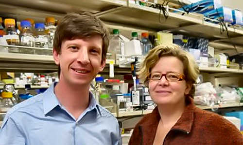
From left to right: Aaron Anselmo, Samir Mitragotri and Sunny Kumar Photo Credit: Sonia Fernandez


“Many diseases result in inflammation,” said Samir Mitragotri, professor of chemical engineering at UC Santa Barbara and director of the campus’s Center for Bioengineering. Whether inflammation is a byproduct of the disease or the inflammation is the disease, it is a common indicator of a problem with the system. “If we could target the common denominator, whether the inflammation is coming from cancer or arthritis, we could deliver the drug there,” said Mitragotri, who specializes in targeted drug delivery.


By taking advantage of natural body processes, researchers at UC Santa Barbara and MIT have developed a method of targeting inflamed tissues, creating a way to treat both the inflammation and its underlying cause.


“It’s a cell-mediated approach to targeted drug delivery,” said UCSB grad student researcher Aaron Anselmo, lead author of a study in the current issue of the Journal of Controlled Release.


Key to this technology is the utilization of monocytes, the type of white blood cell known for its ability to penetrate into deep sections of tissue. Under normal circumstances, the job of these monocytes is to circulate in the blood and respond to biochemical signals that indicate inflammation — a sign of injury or infection. Once at the site, these monocytes transform into macrophages, cells that reside in the affected tissues to engulf and digest foreign material.


Working with the expertise of chemical engineering and materials science researchers at MIT, including graduate researcher Jonathan Gilbert and professors Robert Cohen and Michael Rubner, the UCSB researchers developed an approach based on “cellular backpacks” — flat, disc-shaped polymeric particles that could, in the near future, hold therapeutic agents that can be released at the site of the inflammation. These polymeric discs are coated on one side with a single layer of an antibody that can bind to receptors on the monocyte’s surface.


To prevent the cellular backpack from being engulfed and devoured by the very cell that is transporting it, the researchers chose a flexible particle that is nonspherical in shape, which, according to the study, has proved to be more durable and resistant to phagocytosis than a rigid spherical particle. The shape and flexibility gives the backpack the ability to bind strongly while resisting phagocytosis to hitchhike onto monocytes and reach the inflamed tissue.


In-vitro and in-vivo tests have proved that cellular backpacks are successful in attaching to and being transported by monocytes to target areas without impairing the monocytes’ natural functions, said Anselmo. Further studies will include research into how much drug can be loaded into the cellular backpacks. Ideally, Anselmo said, the cellular backpacks loaded with drugs would be injected into the bloodstream, whereupon they would attach to these traveling monocytes and hitchhike to the target region. At the inflamed site, the particles would simultaneously degrade and release their drugs.


The development of effective cellular backpacks has broad potential, say the researchers.


“Basically the main benefit is that you can deliver the drug in a more effective dose,” Mitragotri said. Take for example the case of chemotherapy, which often has a narrow therapeutic range: Too little and the treatment is not effective, too much and it can be lethal. Because chemo travels through the bloodstream and affects all the tissues it comes in contact with, dosages are restricted at least in part based on the deleterious effect it has on other, unafflicted organs and their functions. Not only can targeted therapy ensure other body systems remain unaffected, Mitragotri explained, but it could allow for higher doses of drug to the site, which could decrease treatment time.


Story Source:


The above story is based on materials provided by University of California – Santa Barbara.


11 Ocak 2015 Pazar

Human brain keeps memories tidy by pruning inaccurate ones

from

BIOENGINEER.ORG http://bioengineer.org/human-brain-keeps-memories-tidy-by-pruning-inaccurate-ones/


Your brain is a memory powerhouse, constantly recording experiences in long-term memory. Those memories help you find your way through the world: Who works the counter each morning at your favorite coffee shop? How do you turn on the headlights of your car? What color is your best friend’s house?


But then your barista leaves for law school, you finally buy a new car and your buddy spends the summer with a paint brush in hand. Suddenly, your memories are out of date.


What happens next?


An experiment conducted by researchers from Princeton University and the University of Texas-Austin shows that the human brain uses memories to make predictions about what it expects to find in familiar contexts. When those subconscious predictions are shown to be wrong, the related memories are weakened and are more likely to be forgotten. And the greater the error, the more likely you are to forget the memory.


“This has the benefit ultimately of reducing or eliminating noisy or inaccurate memories and prioritizing those things that are more reliable and that are more accurate in terms of the current state of the world,” said Nicholas Turk-Browne, an associate professor of psychology at Princeton and one of the researchers.


Nicholas Turk-Browne, an associate professor of psychology, uses the example of a trip to a coffee shop to explain new research about the way the brain responds to inaccurate memories. (Animation by Kyle McKernan, Office of Communications)


The research was featured in an article, “Pruning of memories by context-based prediction error,” that appeared in 2014 in the Proceedings of the National Academy of Sciences. The other co-authors are Ghootae Kim, a Princeton graduate student; Jarrod Lewis-Peacock, an assistant professor of psychology at the University of Texas-Austin; and Kenneth Norman, a Princeton professor of psychology and the Princeton Neuroscience Institute.


The researchers’ experiment involved 24 adults, who were shown a series of photos one at a time while their brain activity was monitored by a functional magnetic resonance imaging (fMRI) machine. The participants were asked a question about each photo, but the real purpose of the exercise was to monitor their brain activity as the photos were shown.


The photos included three-photo sequences, such as two photos of faces followed by a photo of a scene. In this example, the first two photos would appear again later in the series, but this time would be followed by a new face rather than the scene. The researchers measured how strongly participants were expecting to see a photo of the scene the second time by looking for the pattern of brain activity associated with that scene, around the time when it should have appeared in the sequence.


“We wanted to get direct access to what things people are predicting without asking them,” Turk-Browne said. “These kinds of predictions are not necessarily consciously accessible, and if we ask about them, it will change the behavior.”


Later, participants were shown photos and asked whether they recognized them from the fMRI portion.


By analyzing the fMRI data and the memory test results, the researchers found that the more strongly participants’ brains predicted — incorrectly — the next photo in the sequence, the more likely they were to forget the predicted photo.


“Our specific hypothesis in the context of the experiment is that the bigger the prediction, the more the error and the more likely you are to forget the thing you were predicting,” Turk-Browne said. “We think it’s the error causing the forgetting. How can we measure the error? That’s difficult. But we know the error is proportional to the strength of the prediction. So we use the strength of the prediction as a measure of the prediction error.”


Human brain keeps memories tidy by pruning inaccurate ones


Princeton graduate student Ghootae Kim discusses the memory research with Turk-Browne and Kenneth Norman, a Princeton professor of psychology and the Princeton Neuroscience Institute. The researchers’ experiment involved 24 adults whose brain activity was monitored by a functional magnetic resonance imaging (fMRI) machine.


The researchers say the findings fit a model for how the brain handles memories called the nonmonotonic plasticity hypothesis. The model claims that strong activation of a memory — such as when a remembered object or event is encountered again — will strengthen the memory.


But moderate activation of a memory — such as the activation that occurs when your brain makes an unconfirmed prediction using the memory — can degrade the memory.


So, Turk-Browne said, if your brain activates a memory in forming a prediction and then doesn’t re-experience the remembered object or event, the memory can begin to fade.


“This is a very general mechanism for influencing what people remember and forget on the basis of whether it can be reliably predicted given the situation in which it occurs or not,” Turk-Browne said. “People remember and forget a lot of things. This isn’t going to explain all of remembering or all of forgetting, but this is an automatic, unconscious way the brain has to figure out when things turn out not to be really reliable in terms of how the world is structured.”


Morgan Barense, an assistant professor of psychology at the University of Toronto, said the researchers have found a “powerful learning mechanism” in the brain.


“I think it’s an incredibly compelling story that this automatic prediction mechanism is constantly operating under the radar of our conscious experience, optimizing what elements of a memory we should remember based on how likely they are to occur in our environment,” said Barense, who studies how memory functions are organized in the brain. “It makes sense that the brain would operate in this way, yet it was only with the development of modern neuroimaging analyses that we were able to observe this mechanism in action.”


Turk-Browne said the research opens the door to several avenues of additional research, including working to better understand the brain’s predictions.


“What are the physiological mechanisms of generating predictions in the first place? We’re investigating that,” he said.


Story Source:


The above story is based on materials provided by Princeton University.


Pioneering Method to Define Stages of Stem Cell Reprogramming

from

BIOENGINEER.ORG http://bioengineer.org/pioneering-method-to-define-stages-of-stem-cell-reprogramming/


In a groundbreaking study that provides scientists with a critical new understanding of stem cell development and its role in disease, UCLA researchers at the Eli and Edythe Broad Center of Regenerative Medicine and Stem Cell Research led by Dr. Kathrin Plath, professor of biological chemistry, have established a first-of-its-kind methodology that defines the unique stages by which specialized cells are reprogrammed into stem cells that resemble those found in the embryo.


Pioneering Method to Define Stages of Stem Cell Reprogramming


Drs. Vincent Pasque and Kathrin Plath Photo Credit: UCLA Broad Stem Cell Research Center/Plath Lab


The study was published online ahead of print in the journal Cell.


Induced pluripotent stem cells (known as iPSCs) are similar to human embryonic stem cells in that both cell types have the unique ability to self-renew and have the flexibility to become any cell in the human body. iPSC cells, however, are generated by reprogramming skin or blood cells and do not require an embryo.

Reprogramming is a long process (about one to two weeks) and largely inefficient, with typically less than one percent of the primary skin or blood cells successfully completing the journey to becoming an iPSC. The exact stages a cell goes through during the reprogramming process are also not well understood. This knowledge is important, as iPSCs hold great promise in the field of regenerative medicine, as they can provide a single source of patient-specific cells to replace those lost to injury or disease. They can also be used to create novel disease models from which new drugs and therapies can be developed.


“This research has broad impact, because by deepening our understanding of cell reprogramming we have the potential to improve disease modeling and the generation of better sources of patient-specific specialized cells suitable for replacement therapy,” said Plath. “This can ultimately benefit patients with new and better treatments for a wide range of diseases.


Drs. Vincent Pasque and Jason Tchieu, postdoctoral fellows in the lab of Dr. Plath and co-first authors of the study, developed a roadmap of the reprogramming process using detailed time-course analyses. They induced the reprogramming of skin cells into iPSC, then observed and analyzed on a daily basis or every other day the process of transformation at the single-cell level. The data were collected and recorded over a period of up to two weeks.


Plath’s team found that the changes that happen in cells during reprogramming occur in a sequential stage-by-stage manner, and that importantly, the stages were the same across all the different reprogramming systems and different cell types analyzed.


“The exact stage of reprogramming of any cell can now be determined,” said Pasque. “This study signals a big change in thinking, because it provides simple and efficient tools for scientists to study stem cell creation in a stage-by-stage manner. Most studies to date ignore the stages of reprogramming, but we can now seek to better understand the entire process on both a macro and micro level.”


Plath’s team further discovered that the stages of reprogramming to iPSC are different from what was expected. They found that it is not simply the reversed sequence of stages of embryo development. Some steps are reversed in the expected order; others do not actually happen in the exact reverse order and resist a change until late during reprogramming to iPSCs.


“This reflects how cells do not like to change from one specialized cell type to another and resist a change in cell identity,” said Pasque. “Resistance to reprogramming also helps to explain why reprogramming takes place only in a very small proportion of the starting cells.”

With these findings, Plath’s lab plans future studies to actively isolate specific cell types during specific stages of reprogramming. They also hope the research will encourage further investigation into the characteristics of iPSC development.


Story Source:

The above story is based on materials provided by UCLA Eli and Edythe Broad Center of Regenerative Medicine and Stem Cell Research.


9 Ocak 2015 Cuma

Researchers Create Artificial Organs That Fit In Your Hand

from

BIOENGINEER.ORG http://bioengineer.org/researchers-create-artificial-organs-that-fit-in-your-hand/


Great balls of cells! Scientists are developing mock human organs that can fit in the palm of your hand.These organs-on-a-chip are designed to test drugs and help understand the basics of how organs function when they are healthy and when they are diseased. For instance, you have your gut-on-a-chip being developed at the Johns Hopkins School of Medicine. It’s a high-tech approach to dealing with a scourge of the low-tech world.


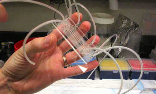
Postdoctoral researcher Jennifer Foulke-Abel holds the gut-on-a-chip inside the lab at Johns Hopkins School of Medicine


Postdoctoral researcher Jennifer Foulke-Abel holds the gut-on-a-chip inside the lab at Johns Hopkins School of Medicine. Photo Credits: Richard Harris/NPR


“I’m interested in solving a worldwide problem of diarrheal diseases,” says Dr. Mark Donowitz, who runs this lab. He says 800,000 children a year die from these diseases — notably cholera, rotavirus and certain strains of E. coli.


“We’ve failed so far to find drugs to treat diarrhea using cell culture models and mouse intestine,” Donowitz says. Mice simply don’t react the way we do to these germs, so they aren’t very helpful for studying diseases of the gut.


So Donowitz’s team is building what it hopes will be a much better way to study these diseases: the gut-on-a-chip. Truth be told, there’s not a lot to see.


Postdoctoral researcher Jennifer Foulke-Abel holds one in the palm of her hand. It’s a thin sheet of glass, topped with a plastic microscope slide and a tiny cavity inside. Half a dozen spaghetti-size tubes bristle from the device.


“The reason there are so many tubes is we have a vacuum chamber that will cause the membrane to stretch, the way the intestine stretches as it moves food along,” Fouke-Abel explains.


Cells plucked from a human intestine will be put into a tiny chamber around that membrane, and they will divide, grow and even organize themselves much as you would find them in human guts. The device, when operating, might hold 50,000 gut cells.


Step 1 in this research is to see whether cells in the ersatz organ react the same way to diseases as do cells in the human gut.


“And in all three of the diseases I mentioned, we’ve been able to take that first step,” Donowitz says. “So we know that these appear to be really good models of the human disease.”


Still, it’s a work in progress. The guts-on-a-chip produce digestive enzymes, hormones and mucus, but they don’t yet incorporate other parts of the human intestine, such as blood vessels or nerve cells.


“They all have to be incorporated if you want to move from a simple to a more complex system, which I think you need to do if you are going to reproduce intestinal biology,” Donowitz says.


This lab is moving in that direction. And once it has a complete system built, one use will be to test potential drugs for the diseases being studied. “We think this could be a real step forward in terms of reducing waste-of-time drug development,” Donowitz says.


While this lab at Johns Hopkins is working to develop the gut, other labs scattered around the country are working on other organ systems.


“There’s going to be a brain-on-a-chip, liver, heart and so on,” says Danilo Tagle, who coordinates this overall effort at the National Center for Advancing Translational Sciences, which is part of the National Institutes of Health. It is funding development of 10 organ systems in all.


“The goal is actually to tie them in all together,” Tagle says. So they will collectively act like an entire human being on a chip — at least from the point of view of a scientist interested in testing drugs. (The brain-on-a-chip will not think, of course.)


And Tagle says in the long run, scientists hope they can build many of these systems, each one based on the cells from an individual person. Imagine a small army of cell-based stand-ins for research.


“And so you can identify which part of the population might be more responsive to particular drugs, or identify a subset of the population that might be more vulnerable to the harmful effects of a particular drug,” Tagle says.


He says this $75 million, five-year project took off thanks to pioneering work at the Wyss Institute for Biologically Inspired Engineering at Harvard. The research has been so promising, Wyss spun off a private company to pursue it.


“It’s called Emulate,” says Donald Ingber, founding director of the Wyss Institute. “It’s just getting its feet on the ground. We have almost 20 people out of the Wyss Institute who are moving out with it.”


Ingber says it would be too much to expect this technology to replace mice in medical research anytime soon. But he is hoping that this will speed up drug development and make it less expensive, “because if we can identify things that are more likely to work in humans, that’s going to have major impact.”


And there are so many avenues to pursue, he says, there’s plenty of room for both industry and academics to work on building and improving these organs-on-a-chip.


Stretchy Electrodes Enable Long-Lasting Brain Implants

from

BIOENGINEER.ORG http://bioengineer.org/stretchy-electrodes-enable-long-lasting-brain-implants/


If you need a piece of hardware attached to the delicate tissue of your brain or spinal cord, wouldn’t it be preferable for that piece of hardware to actually be soft, yielding, and flexible?


ieee-spectrum


Bendy electronics mimic the elastic properties of a neural membrane Photo Credit: EPFL


That kind of thinking led researchers at a Swiss technology institute to develop a new material modeled on dura matter, the protective membrane of the brain and spinal cord. Their “e-dura” contains stretchy electrodes that can both stimulate and record from neurons. When implanted in mice, the e-dura caused less damage and inflammation than today’s rigid implants. Researchers say their biocompatible material could be the key to long-lasting neural therapies.


Brain and spinal implants are no longer the stuff of science fiction. Thousands of people with Parkinson’s and epilepsy are walking around with stimulators implanted in their brains, and researchers are exploring the use of deep brain stimulation for depression and other neuro-psychiatric disorders. Spinal cord stimulation experiments have already enabled paralyzed people to stand, and may someday get them walking. Implants can also be used to record neural acitivity and use it to control external devices like prosthetic limbs.


But the body is a mechanically dynamic environment, says Stéphanie Lacour, one of the study’s lead authors and chair of neuroprosthetic technology at the École Polytechnique Fédérale de Lausanne (EPFL).


“The brain pulsates with blood flow, the spine cord stretches and relax with daily movements,” she told Spectrum. “Therefore an implant with a similar mechanical behavior to the neural tissue will provide a more suitable interface than a non-elastic implant.” The e-dura is designed to be placed on the surface of the spinal cord or brain, and isn’t suitable for penetrating implants used in deep brain stimulation.


Lacour and her colleagues started with a transparant silicone substrate, added gold interconnects with microcracks that make them stretchable while maintaining conductivity, and printed on soft electrodes coated with a platinum-silicone composite. Interestingly, they also included what they call a “chemotrode,” a tiny microfluidic component that can deliver drugs directly to the neurons.


To test their material, the researchers implanted the e-dura directly on rats’ spinal cords (beneath the real dura matter), and left it there for six weeks. They did the same with a stiff implant. They found that rats with the stiff implant began to have trouble walking within just a few weeks, and later examination showed both inflammation and deformation of their spinal cords. The rats with the e-dura implant displayed no such motor problems or physiological degradation.


The material must not only prove harmless, but also effective. The researchers showed both that the e-dura accurately recorded from and stimulated neurons in the brain and spinal cord. The stimulation experiment built on co-author Grégoire Courtine’s work at EPFL, in which he has enabled paralyzed rats to walk again through a combination of spinal cord stimulation and drugs. In the new experiment, the e-dura was placed on the spinal cord below the site of injury, and used to provide electric pulses and drug doses. Throughout the six-week implantation, the paralyzed rats walked and walked.


Courtine says the chemotrode element on the e-dura is an important advance, because delivering drugs only to targeted neurons could reduce doses five- or ten-fold. And lower doses mean fewer side effects, Courtine told Spectrum. For example, he pointed to Parkinson’s patients who take the standard L-dopa medication, which controls symptoms but can also cause muscle spasms and other disruptive side effects.


Other labs have produced similar biocompatible and elastic electronics, including Takao Someya of the University of Tokyo, who wrote for Spectrum about his work on e-skin. Someya commends the e-dura for creating an “intimate connection” with the spinal cord, and says it may produce more efficient therapies with its highly targeted delivery of electric pulses and drugs. “The challenge of their future work is to perhaps integrate more functionality into these devices, increase electrode densities, or tailor electrode placement to each specimen,” he says.


Story Source:


The above story is based on materials provided by IEEE Spectrum.


7 Ocak 2015 Çarşamba

Researchers uncover key cancer-promoting gene

from

BIOENGINEER.ORG http://bioengineer.org/researchers-uncover-key-cancer-promoting-gene/


One of the mysteries in cancer biology is how one protein, TGF-beta, can both stop cancer from forming and encourage its aggressive growth. Now, researchers at the University of Michigan Comprehensive Cancer Center have uncovered a key gene that may explain this paradox and provide a potential target for treatment.


cancer


TGF-beta is known as a tumor suppressor, meaning it’s necessary to keep cells in check and growing normally. But at some point, its function flips and it becomes a tumor promoter, fostering aggressive growth and spread of cancer. The researchers identified Bub1 as a key gene involved in regulating TGF-beta receptor.


The study is published in Science Signaling.


“Our data that Bub1 is involved at the receptor level is completely unexpected,” says study director Alnawaz Rehemtulla, Ph.D., Ruth Tuttle Freeman Research Professor of radiation oncology and radiology and co-director of the Center for Molecular Imaging at the University of Michigan Medical School.

“Bub1 is well-known for its role in cell division. But this is the first study that links it to TGF-beta. We think this may explain the paradox of TGF-beta as a tumor promoter and a tumor suppressor,” he adds.


The team of researchers at the University of Michigan, including Shyam Nyati, Ph.D., and Brian D. Ross, Ph.D., developed a way to screen for genes that regulate the TGF-beta receptor. When 720 genes from the human genome were screened against lung cancer and breast cancer cells, Bub1 emerged as playing a strong role in TGF-beta signaling.


Bub1 was shown to bind to the TGF-beta receptor and allows it to turn on aggressive cell growth. When the researchers blocked Bub1, it shut down the TGF-beta pathway completely.


TGF-beta is known to play a role in cells developing characteristics of aggressive cancer cells. Researchers also have known that Bub1 is highly expressed in many different types of cancer.

Because Bub1 is found in many types of cancer, developing a drug to target it could potentially impact multiple cancers. A compound to target Bub1 has been developed but is not ready for testing in patients. Initial lab testing suggests that a Bub1 inhibitor can very specifically target Bub1 without causing damage to other parts of the cell.


“When you look at gene expression in cancer, Bub1 is in the top five. In addition, Bub1 expression levels correlate with outcome in patients with lung and breast cancer. But we never knew why. Now that we have that link, we’re a step closer to shutting down this cycle,” Rehemtulla says.


Story Source:

The above story is based on materials provided by University of Michigan Health System.


Neuroscientists Get Better Feeling About Touch

from

BIOENGINEER.ORG http://bioengineer.org/neuroscientists-get-better-feeling-about-touch/


Our sense of touch is one we often take for granted, until our leg falls asleep and we aren’t able to stand, or when we experience acute pain. The sense of touch also has been taken for granted in neuroscience, where it’s the sense scientists know the least about.


sense-of-touch


Photo Credit: Chepko Danil


An international group of researchers, including Carnegie Mellon University’s Alison Barth, is changing that. For the first time researchers have linked a group of neurons to a specific type of somatosensation, a finding that can open the door for a heightened understanding about our sense of touch. The research is published in the Dec. 3 issue of Neuron.


“Somatosensation is critical. You can somewhat overcome losing your sense of smell, sight, taste, or hearing. But if you lose your sense of touch, you wouldn’t be able to sit up or walk. You wouldn’t be able to feel pain,” said Barth, a professor of biological sciences and a member of Carnegie Mellon’s BrainHubSM research initiative. “We know less about the features that make up our rich tactile experience than we do about any other sense, yet it’s such a critical sense.”


Somatosensation, which is another word for our sense of touch, occurs in a number of forms, like feeling texture, temperature, pressure, pain or vibration. It’s responsible for proprioception, which helps us know where we are within our environment. It tells us if our feet are firmly planted on the floor, or if we’re holding a paper cup tightly enough that it won’t slip out of our hand, but loosely enough that we don’t crush the cup. Scientists know a good deal about the molecular receptors that mediate the different types of somatosensation, but they know little about how touch is represented in the brain.


“When someone gets pricked by a pin, we know how information about that sensation travels from the skin to the spinal cord. But what happens in the brain has been much less clear — it seems like all different sorts of touch information get jumbled together,” said Barth, who also is a member of the joint Carnegie Mellon/University of Pittsburgh Center for the Neural Basis of Cognition (CNBC).


It was a jumble — until now.


In previous studies, Barth had discovered that certain groups of neurons in the brain’s neocortex were reliably more active than others. Using the fos-GFP mouse, a transgenic mouse model Barth created to study activity in live neurons, she and her colleagues set out to see if these neurons were generally more excitable, or if they responded specifically to one tactile stimulus. They found that these neurons reacted much more quickly and strongly when a puff of air was directed at the mouse’s whiskers, while other neurons had little or no response.


“This is the first time we’ve been able to visualize neurons in the somatosensory cortex that ‘like’ a specific tactile stimulus,” Barth said. “It shows that neurons are individuals. They have different jobs to do in the cortex. In this case these neurons had a special feature: they responded when all of the mouse’s whiskers moved at once.”


They also found that the neurons in question received direct synaptic input from the posteromedial nucleus of the brain’s thalamus. This shows that the neurons that react to the puff-of-air stimulus have a dedicated, unique sub-network of connections that enable them to communicate with one another and amplify the information they are receiving from the stimulus.


“Now that we have isolated the neural underpinnings of a certain feature, we can try to manipulate and change the interactions between cells. Can we train the mouse and strengthen the connections between neurons? What happens to perception if we remove the connections? It’s really the frontier of truly understanding somatosensory function,” Barth said.


This research also could lead to work that will identify how somatosensory information is coded, which could be used to incorporate sensory information into brain-machine interfaces. This could allow robotic limbs and prosthetics to actively sense and receive tactile input.


Story Source:


The above story is based on materials provided by Carnegie Mellon University.