8 Nisan 2015 Çarşamba

A digital field guide to cancer cells

from

BIOENGINEER.ORG http://bioengineer.org/a-digital-field-guide-to-cancer-cells/


Scientists are mapping the habits of cancer cells, turn by microscopic turn. Using advanced technology and an approach that merges engineering and medicine, a Yale University-led team has compiled some of the most sophisticated data yet on the elaborate signaling networks directing highly invasive cancer cells. Think of it as a digital field guide for a deadly scourge.

digital


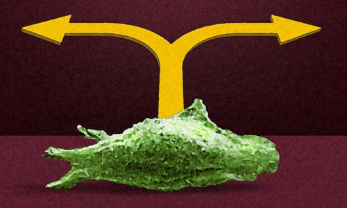
Yale-led research is looking into the directional cues that influence cancer cells. Photo Credit: Yale University/Michael Helfenbein


“This is a very complex set of interactions and processes,” said Andre Levchenko, a Yale systems biologist and biomedical engineer, and director of the Yale Systems Biology Institute. “The systems biology approach acknowledges that complexity by analyzing how cancer cells migrate together and separately in response to complex cues.”


In a study published April 8 in the journal Nature Communications, Levchenko and his colleagues describe the intricate ways breast cancer cells respond to chemical cues in the human body. The idea is to determine which cues cause cancer cells to disperse and metastasize, how these cues are combined with other cues directing the invasion, and which cues hold sway when there are conflicting orders.


Until now, little has been known about how cells decide when and where to turn while traveling through the complex tissues. These cells often encounter contradictory directional cues — begging the questions: Which cues are stronger, and in what situations?


In this study, researchers focused on several cues. One is a protein called Epidermal Growth Factor (EGF), which acts as a strong, directional guidance signal to individual cancer cells. Another cue mediates a poorly understood phenomenon called “contact inhibition of locomotion” (CIL), in which cells act almost like bumper cars, stopping their forward motion on contact and moving away from each other.


Levchenko’s team found that when both EGF and CIL signals act upon a breast cancer cell, the cell acts as a tiny computer, making decisions about which cue dominates. If the EGF cue is weak, the cell can turn around if it encounters another cell; if the EGF cue is strong enough, the two cells will travel together. The researchers unraveled the molecular network that allows the cells to follow these cues and make appropriate decisions. In particular, they found the critical role of proteins called ephrins in mediating the CIL cue. These proteins, also found in other cell types, allow breast cancer cells to be repelled from each other, while ignoring other cells, such as fibroblasts. This knowledge allowed the investigators to suppress CIL, so that even weak EGF inputs could lead to coordinated movement of many cells.


“We have shown that migrating cells prioritize certain cues in the presence of others and thus can switch their migration mode, depending on what they see from the environment,” said first author Benjamin Lin, a postdoctoral associate in biomedical engineering at Yale.


Understanding the interplay of these signals may allow researchers to devise strategies for interfering with, or redirecting, cancer cells in motion. For example, if a traveling cancer cell received strong, artificial CIL cues so that its movement became less directed and invasive, and more chaotic, could that slow the onset of metastasis?


Historically, experiments on cancer cell movement have been unable to mimic the dynamic complexity of the human body. Now, using advanced biosensors and other technology, such experiments come much closer to replicating a realistic, biological environment.


“Scientists have studied quite well how individual cues affect cell migration. But in reality, cells are subject to multiple cues at the same time,” said co-author Takanari Inoue, an associate professor at Johns Hopkins University. “Our work is significant because we clearly demonstrated that cells do integrate multiple pieces of information and that the integration occurs at a place fairly upstream of the signal processing. I think we have been underestimating these cells’ capability to integrate different cues.”


Story Source:


The above story is based on materials provided by Yale university.


Spontaneous appearance of primordial DNA

from

BIOENGINEER.ORG http://bioengineer.org/spontaneous-appearance-of-primordial-dna/


The self-organization properties of DNA-like molecular fragments four billion years ago may have guided their own growth into repeating chemical chains long enough to act as a basis for primitive life, says a new study by the University of Colorado Boulder and the University of Milan.


While studies of ancient mineral formations contain evidence for the evolution of bacteria from 3.5 to 3.8 billion years ago — just half a billion years after the stabilization of Earth’s crust — what might have preceded the formation of such unicellular organisms is still a mystery. The new findings suggest a novel scenario for the non-biological origins of nucleic acids, which are the building blocks of living organisms, said CU-Boulder physics Professor Noel Clark, a study co-author.


sponge


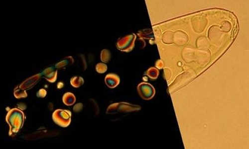
The image shows a droplet of condensed nano-DNA and within it smaller drops of its liquid crystal phase which show up in polarized light on the left. The liquid crystal droplets act as “micro-reactors” where short DNA can join together into long polymer chains without the aid of biological mechanisms. Photo Credit:Noel Clark, University of Colorado


A paper on the subject led by Tommaso Bellini of the University of Milan was published in a recent issue of Nature Communications. Other CU-Boulder co-authors of the study include Professor David Walba, Research Associate Yougwooo Yi and Research Assistant Gregory P. Smith. The study was funded by the Grant PRIN Program of the Italian Ministries of Education, Universities and Research and by the U.S. National Science Foundation.


The discovery in the 1980’s of the ability of RNA to chemically alter its own structure by CU-Boulder Nobel laureate and Distinguished Professor Tom Cech and his research team led to the development of the concept of an “RNA world” in which primordial life was a pool of RNA chains capable of synthesizing other chains from simpler molecules available in the environment. While there now is consensus among origin-of-life researchers that RNA chains are too specialized to have been created as a product of random chemical reactions, the new findings suggest a viable alternative, said Clark.


The new research demonstrates that the spontaneous self-assembly of DNA fragments just a few nanometers in length into ordered liquid crystal phases has the ability to drive the formation of chemical bonds that connect together short DNA chains to form long ones, without the aid of biological mechanisms. Liquid crystals are a form of matter that has properties between those of conventional liquids and those of a solid crystal — a liquid crystal may flow like a liquid, for example, but its molecules may be oriented more like a crystal.


“Our observations are suggestive of what may have happened on the early Earth when the first DNA-like molecular fragments appeared,” said Clark.


For several years the research group has been exploring the hypothesis that the way in which DNA emerged in the early Earth lies in its structural properties and its ability to self-organize. In the pre-RNA world, the spontaneous self-assembly of fragments of nucleic acids (DNA and RNA) may have acted as a template for their chemical joining into polymers, which are substances composed of a large number of repeating units.


“The new findings show that in the presence of appropriate chemical conditions, the spontaneous self assembly of small DNA fragments into stacks of short duplexes greatly favors their binding into longer polymers, thereby providing a pre-RNA route to the RNA world,” said Clark.


Story Source:


The above story is based on materials provided by University of Colorado at Boulder.


7 Nisan 2015 Salı

HIV spreads like internet malware and should be treated earlier

from

BIOENGINEER.ORG http://bioengineer.org/hiv-spreads-like-internet-malware-and-should-be-treated-earlier/


A new model for HIV progression finds that it spreads in a similar way to some computer worms and predicts that early treatment is key to staving off AIDS.


HIV specialists and network security experts at UCL noticed that the spread of HIV through the body using two methods – via the bloodstream and directly between cells – was similar to how some computer worms spread through both the internet and local networks respectively to infect as many computers as possible. They worked together to create a model for this ‘hybrid spreading’, which accurately predicted patients’ progression from HIV to AIDS in a major clinical trial.


Detailed sample data from 17 HIV patients from London were used to verify the model, showing that hybrid spreading provides the best explanation for HIV progression and highlighting the benefits of early treatment.


HIV infects CD4+ T-cells, which play a vital role in the immune system and protect us from diseases. As HIV progresses, it reduces the number of active T-cells in the body until the immune system cannot function correctly, a state known as ‘acquired immune deficiency syndrome’ or AIDS.


Current World Health Organisation guidelines, which the UK government follows, recommend only beginning HIV treatment when the number of T-cells in the bloodstream falls below a certain level. However, the new model predicts that treatment should start as soon as possible after infection to prevent AIDS from developing in the long term.


“The number of HIV cells in the bloodstream is always relatively low, and our model shows that HIV spread through the bloodstream alone would not be enough to cause AIDS,” explains co-senior author Professor Benny Chain (UCL Infection & Immunity). “It is likely that when HIV gains a foothold somewhere with a high T-cell population, such as the gut, it uses a cell-to-cell transfer mechanism to efficiently spread directly between them. As such, if HIV has already spread to an area rich in T-cells by the time treatment begins, preventing its spread through the bloodstream will not stop AIDS. Our model suggests that completely blocking cell-to-cell transfer would prevent progression to AIDS, highlighting the need to develop new treatments.”


The model was inspired by similarities between HIV and computer worms such as the highly damaging ‘Conficker’ worm, first detected in 2008, which has infected military and police computer networks across Europe and is still active today.


“HIV and Conficker have a lot in common,” says lead author Changwang Zhang (UCL Computer Science). “They both use hybrid spreading mechanisms, persist for a very long time and are incredibly difficult to eradicate. Our model enables us to explain these important properties and to predict the infection process.”


Changwang’s supervisor, co-author Dr Shi Zhou (UCL Computer Science) says: “Although the cybersecurity community organised an unprecedented collaboration to tackle Conficker, they still failed to eliminate Conficker from the Internet. HIV researchers face a similar problem. We hope that our new understanding of hybrid epidemics will help us to fight against Conficker and HIV.”


Laboratory research led by co-senior author Dr Clare Jolly (UCL Infection & Immunity) has previously shown that some drugs are better than others at stopping HIV from spreading directly between cells. However, it is not possible to directly measure cell-to-cell spread in patients because it takes place inside internal organs.


“With this new model, we should be able to assess the effectiveness of drugs against different modes of HIV spread in real patients,” explains Dr Jolly. “This could prove invaluable when interpreting the results of drug trials to understand what works and why. Using computer models to understand processes that we cannot directly observe is common in the physical sciences and supports many fundamental theories. Our model provides strong evidence that cell-to-cell spread is an important part of HIV spread, and we hope to show this directly in future animal studies.”


Story Source:


The above story is based on materials provided by University College London.


Researchers Build Brain-Machine Interface to Control Prosthetic Hand

from

BIOENGINEER.ORG http://bioengineer.org/researchers-build-brain-machine-interface-to-control-prosthetic-hand/


A research team from the University of Houston has created an algorithm that allowed a man to grasp a bottle and other objects with a prosthetic hand, powered only by his thoughts.


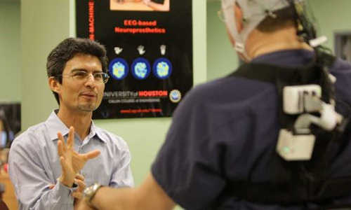
Jose Luis Contreras-Vidal


Jose Luis Contreras-Vidal, a neuroscientist and engineer at UH


The technique, demonstrated with a 56-year-old man whose right hand had been amputated, uses non-invasive brain monitoring, capturing brain activity to determine what parts of the brain are involved in grasping an object. With that information, researchers created a computer program, or brain-machine interface (BMI), that harnessed the subject’s intentions and allowed him to successfully grasp objects, including a water bottle and a credit card. The subject grasped the selected objects 80 percent of the time using a high-tech bionic hand fitted to the amputee’s stump.


Previous studies involving either surgically implanted electrodes or myoelectric control, which relies upon electrical signals from muscles in the arm, have shown similar success rates, according to the researchers.


Jose Luis Contreras-Vidal, a neuroscientist and engineer at UH, said the non-invasive method offers several advantages: It avoids the risks of surgically implanting electrodes by measuring brain activity via scalp electroencephalogram, or EEG. And myoelectric systems aren’t an option for all people, because they require that neural activity from muscles relevant to hand grasping remain intact.


The results of the study were published March 30 in Frontiers in Neuroscience, in the Neuroprosthetics section.


Contreras-Vidal, Hugh Roy and Lillie Cranz Cullen Distinguished Professor of electrical and computer engineering at UH, was lead author of the paper, along with graduate students Harshavardhan Ashok Agashe, Andrew Young Paek and Yuhang Zhang.


The work, funded by the National Science Foundation, demonstrates for the first time EEG-based BMI control of a multi-fingered prosthetic hand for grasping by an amputee. It also could lead to the development of better prosthetics, Contreras-Vidal said.


Beyond demonstrating that prosthetic control is possible using non-invasive EEG, researchers said the study offers a new understanding of the neuroscience of grasping and will be applicable to rehabilitation for other types of injuries, including stroke and spinal cord injury.


T he study subjects – five able-bodied, right-handed men and women, all in their 20s, as well as the amputee – were tested using a 64-channel active EEG, with electrodes attached to the scalp to capture brain activity. Contreras-Vidal said brain activity was recorded in multiple areas, including the motor cortex and areas known to be used in action observation and decision-making, and occurred between 50 milliseconds and 90 milliseconds before the hand began to grasp.


That provided evidence that the brain predicted the movement, rather than reflecting it, he said.


“Current upper limb neuroprosthetics restore some degree of functional ability, but fail to approach the ease of use and dexterity of the natural hand, particularly for grasping movements,” the researchers wrote, noting that work with invasive cortical electrodes has been shown to allow some hand control but not at the level necessary for all daily activities.


“Further, the inherent risks associated with surgery required to implant electrodes, along with the long-term stability of recorded signals, is of concern. … Here we show that it is feasible to extract detailed information on intended grasping movements to various objects in a natural, intuitive manner, from a plurality of scalp EEG signals.”


Until now, this was thought to be possible only with brain signals acquired invasively inside or on the surface of the brain.


Researchers first recorded brain activity and hand movement in the able-bodied volunteers as they picked up five objects, each chosen to illustrate a different type of grasp: a soda can, a compact disc, a credit card, a small coin and a screwdriver. The recorded data were used to create decoders of neural activity into motor signals, which successfully reconstructed the grasping movements.


They then fitted the amputee subject with a computer-controlled neuroprosthetic hand and told him to observe and imagine himself controlling the hand as it moved and grasped the objects.


The subject’s EEG data, along with information about prosthetic hand movements gleaned from the able-bodied volunteers, were used to build the algorithm.


Contreras-Vidal said additional practice, along with refining the algorithm, could increase the success rate to 100 percent.


Story Source:


The above story is based on materials provided by University of Houston.


Computers that mimic the function of the brain

from

BIOENGINEER.ORG http://bioengineer.org/computers-that-mimic-the-function-of-the-brain/


Researchers are always searching for improved technologies, but the most efficient computer possible already exists. It can learn and adapt without needing to be programmed or updated. It has nearly limitless memory, is difficult to crash, and works at extremely fast speeds. It’s not a Mac or a PC; it’s the human brain. And scientists around the world want to mimic its abilities.


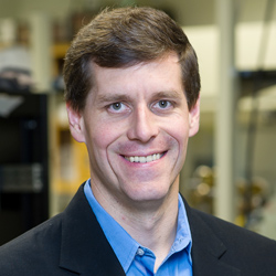
hersam-mark


Mark Hersam


Both academic and industrial laboratories are working to develop computers that operate more like the human brain. Instead of operating like a conventional, digital system, these new devices could potentially function more like a network of neurons.


“Computers are very impressive in many ways, but they’re not equal to the mind,” said Mark Hersam, the Bette and Neison Harris Chair in Teaching Excellence in Northwestern University’s McCormick School of Engineering. “Neurons can achieve very complicated computation with very low power consumption compared to a digital computer.”


A team of Northwestern researchers, including Hersam, has accomplished a new step forward in electronics that could bring brain-like computing closer to reality. The team’s work advances memory resistors, or “memristors,” which are resistors in a circuit that “remember” how much current has flowed through them.


The research is described in the April 6 issue of Nature Nanotechnology. Tobin Marks, the Vladimir N. Ipatieff Professor of Catalytic Chemistry, and Lincoln Lauhon, professor of materials science and engineering, are also authors on the paper. Vinod Sangwan, a postdoctoral fellow co-advised by Hersam, Marks, and Lauhon, served as first author. The remaining co-authors—Deep Jariwala, In Soo Kim, and Kan-Sheng Chen—are members of the Hersam, Marks, and/or Lauhon research groups.


“Memristors could be used as a memory element in an integrated circuit or computer,” Hersam said. “Unlike other memories that exist today in modern electronics, memristors are stable and remember their state even if you lose power.”


Current computers use random access memory (RAM), which moves very quickly as a user works but does not retain unsaved data if power is lost. Flash drives, on the other hand, store information when they are not powered but work much slower. Memristors could provide a memory that is the best of both worlds: fast and reliable. But there’s a problem: memristors are two-terminal electronic devices, which can only control one voltage channel. Hersam wanted to transform it into a three-terminal device, allowing it to be used in more complex electronic circuits and systems.


Hersam and his team met this challenge by using single-layer molybdenum disulfide (MoS2), an atomically thin, two-dimensional nanomaterial semiconductor. Much like the way fibers are arranged in wood, atoms are arranged in a certain direction—called “grains”—within a material. The sheet of MoS2 that Hersam used has a well-defined grain boundary, which is the interface where two different grains come together.


“Because the atoms are not in the same orientation, there are unsatisfied chemical bonds at that interface,” Hersam explained. “These grain boundaries influence the flow of current, so they can serve as a means of tuning resistance.”


When a large electric field is applied, the grain boundary literally moves, causing a change in resistance. By using MoS2 with this grain boundary defect instead of the typical metal-oxide-metal memristor structure, the team presented a novel three-terminal memristive device that is widely tunable with a gate electrode.


“With a memristor that can be tuned with a third electrode, we have the possibility to realize a function you could not previously achieve,” Hersam said. “A three-terminal memristor has been proposed as a means of realizing brain-like computing. We are now actively exploring this possibility in the laboratory.”


Story Source:


The above story is based on materials provided by Northwestern University.


Engineers gain control of gene activity

from

BIOENGINEER.ORG http://bioengineer.org/engineers-gain-control-of-gene-activity/


Duke researchers have developed a new method to precisely control when genes are turned on and active. The new technology allows researchers to turn on specific gene promoters and enhancers—pieces of the genome that control gene activity—by chemically manipulating proteins that package DNA. This web of biomolecules that supports and controls gene activity is known as the epigenome.


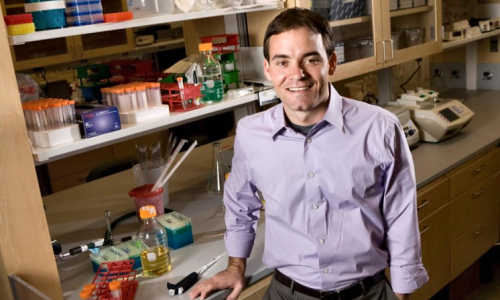
Charles Gersbach


This is Charles Gersbach, assistant professor of biomedical engineering at Duke University. Photo Credit: Duke University


The researchers say having the ability to steer the epigenome will help them explore the roles that particular promoters and enhancers play in cell fate or the risk for genetic disease and it could provide a new avenue for gene therapies and guiding stem cell differentiation.


The study appears online April 6 in Nature Biotechnology.

“The epigenome is everything associated with the genome other than the actual genetic sequence, and is just as important as our DNA in determining cell function in healthy and diseased conditions,” said Charles Gersbach, assistant professor of biomedical engineering at Duke. “That becomes immediately obvious when you consider that we have over 200 cell types, and yet the DNA in each is virtually the same. The epigenome determines which genes each cell activates and to what degree.”


This genetic puppetmaster consists of DNA packaging proteins called histones and a host of chemical modifications—either to these histones or the DNA itself—that help determine whether a gene is on or off.


But Gersbach’s team didn’t have to modify the genes themselves to gain some control.


“Next to every gene is a DNA sequence called a promoter that controls its activity,” explained Gersbach. “But there’s also many other pieces of the genome called enhancers that aren’t next to any genes at all, and yet they play a critical role in influencing gene activity too.”


Timothy Reddy, assistant professor of biostatistics and bioinformatics at Duke, has spent the better part of a decade mapping millions of these enhancers across the human genome. There has not, however, been a good way to find out exactly what each one does. An enhancer might affect a gene next door or several genes across the genome—or maybe none at all.


To activate these enhancers and see what they do, Reddy thought perhaps he could chemically alter the histones at the enhancers to turn them on.


“There are already drugs that will affect enhancers across the whole genome, but that’s like scorching the earth,” said Reddy. “I wanted to develop tools to go in and modify very specific epigenetic marks in very specific places to find out what individual enhancers are doing.”


Reddy found that specificity by teaming up with Gersbach, his neighbor within Duke’s Center for Genomic and Computational Biology, who specializes in a gene-targeting system called CRISPR. Originally discovered as a natural antiviral system in bacteria, researchers have hijacked the system over the past few years and are now using it to cut and paste DNA sequences in the human genome.


For this epigenome editing application, Gersbach silenced the DNA-cutting mechanism of CRISPR and used it solely as a targeting system to deliver an enzyme (acetyltransferase) to specific promoters and enhancers.


“It’s like we use CRISPR to find a genetic address so that we can alter the DNA’s packaging at that specific site,” said Reddy.


Gersbach and Reddy put their artificial epigenetic agent to the test by targeting a few well-studied gene promoters and enhancers. While these histone modifications have long been associated with gene activity, it wasn’t clear if they were enough to turn genes on. And though Gersbach and Reddy had previously used other technologies to activate gene promoters, they had not successfully activated enhancers.


To the duo’s great surprise, not only did the agent activate the gene promoters, it turned on the adjacent genes better than their previous methods. Equally surprising was that it worked on enhancers as well: they could turn on a gene—or even families of genes—by targeting enhancers at distant locations in the genome—something that their previous gene activators could not do.


But the real excitement from their results is an emerging ability to probe millions of potential enhancers in a way never before possible.


“Some genetic diseases are straightforward—if you have a mutation within a particular gene, then you have the disease,” said Isaac Hilton, postdoctoral fellow in the Gersbach Lab and first author of the study. “But many diseases, like cancer, cardiovascular disease or neurodegenerative conditions, have a much more complex genetic component. Many different variations in the genome sequence can affect your risk of disease, and this genetic variation can occur in these enhancers that Tim has identified, where they can change the levels of gene expression. With this technology, we can explore what exactly it is that they’re doing and how it relates to disease or response to drug therapies.”


Story Source:


The above story is based on materials provided by Duke University.


Sound separates cancer cells from blood samples

from

BIOENGINEER.ORG http://bioengineer.org/sound-separates-cancer-cells-from-blood-samples/


Separating circulating cancer cells from blood cells for diagnostic, prognostic and treatment purposes may become much easier using an acoustic separation method and an inexpensive, disposable chip, according to a team of engineers.


“Looking for circulating tumor cells in a blood sample is like looking for a needle in a haystack,” said Tony Jun Huang, professor of engineering science and mechanics. “Typically, the CTCs are about one in every one billion blood cells in the sample.”


Fig 1 revised v3


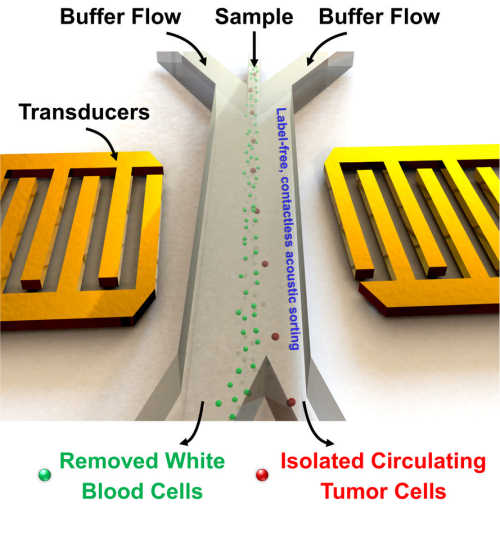
Schematic of acoustic tweezer separating cancer cells from blood cells. Yellow transducer supply sound to move the cells as they pass through in a stream. Photo Credits: Tony Jun Huang, Penn State


Existing methods of separation use tumor-specific antibodies to bind with the cancer cells and isolate them, but require that the appropriate antibodies be known in advance. Other methods rely on size, deformability or electrical properties. Unlike conventional separation methods that centrifuge for 10 minutes at 3000 revolutions per minute, surface acoustic waves can separate cells in a much gentler way with a simple, low-cost device.


Acoustic-based separations are potentially important because they are non-invasive and do not alter or damage cells. However, in order to be effective for clinical use, they also need to be rapidly and easily applicable.


“In order to significantly increase the throughput for capturing those rare CTCs, device design has to be optimized for much higher flow rates and longer acoustic working length,” said Ming Dao, principal research scientist, materials science and engineering, Massachusetts Institute of Technology. “With an integrated experimental/modeling approach, the new generation of the device has improved cell sorting throughput more than 20 times higher than previously achieved and made it possible for us to work with patient samples.”


The researchers worked both experimentally and with models to optimize the separation of CTCs from blood. They used an acoustic-based microfluidic device so that the stream of blood could continuously pass through the device for separation. Using the differential size and weight of the different cells they chose appropriate acoustic pressures that would push the CTCs out of the fluid stream and into a separate channel for collection. They report their results today (Apr. 6) in the Proceedings of the National Academy of Sciences.


Tilted-angle standing surface acoustic waves can separate cells using very small amounts of energy. The power intensity and frequency used in this study are similar to those used in ultrasonic imaging, which has proven to be extremely safe, even for fetuses. Also, each cell experiences the acoustic wave for only a fraction of a second. In addition, cells do not require labeling or surface modification. All these features make the acoustic separation method, termed acoustic tweezers, extremely biocompatible and maximize the potential of CTCs to maintain their functions and native states.


If two sound sources are placed opposite each other and each emits the same wavelength of sound, there will be a location where the opposing sounds cancel each other. Because sound waves have pressure, they can push very small objects, so a cell or nanoparticle will move with the sound wave until it reaches the location where there is no longer lateral movement, in this case, into the fluid stream that moves the separated cells along.


The researchers used two types of human cancer cells to optimize the acoustic separation — HELA cells and MCF7 cells. These cells are similar in size. They then ran an experiment separating these cells and had a separation rate of more than 83 percent. They then did the separation on other cancer cells, ones for which the device had not been optimized, and again had a separation rate of more than 83 percent.


“Because these devices are intended for use with human blood, they need to be disposable,” said Huang. “We are currently figuring out manufacturing and mass production possibilities.”


Physicians could use the devices to monitor how patients reacted to chemotherapy, for initial diagnosis and for determining treatment and prognosis.


“This work, involving a highly cross-disciplinary group of medical doctors, engineers, computational biologists, and device experts, has led to the design and development of a label-free platform for identifying and separating CTCs while preserving the integrity of the cell,” said Subra Suresh, president, Carnegie Mellon University and part of the research team. “It promises to offer new avenues for basic research into the pathology and metastasis, and for clinical diagnosis of rare tumor cells.”


Story Source:


The above story is based on materials provided by Penn State.