26 Nisan 2015 Pazar

Beyond genes: are centrioles carriers of biological information?

from
BIOENGINEER.ORG http://bioengineer.org/beyond-genes-are-centrioles-carriers-of-biological-information/

EPFL scientists discover that certain cell structures, the centrioles, could act as information carriers throughout cell generations. The discovery raises the possibility that transmission of biological information could involve more than just genes.

Electron micrograph of a centriole

Centrioles are barrel-shaped structures inside cells, made up of multiple proteins. They are currently the focus of much research, since mutations in the proteins that make them up can cause a broad range of diseases, including developmental abnormalities, respiratory conditions, male sterility and cancer. Publishing in the Nature journal Cell Research, EPFL scientists show that the original centrioles of a fertilized egg, which only come from the father, persist across tens of cell divisions in the developing embryo. The surprising finding raises the possibility that centrioles may actually be carriers of information, with profound implications for biology and disease treatment.

Perhaps best known for their role in cell division, centrioles ensure that chromosomes are properly passed on to the new daughter cells. However, they are also found in cilia, the long eyelash-like structures that allow many cells in the body to signal to their neighbors and other cells to exhibit motility, e.g. in cells that line the respiratory tracts. During reproduction, both parents equally contribute genetic material, while the female egg donates most of the cell organelles, such as mitochondria. However, the centrioles of the newly fertilized embryo come exclusively from the male’s sperm, bringing with them any malfunctions to the first embryo cells.

Passing information across generations

The lab of Pierre Gönczy at EPFL’s Swiss Institute for Experimental Cancer Research has found that centrioles can carry such information beyond the first cells to many of a developing embryo to several cell generations. The study focused on the worm C. elegans, which is commonly used as a model organism for embryonic development and human genetic diseases. As in other species, including humans, centrioles in C. elegans are only contributed by sperm cells. Gönczy’s team wanted to know how far do these “original” centrioles last across the cell divisions that turn a fertilized egg into a fully formed embryo.

In order to track the fate of the centrioles, the scientists used genetically modified versions of C. elegans, in which they could tag three different centriole proteins with a fluorescent signal. Tagged male worms were mated to untagged females, so that the scientists could specifically track centriole components that were contributed from the father during the course of embryogenesis.

Gönczy’s team imaged the fluorescent signals at different cell divisions of the developing embryos, and discovered that paternally contributed centriole proteins can actually persist up to ten cell generations. The data show for the first time that centrioles are remarkably persistent in the developing embryo.

Even more intriguing are the implications the study has for biology at large, as it raises the possibility that centrioles, persisting across several cell cycles, could effectively be a non-genetic information carrier. If this were confirmed, it could represent a paradigm shift in the way we think and understand the biology of an organelle that has been present across eukaryotic evolution.

Nonetheless, this does not detract from the value this study holds for medicine. Considering the number of diseases associated with centrioles, this could open the way for innovative treatment approaches. In particular, the study demonstrates how malfunctioning centrioles can pass directly from the father and well into the life of the embryo. This can have serious implications for the way we understand centriole diseases.

“Centrioles have always been seen as something that just jumpstarts the development of the embryo,” says Pierre Gönczy. “Here we show that centrioles could be the means of a unidirectional inheritance of information, with considerable impact in early development.” His team will next investigate if the exceptional persistence of centrioles extends to other systems, including human cells.

Story Source:

The above story is based on materials provided by Ecole Polytechnique Fédérale de Lausanne

11 Nisan 2015 Cumartesi

Touch-sensing neurons are multitaskers

from

BIOENGINEER.ORG http://bioengineer.org/touch-sensing-neurons-are-multitaskers/


Two types of touch information — the feel of an object and the position of an animal’s limb — have long been thought to flow into the brain via different channels and be integrated in sophisticated processing regions. Now, with help from a specially devised mechanical exoskeleton that positioned monkeys’ hands in different postures, Johns Hopkins researchers have challenged that view. In a paper published in the April 22 issue of Neuron, they present evidence that the two types of information are integrated as soon as they reach the brain by sense-processing brain cells once thought to be incapable of such higher-order thought.


Past studies have indicated that the feel of an object against the skin and position information of the hands and fingers were processed separately in the sensory system’s first-line sense processors, and then passed along to more sophisticated brain regions to be integrated. But it was a challenge to reliably differentiate brain activity caused by the two inputs from one another and from the brain’s commands to muscles, says Manuel Gomez-Ramirez, Ph.D., an assistant research scientist at The Johns Hopkins University. To solve that problem, Steven Hsiao, Gomez-Ramirez’s late mentor, and his colleagues developed a machine that positions a monkey’s hand and delivers stimuli to its fingers.


In this experiment, then-graduate student Sung Soo Kim, Ph.D., now a research specialist at the Howard Hughes Medical Institute, trained monkeys to perform an unrelated visual task while their hands were manipulated by the machine, which moved their fingers slightly from side to side and up and down at precise angles. The machine also pressed a plastic bar to the monkeys’ fingertips in different orientations. By monitoring the monkeys’ brains in real time, the research group saw that the position and touch information were conveyed through the same cells in the somatosensory cortex.


“This study changes our understanding of how position and touch signals are combined in the brain,” says Gomez-Ramirez. This information could be used in efforts to better integrate prostheses with patients’ brains so that they behave more like natural limbs, he notes.


“Holding objects and exploring the world with our hands requires integrating many sensory signals in the brain and continuously supplying this information to motor areas so that it can issue the appropriate commands for holding objects,” Gomez-Ramirez says. “Our understanding of how these processes occur is very limited, and Steve Hsiao spent a lot of time thinking about the problem and figuring out how to test it.”


Story Source:


The above story is based on materials provided by Johns Hopkins Medicine.


Synthetic muscle ready for launch

from

BIOENGINEER.ORG http://bioengineer.org/synthetic-muscle-ready-for-launch/


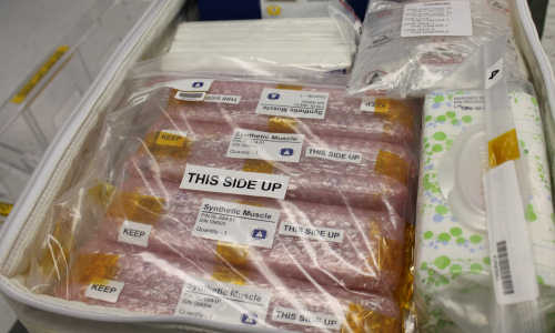
Lenore Rasmussen’s dream of developing a synthetic muscle that could be used to make better prosthetic limbs and more responsive robots will literally become airborne on Monday April 13 at 4:33 p.m. when her experiment will rocket off to the International Space Station from Cape Canaveral in Florida.


synthetic muscle


Rasmussen developed the material at RAS Labs and has worked closely with researchers and engineers at the U.S. Department of Energy’s Princeton Plasma Physics Laboratory (PPPL) to develop the material’s ability to adhere to metal. The Synthetic Muscle™ could be used in robotics in deep space travel such as travel to Mars because of its radiation resistance.


“Based on the good results we had on planet Earth, the next step is to see how it behaves in a space environment,” said Charles Gentile, an engineer at PPPL who has worked closely with Rasmussen. “From there the next step might be to use it on a mission to Mars.”


Early Connection with PPPL


Rasmussen began working with PPPL in 2007 just four years after she started Ras Labs. She received her first patent for a synthetic muscle in 1998. It is a gel-like material called an electroactive polymer that can potentially mimic human movement because it can expand and contract to simulate the movement of muscles in humans. That ability would make it very useful in robotics and in developing better prosthetic limbs.


“We can’t explore space without robots,” Rasmussen said. “Humans can only withstand a certain amount of radiation so that limits the time that people can be in space, whereas robots particularly if they’re radiation-resistant can be up there for long periods of time without being replaced.”


Lew Meixler, the long-time head of Technology Transfer at PPPL, who retired in March, said he has enjoyed helping Rasmussen follow her quest. “That’s what entrepreneurs are,” he said. “They’re the dreamers who devote all their time, energy and resources to following their dreams.”


Rasmussen credits PPPL with providing help and support during critical points in her project. “It was and continues to be a wonderful resource not just because of the plasma physics but the people,” she said. “Charlie and Lew found ways to make things happen.”


At PPPL, Rasmussen solved a crucial problem: getting the gel, which can be as soft as jelly or as hard as rubber, to adhere to the metal electrodes. Initially working with Lew Meixler on a federal Cooperative Research and Development Agreement in the Plasma Surface Laboratory, she solved the problem by treating the metal (steel or titanium) with a plasma. This changed the metal’s surface and made the gel adhere more closely to the metal


PPPL was also involved with tests of the material last summer, when the material was exposed to over 300,000 RADs of gamma radiation. That is 20 times the amount that would be lethal to a human and was equivalent to a trip from earth to Mars and back. A second test of 45 hours was enough to be equivalent to a trip to Jupiter and beyond


Rasmussen and Gentile found that there was no change in the strength, electroacivity, or durability of the material due to the radiation, although there was a slight change in color. Tests on selected samples of the material found it was not affected by extreme temperatures down to -271 degrees Celsius, which is close to absolute zero, the coldest temperature possible in the universe.


Preparing for launch


Since then, PPPL staff members have been involved in planning for the launch. This involves mapping out each detail with military precision. Several PPPL staff members, along with Rasmussen and her staff, signed the back of the metal container or coupon holding the material. “All of the people who worked on the lab signed it and the coupon will go into space,” said Gentile. “So I’ll be up there with Gene Roddenberry.”


The Synthetic Muscle™ material will be launched on the Falcon 9, a rocket carrying the Dragon spacecraft, both produced by Space X, which will carry 4,300 pounds of supplies and payloads, including material for research experiments, to the International Space Station’s U.S. National Laboratory. The nine-engine rocket will propel the Dragon into orbit where it will meet with the Space Station 33 hours after the rocket is launched. Astronauts will use the station’s 57-foot arm to reach out and capture Dragon at 7:15 a.m. on April 15. Additional information about the launch is available at https://blogs.nasa.gov/spacex/2015/03/31/spacex-targeting-april-13-for-station-resupply-launch/ (link is external).


The material will be kept in a zero gravity storage rack in the U.S. National Laboratory on the space station for 90 days. The astronauts will photograph the materials every three weeks. When the material returns to Earth in July, it will be tested and compared with identical materials that remained on Earth.


The International Space Station is an international science laboratory in low Earth orbit where astronauts conduct scientific research in biology, human biology, astronomy, meteorology and other fields in a gravity-free environment. It has operated since November of 2000 with the cooperation of the U.S., Russia, many European nations, Japan, Canada, and Brazil. It is currently staffed by two astronauts from NASA, three cosmonauts from Russia and an astronaut from the European Space Agency.


Use as a prosthetic


Rasmussen is also exploring whether Synthetic Muscle™ could be used as a prosthetic liner. The vestigial limbs of amputees can expand and contract during the day and the Ras Labs material is designed to expand and contract so it could make prosthetics more comfortable. She recently received a grant from the Pediatric Medical Device Consortium at the Children’s Hospital of Philadelphia to research this possibility.


Story Source:


The above story is based on materials provided by Princeton Plasma Physics Laboratory.


9 Nisan 2015 Perşembe

What can brain-controlled prosthetics tell us about the brain?

from

BIOENGINEER.ORG http://bioengineer.org/what-can-brain-controlled-prosthetics-tell-us-about-the-brain/


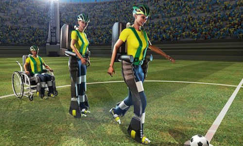
The ceremonial opening kick of the 2014 FIFA World Cup in Sao Paolo, Brazil, which was performed—with the help of a brain-controlled exo-skeleton—by a local teen who had been paralyzed from the waste down due to a spinal cord injury, was a seminal moment for the area of neuroscience that strives to connect the brain with functional prosthetics.


prostetics


A graphic illustration of the brain-controlled exoskeleton used for the opening kick of the 2014 FIFA World Cup. Technology like this could help neuroscientists better understand brain activity. Photo Credit: Drexel University


The public display was a representative of thousands of such neuroprosthetic advances in recent years, and the tens of years of brain research and technological development that have gone into them. And while this display was quite an achievement in its own right, a Drexel University biomedical engineer working at the leading edge of the field contends that these devices are also opening a new portal for researchers to understand how the brain functions.


Karen Moxon, PhD, a professor in Drexel’s School of Biomedical Engineering Science and Health Systems, was a postdoctoral researcher in Drexel’s medical school when she participated in the first study ever to examine how the brain could be connected to operate a prosthetic limb. More than 15 years after that neuroscience benchmark, Moxon’s lab is showing that it’s now possible to glean new insight about how the brain stores and accesses information, and into the causes of pathologies like epilepsy and Parkinson’s disease.


In a perspective published in the latest edition of the neuroscience journal Neuron Moxon and her colleague, Guglielmo Foffani from San Pablo University in Spain, build a framework for how researchers can use neuroprosthetics as a tool for examining how and where the brain encodes new information. The duo highlights three examples from their own research where the brain-machine-interface technology allowed them to isolate and study new areas of brain function.


“We believe neuroprosthetics can be a powerful tool to address fundamental questions of neuroscience,” Moxon said. “These subjects can provide valuable data as indirect observers of their own neural activity that are modulated during the experiments they are taking part in. This allows researchers to pinpoint a causal relationship between neural activity and the subject’s behavior rather than one that is indirectly correlative.”


The challenge faced by all scientists who study the brain is proving a direct relationship between the action of the subject and the behavior of brain cells. Each experiment is designed to chisel away at the uncertainty in this relationship with the goal of establishing causality—proof that the behavior of neurons in the brain is actually what is causing a subject to perform a certain action. Or, conversely, that a certain neural behavior is the direct result of an external stimulus.


Neuroprosthetics, according to Moxon, could be the way around this obstacle. This is because the prosthetic, as a stand-in for an actual body part or set of them, is also a vehicle for getting real-time feedback from the brain.


“Subjects can be viewed as indirect observers of their own neurophysiological activity during neuroprosthetic experiments,” Moxon said. “To move the prosthesis they must think both about the motor functions involved and the goal of the movement. As they see the movement of the prosthetic their brain adjusts in real time to continue planning the movement, but doing it without the normal feedback from the moving body part—as the prosthetic technology is standing in for that part of the body.”


This separation of planning and movement control was pivotal to Moxon’s research on how the brain encodes for the passage of time, which she recently reported in the Journal of Neuroscience. But this is just one example of how brain-machine-interface technology can be used to experimentally tease out and observe new certainties about the brain.


Moxon, who was recently elected a fellow of the American Association for the Advancement of Science and the American Institute for Medical and Biological Engineers, suggests that in addition to the study of how neurons encode and decode information in real time, incorporating neuroprosthetics into experiments could also show how this coding process changes with learning and is altered in pathological conditions like the ones that cause epilepsy and Parkinson’s disease.


“While the past 15 years have witnessed tremendous advancements in neuroprosthetic technology and our basic understanding of brain function, the brain-machine-interface approach is still expanding the landscape of neuroscienctific inquiry,” Moxon said. “By circumventing classical object-observer duality, the BMI research paradigm opens doors for a new understanding of how we control our own brain function including neural plasticity—and this has the potential to lead to new treatments and therapies for epilepsy, Parkinson’s and other pathologies.”


Story Source:


The above story is based on materials provided by Drexel University.


Fountain of youth uncovered in mammary glands of mice

from

BIOENGINEER.ORG http://bioengineer.org/fountain-of-youth-uncovered-in-mammary-glands-of-mice/


The Fountain of Youth has been discovered and it’s not in Florida as Ponce de Leon claimed. Instead, it was found in the mammary glands of genetically modified mice. A research team led by Professor Rama Khokha has found that when two factors that control tissue development are removed, you can avoid the impact of aging.


youth


Professor Rama Khokha and Dr. Hartland Jackson Photo Credits: University Health Network.


Think of tissue as a building that is constantly under renovation. The contractors would be “metalloproteinases,” which are constantly working to demolish and reconstruct the tissue. The architects in this case, who are trying to reign in and direct the contractors, are known as “tissue inhibitors of metalloproteinases” — or TIMPs. When the architect and the contractors don’t communicate well, a building can fall down. In the case of tissue, the result can be cancer.


To understand how metalloproteinases and TIMPs interact, medical researchers breed mice that have one or more of the four different types of TIMPs removed. Khokha’s team examined the different combinations and found that when TIMP1 and TIMP3 were removed, breast tissue remained youthful in aged mice. The results are presented in Nature Cell Biology.


In the normal course of aging, your tissue losses its ability to develop and repair as fast as it did when you were young. That’s because stem cells, which are abundant in your youth, decline with the passing of time. The U of T team found that with the TIMP1 and TIMP3 architects missing, the pool of stem cells expanded and remained functional throughout the lifetime of these mice.


“Normally you would see these pools of stem cells, which reach their peak at six months in the mice, start to decline. As a result, the mammary glands start to degenerate, which increases the risk of breast cancer occurring,” explains Khokha. “However, we found that in these particular mice, the stem cells remained consistently high when we measured them at every stage of life.”


The team also found that despite large number of stem cells, there was no increased risk of cancer.


“It’s generally assumed that the presence of a large number of stem cells can lead to an increased cancer risk,” says Khokha. “However, we found these mice had no greater predisposition to cancer.”


The next step in this research is to understand why this is happening. Khokha is also working with her colleagues at Princess Margaret to see how altered tissue remodeling might prevent cancer development or lead to a new therapeutic treatment for patients.


Khokha is a Professor in the departments of Medical Biophysics and Laboratory Medicine and Pathobiology, as well as a Senior Scientist at the Princess Margaret Cancer Centre. Her work is supported by the Canadian Breast Cancer Foundation and the Canadian Cancer Society Research Institute.


She was drawn to this research by the complexity of breast tissue.


“It’s a fundamental tissue that is constantly reorganizing. It develops at puberty. It goes through cycles of change in the adult female. New structures appear and regress,” she explains. “It is therefore a good system to explore in order to understand tissue maintenance and epithelial cell turnover – the cells that underlie carcinomas, the most frequent type of cancer.”


She worked closely with the paper’s lead author, Dr. Hartland Jackson, who earned his PhD under her supervision. He is now completing a post-doctoral fellowship at the University of Zurich’s Institute of Molecular Life Sciences.


“He’s continuing his work in breast cancer and learning some really interesting techniques that, I hope, he’ll bring back here,” says Khokha, who beams with pride at her former student’s success.


Story Source:


The above story is based on materials provided by University of Toronto.


8 Nisan 2015 Çarşamba

Purging a virus from organ transplants

from

BIOENGINEER.ORG http://bioengineer.org/purging-a-virus-from-organ-transplants/


EPFL scientists have discovered the switch that can awake a dormant cytomegalovirus, a dreadful pathogen in immuno-compromised patients. The switch can be controlled with common drugs, opening a new strategy for purging the virus from organ transplants.


infect


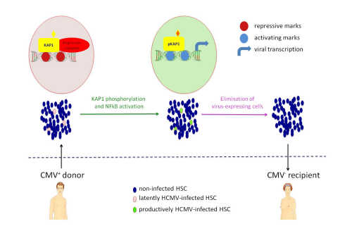
Human cytomegalovirus (HCMV) is an extremely common herpes-family virus that infects people for life. It infects 60% of the population in industrialized countries, and almost everybody in poorer regions. Its symptoms are easily fought off by a healthy immune system, but can be devastating to individuals with defective immunity, e.g. newborn babies, people with AIDS, or those taking immunosuppressive drugs after receiving organ transplants. After infection, HCMV hides (“lies dormant”) in blood-making stem cells, occasionally reactivating as these cells mature. Scientists at EPFL have now discovered the molecular switch that allows HCMV to either lie dormant in these cells or reactivate and start infecting again. The switch can be manipulated with simple drugs to force HCMV reactivation, making the virus easy to target and kill. Published in eLife, the study shows how HCMV could be fought in high-risk patients and purged from organs before transplantation.


Throwing the switch


The lab of Didier Trono at EPFL discovered a protein that switches HCMV between dormancy and reactivation. They found this protein to be bound to the HCMV genome in latently infected hematopoietic stem cells and, upon a variety of external stimuli, to undergo a modification that allows for viral activation. The researchers were able to control this switch with a drug called chloroquine, usually used against malaria. When they treated blood-making stem cells that contained dormant HCMV with chloroquine, the virus reactivated and became exposed, opening the door to maneuvers aimed at eliminating virus-infected cells.


The simplicity of the study’s design underlies its enormous significance. On one hand, it sheds light on the molecular mechanism by which HCMV becomes dormant in hematopoietic stem cells, possibly offering insights into similar infections by other herpes viruses. On the other hand, the study provides a straightforward method for forcing HCMV out of dormancy in infected tissue. Coupled with a simultaneous dose of an antiviral, this could become a standard regimen for eradicating HCMV from high-risk patients and purging it from tissue before transplantation.


Trono’s team is now testing the method’s efficiency in purging HCMV from cells to be used for bone marrow transplantation. Following that step, the group will be developing the first trials in humans.


Story Source:


The above story is based on materials provided by Ecole Polytechnique Fédérale de Lausanne.


Photo credit: Ben Edwards


A Tiny Hair Follicle Holds Big Clues About Stem Cells

from

BIOENGINEER.ORG http://bioengineer.org/a-tiny-hair-follicle-holds-big-clues-about-stem-cells/


Inside the microscopic world of the mouse hair follicle, Yale Cancer Center researchers have discovered big clues about how stem cells regenerate and die. These findings, reported in the journal Nature, could lead to a better understanding of how the stem cell pool is maintained or altered in tissues throughout the body.


Stem cells are undifferentiated cells that replenish themselves and based on their tissue location can become specialized cells such as blood or skin cells. The hair follicle is an ideal site for exploring stem cell behavior because it has distinct and predictable oscillations in the number and behavior of stem cells, said the study’s lead author Kailin R. Mesa, a third-year doctoral student in the lab of Valentina Greco, associate professor of genetics, cell biology and dermatology.


Using live microscopic imaging to track stem cell behavior in the skin of living mice, researchers observed that the stem cell niche, or surrounding area, played a critical role in whether stem cells grow or die.


“Prior to this, it wasn’t clear whether stem cell regulation was intrinsic or extrinsic, and now we know it is external in that the niche instructs the stem cells,” Mesa said. “In terms of cancer, we can next explore how we might perturb or change the niche in hopes of affecting the growth of cancer stem cells.”


Also, researchers were surprised to find that the stem cells within the pool fed on other dying stem cells. This reveals a mechanism for removing dead cells, a process previously observed in mammary glands but never in the skin.


Story Source:


The above story is based on materials provided by Yale Cancer Center.


Photo Credits: Shutterstock via Yale Cancer Center