1 Temmuz 2014 Salı

Noninvasive brain control

from

Bioengineer.org http://bioengineer.org/noninvasive-brain-control/


Optogenetics, a technology that allows scientists to control brain activity by shining light on neurons, relies on light-sensitive proteins that can suppress or stimulate electrical signals within cells. This technique requires a light source to be implanted in the brain, where it can reach the cells to be controlled.


Noninvasive brain control


Illustration: Jose-Luis Olivares/MIT


MIT engineers have now developed the first light-sensitive molecule that enables neurons to be silenced noninvasively, using a light source outside the skull. This makes it possible to do long-term studies without an implanted light source. The protein, known as Jaws, also allows a larger volume of tissue to be influenced at once.


This noninvasive approach could pave the way to using optogenetics in human patients to treat epilepsy and other neurological disorders, the researchers say, although much more testing and development is needed. Led by Ed Boyden, an associate professor of biological engineering and brain and cognitive sciences at MIT, the researchers described the protein in the June 29 issue of Nature Neuroscience.

Optogenetics, a technique developed over the past 15 years, has become a common laboratory tool for shutting off or stimulating specific types of neurons in the brain, allowing neuroscientists to learn much more about their functions.


The neurons to be studied must be genetically engineered to produce light-sensitive proteins known as opsins, which are channels or pumps that influence electrical activity by controlling the flow of ions in or out of cells. Researchers then insert a light source, such as an optical fiber, into the brain to control the selected neurons.


Such implants can be difficult to insert, however, and can be incompatible with many kinds of experiments, such as studies of development, during which the brain changes size, or of neurodegenerative disorders, during which the implant can interact with brain physiology. In addition, it is difficult to perform long-term studies of chronic diseases with these implants.


Mining nature’s diversity


To find a better alternative, Boyden, graduate student Amy Chuong, and colleagues turned to the natural world. Many microbes and other organisms use opsins to detect light and react to their environment. Most of the natural opsins now used for optogenetics respond best to blue or green light.


Boyden’s team had previously identified two light-sensitive chloride ion pumps that respond to red light, which can penetrate deeper into living tissue. However, these molecules, found in the bacteria Haloarcula marismortui and Haloarcula vallismortis, did not induce a strong enough photocurrent — an electric current in response to light — to be useful in controlling neuron activity.


Chuong set out to improve the photocurrent by looking for relatives of these proteins and testing their electrical activity. She then engineered one of these relatives by making many different mutants. The result of this screen, Jaws, retained its red-light sensitivity but had a much stronger photocurrent — enough to shut down neural activity.


“This exemplifies how the genomic diversity of the natural world can yield powerful reagents that can be of use in biology and neuroscience,” says Boyden, who is a member of MIT’s Media Lab and the McGovern Institute for Brain Research.


Using this opsin, the researchers were able to shut down neuronal activity in the mouse brain with a light source outside the animal’s head. The suppression occurred as deep as 3 millimeters in the brain, and was just as effective as that of existing silencers that rely on other colors of light delivered via conventional invasive illumination.


A key advantage to this opsin is that it could enable optogenetic studies of animals with larger brains, says Garret Stuber, an assistant professor of psychiatry and cell biology and physiology at the University of North Carolina at Chapel Hill.


“In animals with larger brains, people have had difficulty getting behavior effects with optogenetics, and one possible reason is that not enough of the tissue is being inhibited,” he says. “This could potentially alleviate that.”


Restoring vision


Working with researchers at the Friedrich Miescher Institute for Biomedical Research in Switzerland, the MIT team also tested Jaws’s ability to restore the light sensitivity of retinal cells called cones. In people with a disease called retinitis pigmentosa, cones slowly atrophy, eventually causing blindness.


Friedrich Miescher Institute scientists Botond Roska and Volker Busskamp have previously shown that some vision can be restored in mice by engineering those cone cells to express light-sensitive proteins. In the new paper, Roska and Busskamp tested the Jaws protein in the mouse retina and found that it more closely resembled the eye’s natural opsins and offered a greater range of light sensitivity, making it potentially more useful for treating retinitis pigmentosa.


This type of noninvasive approach to optogenetics could also represent a step toward developing optogenetic treatments for diseases such as epilepsy, which could be controlled by shutting off misfiring neurons that cause seizures, Boyden says. “Since these molecules come from species other than humans, many studies must be done to evaluate their safety and efficacy in the context of treatment,” he says.


Boyden’s lab is working with many other research groups to further test the Jaws opsin for other applications. The team is also seeking new light-sensitive proteins and is working on high-throughput screening approaches that could speed up the development of such proteins.


Story Source:


The above story is based on materials provided by MIT News Office, Anne Trafton.


The post Noninvasive brain control appeared first on Bioengineer.org.


23 Haziran 2014 Pazartesi

Seeing the inner workings of brain made easier by new technique

from

Bioengineer.org http://bioengineer.org/seeing-inner-workings-brain-made-easier-new-technique/


Last year Karl Deisseroth, a Stanford professor of bioengineering and of psychiatry and behavioral sciences, announced a new way of peering into a brain – removed from the body – that provided spectacular fly-through views of its inner connections. Since then laboratories around the world have begun using the technique, called CLARITY, with some success, to better understand the brain’s wiring.


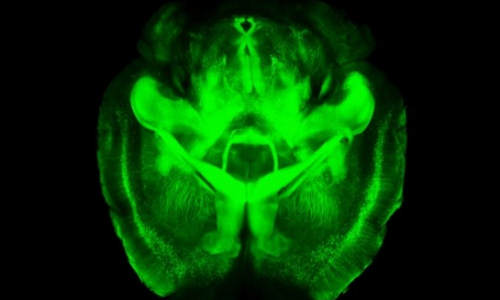
A three-dimensional rendering of clarified brain imaged from below


A three-dimensional rendering of clarified brain imaged from below. Photo Credit: Deisseroth lab


However, Deisseroth said that with two technological fixes CLARITY could be even more broadly adopted. The first problem was that laboratories were not set up to reliably carry out the CLARITY process. Second, the most commonly available microscopy methods were not designed to image the whole transparent brain. “There have been a number of remarkable results described using CLARITY,” Deisseroth said, “but we needed to address these two distinct challenges to make the technology easier to use.”


In a Nature Protocols paper published June 19, Deisseroth presented solutions to both of those bottlenecks. “These transform CLARITY, making the overall process much easier and the data collection much faster,” he said. He and his co-authors, including postdoctoral fellows Raju Tomer and Li Ye and graduate student Brian Hsueh, anticipate that even more scientists will now be able to take advantage of the technique to better understand the brain at a fundamental level, and also to probe the origins of brain diseases.


This paper may be the first to be published with support of the White House BRAIN Initiative, announced last year with the ambitious goal of mapping the brain’s trillions of nerve connections and understanding how signals zip through those interconnected cells to control our thoughts, memories, movement and everything else that makes us us.


“This work shares the spirit of the BRAIN Initiative goal of building new technologies to understand the brain – including the human brain,” said Deisseroth, who is also a Stanford Bio-X affiliated faculty member.


Eliminating fat


When you look at the brain, what you see is the fatty outer covering of the nerve cells within, which blocks microscopes from taking images of the intricate connections between deep brain cells. The idea behind CLARITY was to eliminate that fatty covering while keeping the brain intact, complete with all its intricate inner wiring.


The way Deisseroth and his team eliminated the fat was to build a gel within the intact brain that held all the structures and proteins in place. They then used an electric field to pull out the fat layer that had been dissolved in an electrically charged detergent, leaving behind all the brain’s structures embedded in the firm water-based gel, or hydrogel. This is called electrophoretic CLARITY.


The electric field aspect was a challenge for some labs. “About half the people who tried it got it working right away,” Deisseroth said, “but others had problems with the voltage damaging tissue.” Deisseroth said that this kind of challenge is normal when introducing new technologies. When he first introduced optogenetics, which allows scientists to control individual nerves using light, a similar proportion of labs were not initially set up to easily implement the new technology, and ran into challenges.


To help expand the use of CLARITY, the team devised an alternate way of pulling out the fat from the hydrogel-embedded brain – a technique they call passive CLARITY. It takes a little longer, but still removes all the fat, is much easier and does not pose a risk to the tissue. “Electrophoretic CLARITY is important for cases where speed is critical, and for some tissues,” said Deisseroth, who is also the D.H. Chen Professor. “But passive CLARITY is a crucial advance for the community, especially for neuroscience.” Passive CLARITY requires nothing more than some chemicals, a warm bath and time.


Many groups have begun to apply CLARITY to probe brains donated from people who had diseases like epilepsy or autism, which might have left clues in the brain to help scientists understand and eventually treat the disease. But scientists, including Deisseroth, had been wary of trying electrophoretic CLARTY on these valuable clinical samples with even a very low risk of damage. “It’s a rare and precious donated sample, you don’t want to have a chance of damage or error,” Deisseroth said. “Now the risk issue is addressed, and on top of that you can get the data very rapidly.”


Fast CLARITY imaging in color


The second advance had to do this rapidity of data collection. In studying any cells, scientists often make use of probes that will go into the cell or tissue, latch onto a particular molecule, then glow green, blue, yellow or other colors in response to particular wavelengths of light. This is what produces the colorful cellular images that are so common in biology research. Using CLARITY, these colorful structures become visible throughout the entire brain, since no fat remains to block the light.


But here’s the hitch. Those probes stop working, or get bleached, after they’ve been exposed to too much light. That’s fine if a scientist is just taking a picture of a small cellular structure, which takes little time. But to get a high-resolution image of an entire brain, the whole tissue is bathed in light throughout the time it takes to image it point by point. This approach bleaches out the probes before the entire brain can be imaged at high resolution.


The second advance of the new paper addresses this issue, making it easier to image the entire brain without bleaching the probes. “We can now scan an entire plane at one time instead of a point,” Deisseroth said. “That buys you a couple orders of magnitude of time, and also efficiently delivers light only to where the imaging is happening.” The technique is called light sheet microscopy and has been around for a while, but previously didn’t have high enough resolution to see the fine details of cellular structures. “We advanced traditional light sheet microscopy for CLARITY, and can now see fine wiring structures deep within an intact adult brain,” Deisseroth said. His lab built their own microscope, but the procedures are described in the paper, and the key components are commercially available. Additionally, Deisseroth’s lab provides free training courses in CLARITY, modeled after his optogenetics courses, to help disseminate the techniques.


Brain imaging to help soldiers


The BRAIN Initiative is being funded through several government agencies including the Defense Advanced Research Projects Agency (DARPA), which funded Deisseroth’s work through its new Neuro-FAST program. Deisseroth said that like the National Institute of Mental Health (NIMH, another major funder of the new paper), DARPA “is interested in deepening our understanding of brain circuits in intact and injured brains to inform the development of better therapies.” The new methods Deisseroth and his team developed will accelerate both human- and animal-model CLARITY; as CLARITY becomes more widely used, it will continue to help reveal how those inner circuits are structured in normal and diseased brains, and perhaps point to possible therapies.


Other arms of the BRAIN Initiative are funded through the National Science Foundation (NSF) and the National Institutes of Health (NIH). A working group for the NIH arm was co-led by William Newsome, professor of neurobiology and director of the Stanford Neurosciences Institute, and also included Deisseroth and Mark Schnitzer, associate professor of biology and of applied physics. That group recently recommended a $4.5 billion investment in the BRAIN Initiative over the next 12 years, which NIH Director Francis Collins approved earlier this month.


Story Source:


The above story is based on materials provided by Stanford University, Amy Adams.


The post Seeing the inner workings of brain made easier by new technique appeared first on Bioengineer.org.


15 Haziran 2014 Pazar

‘Trust hormone’ oxytocin helps old muscle work like new

from

Bioengineer.org http://bioengineer.org/trust-hormone-oxytocin-helps-old-muscle-work-like-new/


UC Berkeley researchers have discovered that oxytocin — a hormone associated with maternal nurturing, social attachments, childbirth and sex — is indispensable for healthy muscle maintenance and repair, and that in mice it declines with age.


‘Trust hormone’ oxytocin helps old muscle work like new


The new study, published today (June 10) in the journal Nature Communications, presents oxytocin as the latest treatment target for age-related muscle wasting, or sarcopenia.


A few other biochemical factors in blood have been connected to aging and disease in recent years, but oxytocin is the first anti-aging molecule identified that is approved by the Food and Drug Administration for clinical use in humans, the researchers said. Pitocin, a synthetic form of oxytocin, is already used to help with labor and to control bleeding after childbirth. Clinical trials of an oxytocin nasal spray are also underway to alleviate symptoms associated with mental disorders such as autism, schizophrenia and dementia.


“Unfortunately, most of the molecules discovered so far to boost tissue regeneration are also associated with cancer, limiting their potential as treatments for humans,” said study principal investigator Irina Conboy, associate professor of bioengineering. “Our quest is to find a molecule that not only rejuvenates old muscle and other tissue, but that can do so sustainably long-term without increasing the risk of cancer.”


Conboy and her research team say that oxytocin, secreted into the blood by the brain’s pituitary gland, is a good candidate because it is a broad range hormone that reaches every organ, and it is not known to be associated with tumors or to interfere with the immune system.


A happy hormone


Oxytocin is sometimes referred to as the “trust hormone” because of its association with romance and friendship. It is released with a warm hug, a grasped hand or a loving gaze, and it increases libido. The hormone kicks into high gear during and after childbirth, helping new mothers bond with and breastfeed their new babies.


“This is the hormone that makes your heart melt when you see kittens, puppies and human babies,” said Conboy, who is also a member of the Berkeley Stem Cell Center and of the California Institute for Quantitative Biosciences (QB3). “There is an ongoing joke among my research team that we’re all happy, friendly and trusting because oxytocin permeates the lab.”


The researchers pointed out that while oxytocin is found in both young boys and girls, it is not yet known when levels of the hormone start to decline in humans, and what levels are necessary for maintaining healthy tissues.


Christian Elabd and Wendy Cousin, both senior scientists in Conboy’s lab, were co-lead authors on this study.


Previous research by Elabd found that administering oxytocin helped prevent the development of osteoporosis in mice that had their ovaries removed to mimic menopause.


Extra oxytocin more beneficial for the old


The new study determined that in mice, blood levels of oxytocin declined with age. They also showed that there are fewer receptors for oxytocin in muscle stem cells in old versus young mice.


To tease out oxytocin’s role in muscle repair, the researchers injected the hormone under the skin of old mice for four days, and then for five days more after the muscles were injured. After the nine-day treatment, they found that the muscles of the mice that had received oxytocin injections healed far better than those of a control group of mice without oxytocin.


“The action of oxytocin was fast,” said Elabd. “The repair of muscle in the old mice was at about 80 percent of what we saw in the young mice.”


Interestingly, giving young mice an extra boost of oxytocin did not seem to cause a significant change in muscle regeneration.


“This is good because it demonstrates that extra oxytocin boosts aged tissue stem cells without making muscle stem cells divide uncontrollably,” Cousin added.


The researchers also found that blocking the effects of oxytocin in young mice rapidly compromised their ability to repair muscle, which resembled old tissue after an injury.


The researchers also studied mice whose gene for oxytocin was disabled, and compared them with a group of control mice. At a young age, there was no significant difference between the two groups in muscle mass or repair efficiency after an injury. It wasn’t until the mice with the disabled oxytocin gene reached adulthood that signs of premature aging began to appear.


“When disabling other types of genes associated with tissue repair, defects appear right away either during embryonic development, or early in life,” said Conboy. “To our knowledge, the oxytocin gene is the only one whose impact is seen later in life, suggesting that its role is closely linked to the aging process.”


Future treatment options


Cousin noted that oxytocin could become a viable alternative to hormone replacement therapy as a way to combat the symptoms of both female and male aging, and for long-term health. Hormone therapy did not show improvements in agility or muscle regeneration ability, and it is no longer recommended for disease prevention because research has found that the therapy’s benefits did not outweigh its health risks.


In addition to healthy muscle, oxytocin is predicted to improve bone health, and it might be important in combating obesity.


Conboy said her lab plans to examine oxytocin’s role in extending a healthy life in animals, and in conserving its beneficial anti-aging effects in humans.


She noted that there is a growing circle of scientists who believe that aging is the underlying cause of a number of chronic diseases, including Parkinson’s and Type 2 diabetes.


“If you target processes associated with aging, you may be tackling those diseases at the same time,” said Conboy. “Aging is a natural process, but I believe that we can meaningfully intervene with age-imposed organ degeneration, thereby slowing down the rate at which we become progressively unhealthy.”


Story Source:


The above story is based on materials provided by University of California – Berkeley.


The post ‘Trust hormone’ oxytocin helps old muscle work like new appeared first on Bioengineer.org.

Unexpected origin for important parts of the nervous system

from

Bioengineer.org http://bioengineer.org/unexpected-origin-important-parts-nervous-system/


A new study from Karolinska Institutet shows that a part of the nervous system, the parasympathetic nervous system, is formed in a way that is different from what researchers previously believed. In this study, which is published in the journal Science, a new phenomenon is investigated within the field of developmental biology, and the findings may lead to new medical treatments for congenital disorders of the nervous system.


Unexpected origin for important parts of the nervous system


Almost all of the body’s functions are controlled by the autonomous, involuntary nervous system, for example the heart and blood vessels, liver and gastrointestinal system. At rest, the body is set up for energy saving functions, which is regulated by the parasympathetic part of the autonomous nervous system.


Current understanding is that many important types of cells, including the parasympathetic nerve cells in various organs, originate in early progenitor cells that move short distances while the embryo is still small. But this model does not explain how many of our organs – which develop relatively late, when the embryo is large – are furnished with cells that form the parasympathetic neurons.


This study alters a fundamental principal of our understanding of how the peripheral nervous system develops in the body. Researchers at Karolinska Institutet have made three-dimensional reconstructions of mouse embryos. These show that the parasympathetic neurons are formed from immature glial cells known as Schwann cell precursors that travel along the peripheral nerves out to the body’s tissues and organs. The immature cells have the properties of stem cells and may be the origin of several different types of cells. For example, the researchers behind this new study have previously demonstrated that the majority of our melanocytes (pigment cells) are born from these cells.


New principal of developmental biology


“Our study focuses on a new principal of developmental biology, a targeted recruitment of cells that are probably also used in the reconstruction of tissue. Despite the elegance, simplicity and beauty of this principal, it is still unclear how the number of parasympathetic neurons is controlled and why only some of the cells transported by nerves are transformed into that which becomes an important part of the nervous system”, says Igor Adameyko at the Department of Physiology and Pharmacology who, together with Patrik Ernfors at the Department of Medical Biochemistry and Biophysics, is responsible for the study.


Somewhat surprisingly, the researchers found that the entire parasympathetic nervous system arises from these progenitor cells that travel along the peripheral nerves. The researchers hope that this discovery will open up the possibility of new ways to treat congenital disorders of the autonomous nervous system using regenerative medicine.


The study has been financed with funds from, among others, the Swedish Research Council, the Swedish Cancer Society, the European Research Council and Hjärnfonden.


Story Source:


The above story is based on materials provided by Karolinska Institutet.


The post Unexpected origin for important parts of the nervous system appeared first on Bioengineer.org.


New computer program aims to teach itself everything about anything

from

Bioengineer.org http://bioengineer.org/new-computer-program-aims-teach-everything-anything/


In today’s digitally driven world, access to information appears limitless. But when you have something specific in mind that you don’t know, like the name of that niche kitchen tool you saw at a friend’s house, it can be surprisingly hard to sift through the volume of information online and know how to search for it. Or, the opposite problem can occur – we can look up anything on the Internet, but how can we be sure we are finding everything about the topic without spending hours in front of the computer?


New computer program aims to teach itself everything about anything


Some of the many variations the new program has learned for three different concepts. Photo Credit: Image courtesy of University of Washington


Computer scientists from the University of Washington and the Allen Institute for Artificial Intelligence in Seattle have created the first fully automated computer program that teaches everything there is to know about any visual concept. Called Learning Everything about Anything, or LEVAN, the program searches millions of books and images on the Web to learn all possible variations of a concept, then displays the results to users as a comprehensive, browsable list of images, helping them explore and understand topics quickly in great detail.


“It is all about discovering associations between textual and visual data,” said Ali Farhadi, a UW assistant professor of computer science and engineering. “The program learns to tightly couple rich sets of phrases with pixels in images. This means that it can recognize instances of specific concepts when it sees them.”


The research team will present the project and a related paper this month at the Computer Vision and Pattern Recognition annual conference in Columbus, Ohio.


The program learns which terms are relevant by looking at the content of the images found on the Web and identifying characteristic patterns across them using object recognition algorithms. It’s different from online image libraries because it draws upon a rich set of phrases to understand and tag photos by their content and pixel arrangements, not simply by words displayed in captions.


Users can browse the existing library of roughly 175 concepts. Existing concepts range from “airline” to “window,” and include “beautiful,” “breakfast,” “shiny,” “cancer,” “innovation,” “skateboarding,” “robot,” and the researchers’ first-ever input, “horse.”


If the concept you’re looking for doesn’t exist, you can submit any search term and the program will automatically begin generating an exhaustive list of subcategory images that relate to that concept. For example, a search for “dog” brings up the obvious collection of subcategories: Photos of “Chihuahua dog,” “black dog,” “swimming dog,” “scruffy dog,” “greyhound dog.” But also “dog nose,” “dog bowl,” “sad dog,” “ugliest dog,” “hot dog” and even “down dog,” as in the yoga pose.


The technique works by searching the text from millions of books written in English and available on Google Books, scouring for every occurrence of the concept in the entire digital library. Then, an algorithm filters out words that aren’t visual. For example, with the concept “horse,” the algorithm would keep phrases such as “jumping horse,” “eating horse” and “barrel horse,” but would exclude non-visual phrases such as “my horse” and “last horse.”


Once it has learned which phrases are relevant, the program does an image search on the Web, looking for uniformity in appearance among the photos retrieved. When the program is trained to find relevant images of, say, “jumping horse,” it then recognizes all images associated with this phrase.


“Major information resources such as dictionaries and encyclopedias are moving toward the direction of showing users visual information because it is easier to comprehend and much faster to browse through concepts. However, they have limited coverage as they are often manually curated. The new program needs no human supervision, and thus can automatically learn the visual knowledge for any concept,” said Santosh Divvala, a research scientist at the Allen Institute for Artificial Intelligence and an affiliate scientist at UW in computer science and engineering.


The research team also includes Carlos Guestrin, a UW professor of computer science and engineering. The researchers launched the program in March with only a handful of concepts and have watched it grow since then to tag more than 13 million images with 65,000 different phrases.


Right now, the program is limited in how fast it can learn about a concept because of the computational power it takes to process each query, up to 12 hours for some broad concepts. The researchers are working on increasing the processing speed and capabilities.


The team wants the open-source program to be both an educational tool as well as an information bank for researchers in the computer vision community. The team also hopes to offer a smartphone app that can run the program to automatically parse out and categorize photos.


Story Source:


The above story is based on materials provided by University of Washington.


The post New computer program aims to teach itself everything about anything appeared first on Bioengineer.org.


13 Haziran 2014 Cuma

New insight into how brain regulates its blood flow

from

Bioengineer.org http://bioengineer.org/new-insight-into-how-brain-regulates-its-blood-flow/


In a new study published online in the Journal of the American Heart Association June 12, 2014, researchers at Columbia Engineering report that they have identified a new component of the biological mechanism that controls blood flow in the brain. Led by Elizabeth M. C. Hillman, associate professor of biomedical engineering, the team has demonstrated, for the first time, that the vascular endothelium plays a critical role in the regulation of blood flow in response to stimulation in the living brain.


Blush of the brain


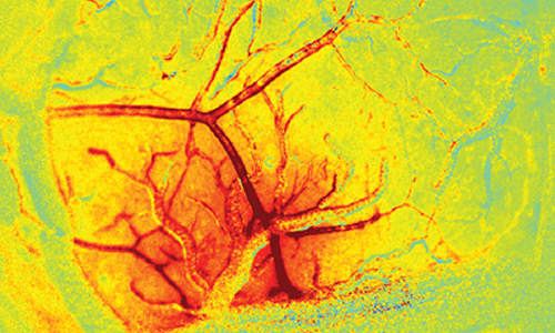
The blush of the brain. Red hue shows the increase in the amount of blood in the brain (total hemoglobin concentration) in response to stimulation. This response extends up along surface arteries in a wave of dilation that travels over a millimeter in less than half a second. This ‘brain blush’ provides the contrast seen in fMRI images, and is essential for normal brain function. The green vessel crossing the responding region is a draining vein. Veins exhibit very little diameter change in response to normal stimulation, acting as passive drains of increased blood flow. Photo Credit: Elizabeth Hillman


“We think we’ve found a missing link in our understanding of how the brain dynamically tunes its blood flow to stay in sync with the activity of neurons,” says Hillman, who has a joint appointment in Radiology. She is also a member of the Zuckerman Mind Brain Behavior Institute and the Kavli Institute for Brain Science at Columbia. Hillman has spent more than 10 years using advanced imaging tools to study how blood flow is controlled in the brain. “Earlier studies identified small pieces of the puzzle, but we didn’t believe they formed a cohesive ‘big picture’ that unified everybody’s observations. Our new finding seems to really connect the dots.”


Understanding how and why the brain regulates its blood flow could provide important clues to understanding early brain development, disease, and aging. The brain increases local blood flow when neurons fire, and this increase is what is detected by a functional magnetic resonance imaging (fMRI) scan. Hillman found that the vascular endothelium, the inner layer of blood vessels, plays a critical role in propagating and shaping the blood flow response to local neuronal activity. While the vascular endothelium is known to do this in other areas of the body, until now the brain was thought to use a different, more specialized mechanism and researchers in the field were focused on the cells surrounding the vessels in the brain.


“Once we realized the importance of endothelial signaling in the regulation of blood flow in the brain,” Hillman adds, “we wondered whether overlooking the vascular endothelium might have led researchers to misinterpret their results.”


“As we identified this pathway, so many things fell into place,” she continues, “We really hope that our work will encourage others to take a closer look at the vascular endothelium in the brain. So far, we think that our findings have far-reaching and really exciting implications for neuroscience, neurology, cardiovascular medicine, radiology, and our overall understanding of how the brain works.”


This research was carried out in Hillman’s Laboratory for Functional Optical Imaging, led by PhD student and lead author on the study, Brenda Chen. Other lab members who assisted with the study included PhD and MD/PhD students from Columbia Engineering, Neurobiology and Behavior, and Columbia University Medical Center. The group combined their engineering skills with their expertise in neuroscience, biology, and medicine to understand this new aspect of brain physiology.


To tease apart the role of endothelial signaling in the living brain, they had to develop new ways to both image the brain at very high speeds, and also to selectively alter the ability of endothelial cells to propagate signals within intact vessels. The team achieved this through a range of techniques that use light and optics, including imaging using a high-speed camera with synchronized, strobed LED illumination to capture changes in the color, and thus the oxygenation level of flowing blood. Focused laser light was used in combination with a fluorescent dye within the bloodstream to cause oxidative damage to the inner endothelial layer of blood brain arterioles, while leaving the rest of the vessel intact and responsive. The team showed that, after damaging a small section of a vessel using their laser, the vessel no longer dilated beyond the damaged point. When the endothelium of a larger number of vessels was targeted in the same way, the overall blood flow response of the brain to stimulation was significantly decreased.


“Our finding unifies what is known about blood flow regulation in the rest of the body with how it is regulated in the brain,” Hillman explains. “This has wider reaching implications since there are many disease states known to affect blood flow regulation in the rest of the body that, until now, were not expected to directly affect brain health.” For instance, involvement of the endothelium might explain neural deficits in diabetics; a clue that could lead to new diagnostics tests and treatments for neurological conditions associated with broader cardiovascular problems.


“Improving our fundamental understanding of how and why the brain regulates its blood flow is key to understanding how and when this mechanism could be altered or broken,” she says. “We think this could extend to studies of early brain development, aging, and diseases such as Alzheimer’s and dementia.”


The team’s research findings may also explain the effects of some drugs on the brain, and on the fMRI response to stimulation, since the vascular endothelium is exposed to chemicals in the bloodstream. “Overall, this work could dramatically improve our ability to interpret fMRI data collected in humans, perhaps making it a better tool for doctors to understand brain disease,” she adds. Hillman’s work in this area is also featured in an upcoming review in the 2014 edition of the Annual Review of Neuroscience, as well as an article in Scientific American MIND (July/August 2014).


Hillman plans next to address the broad range of implications her latest finding may have. She wants to explore the effects of drugs and disease states on the coupling of blood flow to neuronal activity in the brain, and is now starting studies to explore fMRI data from a range of different disease states to see whether she can find signs of neurovascular dysfunction. She is also working on characterizing the co-evolution of neuronal and hemodynamic activity during brain development and is beginning to develop new imaging tools that will enable non-invasive, inexpensive monitoring of brain hemodynamics in infants and children who cannot be imaged within an MRI scanner.


“Our latest finding gives us a new way of thinking about brain disease—that some conditions assumed to be caused by faulty neurons could actually be problems with faulty blood vessels,” Hillman adds. “This gives us a new target to focus on to explore treatments for a wide range of disorders that have, until now, been thought of as impossible to treat. The brain’s vasculature is a critical partner in normal brain function. We hope that we are slowly getting closer to untangling some of the mysteries of the human brain.”


Story Source:


The above story is based on materials provided by Columbia University School of Engineering and Applied Science.


The post New insight into how brain regulates its blood flow appeared first on Bioengineer.org.


Synchronized brain waves enable rapid learning

from

Bioengineer.org http://bioengineer.org/synchronized-brain-waves-enable-rapid-learning/


The human mind can rapidly absorb and analyze new information as it flits from thought to thought. These quickly changing brain states may be encoded by synchronization of brain waves across different brain regions, according to a new study from MIT neuroscientists.


Synchronized brain waves enable rapid learning


MIT neuroscientists found that brain waves originating from the striatum (red) and from the prefrontal cortex (blue) become synchronized when an animal learns to categorize different patterns of dots. Photo Credits: Jose-Luis Olivares/MIT


The researchers found that as monkeys learn to categorize different patterns of dots, two brain areas involved in learning — the prefrontal cortex and the striatum — synchronize their brain waves to form new communication circuits.


“We’re seeing direct evidence for the interactions between these two systems during learning, which hasn’t been seen before. Category-learning results in new functional circuits between these two areas, and these functional circuits are rhythm-based, which is key because that’s a relatively new concept in systems neuroscience,” says Earl Miller, the Picower Professor of Neuroscience at MIT and senior author of the study, which appears in the June 12 issue of Neuron.


There are millions of neurons in the brain, each producing its own electrical signals. These combined signals generate oscillations known as brain waves, which can be measured by electroencephalography (EEG). The research team focused on EEG patterns from the prefrontal cortex —the seat of the brain’s executive control system — and the striatum, which controls habit formation.


The phenomenon of brain-wave synchronization likely precedes the changes in synapses, or connections between neurons, believed to underlie learning and long-term memory formation, Miller says. That process, known as synaptic plasticity, is too time-consuming to account for the human mind’s flexibility, he believes.


“If you can change your thoughts from moment to moment, you can’t be doing it by constantly making new connections and breaking them apart in your brain. Plasticity doesn’t happen on that kind of time scale,” says Miller, who is a member of MIT’s Picower Institute for Learning and Memory. “There’s got to be some way of dynamically establishing circuits to correspond to the thoughts we’re having in this moment, and then if we change our minds a moment later, those circuits break apart somehow. We think synchronized brain waves may be the way the brain does it.”


The paper’s lead author is former Picower Institute postdoc Evan Antzoulatos, who is now at the University of California at Davis.


Humming together


Miller’s lab has previously shown that during category-learning, neurons in the striatum become active early, followed by slower activation of neurons in the prefrontal cortex. “The striatum learns very simple things really quickly, and then its output trains the prefrontal cortex to gradually pick up on the bigger picture,” Miller says. “The striatum learns the pieces of the puzzle, and then the prefrontal cortex puts the pieces of the puzzle together.”


In the new study, the researchers wanted to investigate whether this activity pattern actually reflects communication between the prefrontal cortex and striatum, or if each region is working independently. To do this, they measured EEG signals as monkeys learned to assign patterns of dots into one of two categories.


At first, the animals were shown just two different examples, or “exemplars,” from each category. After each round, the number of exemplars was doubled. In the early stages, the animals could simply memorize which exemplars belonged to each category. However, the number of exemplars eventually became too large for the animals to memorize all of them, and they began to learn the general traits that characterized each category.


By the end of the experiment, when the researchers were showing 256 novel exemplars, the monkeys were able to categorize all of them correctly.


As the monkeys shifted from rote memorization to learning the categories, the researchers saw a corresponding shift in EEG patterns. Brain waves known as “beta bands,” produced independently by the prefrontal cortex and the striatum, began to synchronize with each other. This suggests that a communication circuit is forming between the two regions, Miller says.


“There is some unknown mechanism that allows these resonance patterns to form, and these circuits start humming together,” he says. “That humming may then foster subsequent long-term plasticity changes in the brain, so real anatomical circuits can form. But the first thing that happens is they start humming together.”


A little later, as an animal nailed down the two categories, two separate circuits formed between the striatum and prefrontal cortex, each corresponding to one of the categories.


“This is the first paper that provides data suggesting that coupling in the beta-band between prefrontal cortex and striatum may play a key role in category-formation. In addition to revealing a novel mechanism involved in category-learning, the results also contribute to better understanding of the significance of coupled beta-band oscillations in the brain,” says Andreas Engel, a professor of physiology at the University Medical Center Hamburg-Eppendorf in Germany.


“Expanding your knowledge”


Previous studies have shown that during cognitively demanding tasks, there is increased synchrony between the frontal cortex and visual cortex, but Miller’s lab is the first to show specific patterns of synchrony linked to specific thoughts.


Miller and Antzoulatos also showed that once the prefrontal cortex learns the categories and sends them to the striatum, they undergo further modification as new information comes in, allowing more expansive learning to take place. This iteration can occur over and over.


“That’s how you get the open-ended nature of human thought. You keep expanding your knowledge,” Miller says. “The prefrontal cortex learning the categories isn’t the end of the game. The cortex is learning these new categories and then forming circuits that can send the categories down to the striatum as if it’s just brand-new material for the brain to elaborate on.”


In follow-up studies, the researchers are now looking at how the brain learns more abstract categories, and how activity in the striatum and prefrontal cortex might reflect that type of abstraction.


Story Source:


The above story is based on materials provided by Massachusetts Institute of Technology.


The post Synchronized brain waves enable rapid learning appeared first on Bioengineer.org.