20 Aralık 2014 Cumartesi

New technique reveals immune cell motion through variety of tissues

from

BIOENGINEER.ORG http://bioengineer.org/new-technique-reveals-immune-cell-motion-through-variety-of-tissues/


Neutrophils, cells recruited by the immune system to fight infection, need to move through a great variety of tissues. New research shows how neutrophils move through confined spaces in the body. A new system can mimic tissues of different densities and stiffness, enabling improved development and testing of drugs.

New technique reveals immune cell motion through variety of tissues


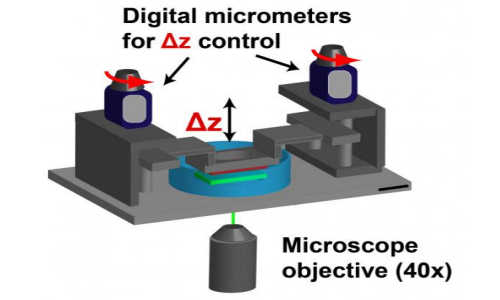
By placing neutrophils between two hydrogel sacks, researchers can mimic cell movement through 3-D tissue. Digital micrometers can change the characteristics — density, stiffness — of the medium through which the cells move. Photo Credit: Frank lab/Brown University


Neutrophils, a type of white blood cell, are the immune system’s all-terrain vehicles. The cells are recruited to fight infections or injury in any tissue or organ in the body despite differences in the cellular and biochemical composition. Researchers from Brown University’s School of Engineering and the Department of Surgery in the Warren Alpert Medical School collaborated to devise a new technique for understanding how neutrophils move in these confined spaces.


The technique involves two hydrogel sacks sandwiched together with a miniscule space in between. Neutrophils could be placed in that space, mimicking the confinement they experience within tissue. Time-lapse cameras measure how fast the cells move, and traction force microscopes determine the forces the cells exert on the surrounding gel.


In a paper published in the Journal of Biological Chemistry, the researchers used the device to reveal new details about the motion of neutrophils. Bodily tissues are highly confined, densely packed, three-dimensional spaces that can vary widely in physical shape and elasticity. The researchers showed that neutrophils are sensitive to the physical aspects of their environment: They behave differently on flat surfaces than in confined three-dimensional space. Ultimately, the team hopes the system can be useful in screening drugs aimed at optimizing neutrophils to fight infection in specific tissue types.


Traditionally, research on neutrophil motion in the lab is often done on two-dimensional, inflexible surfaces composed of plastic or glass. Those studies showed that neutrophils move using arm-like appendages called integrins. The cell extends the integrins, which grab onto to flat surfaces like tiny grappling hooks. By reeling those integrins back in, the cell is able to crawl along.


Scientists thought that by inhibiting integrins, they could greatly reduce the cells’ ability to move through tissue. That, they thought, could be a good strategy for fighting autoimmune diseases in which neutrophils attack and damage healthy tissue.


But in 2008, a landmark paper showed that neutrophils have a second mode of motion. The work showed that cells in which integrins had been disabled were still able to move through dense tissue.


Christian Franck, assistant professor of engineering at Brown, and his colleagues wanted to learn more about this second mode of motion.


“On flat 2-D surfaces there’s integrin-dependent motion, but in complicated 3-D materials there’s integrin-independent motion,” Franck said. “The question we were asking is can we find an in-vitro system that can recreate that integrin-independent motion, because you can’t get it in a regular petri dish.”


Using their gel system and the traction force microscopes, Franck and his colleagues showed that, when confined, neutrophils exert force in several distinct spots. On the bottom of the cell, forces were generated in a way that was consistent with previous imaging of integrin engagement. But on the top of the cell, there was another source of force. The cell pushed on the upper gel surface with its nuclear lobe, the area of the cell where DNA resides.


“It’s like a rock climber pushing against the walls of a canyon,” Franck said.


To see if the force generated by the nuclear lobe was responsible for the cells’ ability to move without integrins, the researchers repeated the experiment with cells in which integrins were chemically inhibited. Sure enough, the cells were still able to move when confined between the gels. In fact, they were able to move faster.


“We showed that physical confinement is the key feature to reproduce integrin-independent motion in a relatively simple setting,” Franck said. “That wasn’t possible previously on a flat surface.”


The fact that confined cells actually move faster without their integrin suggests that even though integrins aren’t essential for the cells motion, they still play a regulatory role.


“What we showed was that [use of integrins] is not black and white,” Franck said. “Even in this integrin-independent motion, integrins remain to regulate motion and force generation.”


Now that they have a means of recreating how neutrophils travel through confined spaces in the lab, Franck and his team plan to do further experiments aimed at fine-tuning that motion. The system they’ve developed enables them to control the stiffness of the gel surfaces between which the cells travel, mimicking the varying stiffness of tissue in the body.


“If motility is specific to a neutrophil being in a specific tissue, maybe we could attenuate its response,” Franck said. “Maybe we could make it move faster in the muscle and slower everywhere else, for example.”


This new system enables testing of drugs aimed at doing just that. Such drugs could be of great benefit to people who have disorders of the immune system.


Story Source:


The above story is based on materials provided by Brown University.


Lost memories might be able to be restored

from

BIOENGINEER.ORG http://bioengineer.org/lost-memories-might-be-able-to-be-restored/


New UCLA research indicates that lost memories can be restored. The findings offer some hope for patients in the early stages of Alzheimer’s disease. For decades, most neuroscientists have believed that memories are stored at the synapses — the connections between brain cells, or neurons — which are destroyed by Alzheimer’s disease. The new study provides evidence contradicting the idea that long-term memory is stored at synapses.


Lost memories might be able to be restored


David Glanzman is holding a marine snail. Photo Credit: Christelle Nahas/UCLA


“Long-term memory is not stored at the synapse,” said David Glanzman, a senior author of the study, and a UCLA professor of integrative biology and physiology and of neurobiology. “That’s a radical idea, but that’s where the evidence leads. The nervous system appears to be able to regenerate lost synaptic connections. If you can restore the synaptic connections, the memory will come back. It won’t be easy, but I believe it’s possible.”


The findings were published recently in eLife, a highly regarded open-access online science journal.


Glanzman’s research team studies a type of marine snail called Aplysia to understand the animal’s learning and memory. The Aplysia displays a defensive response to protect its gill from potential harm, and the researchers are especially interested in its withdrawal reflex and the sensory and motor neurons that produce it.


They enhanced the snail’s withdrawal reflex by giving it several mild electrical shocks on its tail. The enhancement lasts for days after a series of electrical shocks, which indicates the snail’s long-term memory. Glanzman explained that the shock causes the hormone serotonin to be released in the snail’s central nervous system.


Long-term memory is a function of the growth of new synaptic connections caused by the serotonin, said Glanzman, a member of UCLA’s Brain Research Institute. As long-term memories are formed, the brain creates new proteins that are involved in making new synapses. If that process is disrupted — for example by a concussion or other injury — the proteins may not be synthesized and long-term memories cannot form. (This is why people cannot remember what happened moments before a concussion.)


“If you train an animal on a task, inhibit its ability to produce proteins immediately after training, and then test it 24 hours later, the animal doesn’t remember the training,” Glanzman said. “However, if you train an animal, wait 24 hours, and then inject a protein synthesis inhibitor in its brain, the animal shows perfectly good memory 24 hours later. In other words, once memories are formed, if you temporarily disrupt protein synthesis, it doesn’t affect long-term memory. That’s true in the Aplysia and in human’s brains.” (This explains why people’s older memories typically survive following a concussion.)


Glanzman’s team found the same mechanism held true when studying the snail’s neurons in a Petri dish. The researchers placed the sensory and motor neurons that mediate the snail’s withdrawal reflex in a Petri dish, where the neurons re-formed the synaptic connections that existed when the neurons were inside the snail’s body. When serotonin was added to the dish, new synaptic connections formed between the sensory and motor neurons. But if the addition of serotonin was immediately followed by the addition of a substance that inhibits protein synthesis, the new synaptic growth was blocked; long-term memory could not be formed.


The researchers also wanted to understand whether synapses disappeared when memories did. To find out, they counted the number of synapses in the dish and then, 24 hours later, added a protein synthesis inhibitor. One day later, they re-counted the synapses.


What they found was that new synapses had grown and the synaptic connections between the neurons had been strengthened; late treatment with the protein synthesis inhibitor did not disrupt the long-term memory. The phenomenon is extremely similar to what happens in the snail’s nervous system during this type of simple learning, Glanzman said.


Next, the scientists added serotonin to a Petri dish containing a sensory neuron and motor neuron, waited 24 hours, and then added another brief pulse of serotonin — which served to remind the neurons of the original training — and immediately afterward add the protein synthesis inhibitor. This time, they found that synaptic growth and memory were erased. When they re-counted the synapses, they found that the number had reset to the number before the training, Glanzman said. This suggests that the “reminder” pulse of serotonin triggered a new round of memory consolidation, and that inhibiting protein synthesis during this “reconsolidation” erased the memory in the neurons.


If the prevailing wisdom were true — that memories are stored in the synapses — the researchers should have found that the lost synapses were the same ones that had grown in response to the serotonin. But that’s not what happened: Instead, they found that some of the new synapses were still present and some were gone, and that some of the original ones were gone, too.


Glanzman said there was no obvious pattern to which synapses stayed and which disappeared, which implied that memory is not stored in synapses.


When the scientists repeated the experiment in the snail, and then gave the animal a modest number of tail shocks — which do not produce long-term memory in a naive snail — the memory they thought had been completely erased returned. This implies that synaptic connections that were lost were apparently restored.


“That suggests that the memory is not in the synapses but somewhere else,” Glanzman said. “We think it’s in the nucleus of the neurons. We haven’t proved that, though.”


Glanzman said the research could have significant implications for people with Alzheimer’s disease. Specifically, just because the disease is known to destroy synapses in the brain doesn’t mean that memories are destroyed.


“As long as the neurons are still alive, the memory will still be there, which means you may be able to recover some of the lost memories in the early stages of Alzheimer’s,” he said.


Glanzman added that in the later stages of the disease, neurons die, which likely means that the memories cannot be recovered.


The cellular and molecular processes seem to be very similar between the marine snail and humans, even though the snail has approximately 20,000 neurons and humans have about 1 trillion. Neurons each have several thousand synapses.


Glanzman used to believe that traumatic memories could be erased but he has changed his mind. He now believes that, because memories are stored in the nucleus, it may be much more difficult to modify them. He will continue to study how the marine snail’s memories are restored and how synapses re-grow.


Co-authors of the study include Shanping Chen, Diancai Cai and Kaycey Pearce, research associates in Glanzman’s laboratory.


The research was funded by the National Institutes of Health’s National Institute of Neurological Disorders and Stroke, the National Institute of Mental Health and the National Science Foundation.


Almost all the processes that are involved in memory in the snail also have been shown to be involved in memory in the brains of mammals, Glanzman said.


In a 1997 study published in the journal Science, Glanzman and colleagues identified a cellular mechanism in the Aplysia that plays an important role in learning and memory. A protein called N-methyl D-aspartate, or NMDA, receptor enhances the strength of synaptic connections in the nervous system and plays a vital role in memory and in certain kinds of learning in the mammalian brain as well. Glanzman’s demonstration that the NMDA receptor plays a critical role in learning in a simple animal like the marine snail was entirely unexpected at the time.


Story Source:


The above story is based on materials provided by University of California – Los Angeles.


19 Aralık 2014 Cuma

New class of synthetic molecules mimics antibodies

from

BIOENGINEER.ORG http://bioengineer.org/new-class-of-synthetic-molecules-mimics-antibodies/


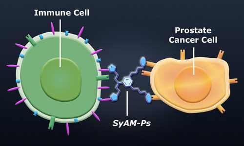
A Yale University lab has crafted the first synthetic molecules that have both the targeting and response functions of antibodies. The new molecules — synthetic antibody mimics (SyAMs) — attach themselves simultaneously to disease cells and disease-fighting cells. The result is a highly targeted immune response, similar to the action of natural human antibodies.


New class of synthetic molecules mimics antibodies


The new molecules — synthetic antibody mimics (SyAMs) — attach themselves simultaneously to disease cells and disease-fighting cells. The result is a highly targeted immune response, similar to the action of natural human antibodies. Photo Credit: Image courtesy of Yale University


“Unlike antibodies, however, our molecules are synthetic organic compounds that are approximately one-twentieth the size of antibodies,” said David A. Spiegel, a professor of chemistry at Yale whose lab developed the molecules. “They are unlikely to cause unwanted immune reactions due to their structure, are thermally stable, and have the potential to be administered orally, just like traditional, small-molecule drugs.”


Spiegel and his team describe the research in a paper published online Dec. 16 by the Journal of the American Chemical Society.


The paper looks specifically at SyAM molecules used to attack prostate cancer. Called SyAM-Ps, they work first by recognizing cancer cells and binding with a specific protein on their surface. Next, they also bind with a receptor on an immune cell. This induces a targeted response that leads to the destruction of the cancer cell.


Spiegel said the process of synthesizing and optimizing the structure of the molecules required considerable time and effort. “We now know that synthetic molecules of intermediate size possess perhaps the most important functional properties of antibodies — targeting and stimulation of immune cells,” he said.


“It’s also noteworthy that molecules of such a small size can bring together two objects as enormous as cells, and trigger a specific functional response, entirely as a result of specific receptor interactions,” Spiegel added.


Beyond their potential for treating prostate cancer, SyAMs may have applications for treating other forms of cancer, HIV and various bacterial diseases.


Story Source:


The above story is based on materials provided by Yale University, Jim Shelton.


Amputee puts limb system through its paces

from

BIOENGINEER.ORG http://bioengineer.org/amputee-puts-limb-system-paces/


A Colorado man made history at the Johns Hopkins University Applied Physics Laboratory (APL) this summer when he became the first bilateral shoulder-level amputee to wear and simultaneously control two of the Laboratory’s Modular Prosthetic Limbs. Most importantly, Les Baugh, who lost both arms in an electrical accident 40 years ago, was able to operate the system by simply thinking about moving his limbs, performing a variety of tasks during a short training period.


apl


APL prosthetist Courtney Moran looks on as Les Baugh tests out the Modular Prosthetic Limbs. Credit: Johns Hopkins University Applied Physics Laboratory


Baugh was in town for two weeks in June as part of an APL-funded research effort to further assess the usability of the MPL, developed over the past decade as part of the Revolutionizing Prosthetics Program. Before putting the limb system through the paces, Baugh had to undergo a surgery at Johns Hopkins Hospital known as targeted muscle reinnervation.


“It’s a relatively new surgical procedure that reassigns nerves that once controlled the arm and the hand,” explained Johns Hopkins Trauma Surgeon Albert Chi, M.D. “By reassigning existing nerves, we can make it possible for people who have had upper-arm amputations to control their prosthetic devices by merely thinking about the action they want to perform.”


After recovery, Baugh visited the Laboratory for training on the use of the MPLs. First, he worked with researchers on the pattern recognition system.


“We use pattern recognition algorithms to identify individual muscles that are contracting, how well they communicate with each other, and their amplitude and frequency,” Chi explained. “We take that information and translate that into actual movements within a prosthetic.”


Then Baugh was fitted for a custom socket for his torso and shoulders that supports the prosthetic limbs and also makes the neurological connections with the reinnervated nerves. While the socket got its finishing touches, the team had him work with the limb system through a Virtual Integration Environment (VIE), a virtual-reality version of the MPL.


The VIE is completely interchangeable with the prosthetic limbs and through APL’s licensing process currently provides 19 groups in the research community with a low-cost means of testing brain–computer interfaces. It’s being used to test novel neural interface methods and study phantom limb pain, and serves as a portable training system.


By the time the socket was finished, Baugh said he was more than ready to get started. When he was fitted with the socket, and the prosthetic limbs were attached, he said “I just went into a whole different world.” He moved several objects, including an empty cup from a counter-shelf height to a higher shelf, a task that required him to coordinate the control of eight separate motions to complete.


“This task simulated activities that may commonly be faced in a day-to-day environment at home,” said APL’s Courtney Moran, a prosthetist working with Baugh. “This was significant because this is not possible with currently available prostheses. He was able to do this with only 10 days of training, which demonstrates the intuitive nature of the control.”


Moran said the research team was floored by what Baugh was able to accomplish.


“We expected him to exceed performance compared to what he might achieve with conventional systems, but the speed with which he learned motions and the number of motions he was able to control in such a short period of time was far beyond expectation,” she said. “What really was amazing, and was another major milestone with MPL control, was his ability to control a combination of motions across both arms at the same time. This was a first for simultaneous bimanual control.”


RP Principal Investigator Michael McLoughlin said “I think we are just getting started. It’s like the early days of the Internet. There is just a tremendous amount of potential ahead of us, and we’ve just started down this road. And I think the next five to 10 years are going to bring phenomenal advancement.”


The next step, McLoughlin said, is to send Baugh home with a pair of limb systems so that he can see how they integrate with his everyday life.


Baugh is looking forward to that day. “Maybe for once I’ll be able to put change in the pop machine and get pop out of it,” he said. He’s looking forward to doing “simple things that most people don’t think of. And it’s re-available to me.”


Story Source:


The above story is based on materials provided by The Johns Hopkins University Applied Physics Laboratory LLC.


The post Amputee puts limb system through its paces appeared first on BIOENGINEER.ORG.


Researchers Help Unlock Mystery of Skin’s Sensory Abilities

from

BIOENGINEER.ORG http://bioengineer.org/researchers-help-unlock-mystery-skins-sensory-abilities/


Humans’ ability to detect the direction of movement of stimuli in their sensory world is critical to survival. Much of this stimuli detection comes from sight and sound, but little is known about how the direction of movement of stimuli on the skin — humans’ largest sensory organ — is detected and processed.Until now.


skin


Photo Credit: Shutterstock via wired


For the past few years, two University of Wyoming researchers experimented with mice to discover some answers. C. Jeffery Woodbury, a UW associate professor in the Department of Zoology and Physiology, and Colleen Cassidy, a UW doctoral student from Cheyenne studying neuroscience, isolated various skin sensory cells from mice and recorded the responses of low-threshold mechanoreceptors, or LTMRs, on the skin. LTMRs are sensitive to innocuous skin indentation, stroking, vibration or stretch of the skin, and the deflection of hair follicles.


The neurological steps leading to the perception of touch begin with the activation of LTMRs, Woodbury says.


“These studies show that there are sensory neurons throughout the skin that are preferentially selective for movement in one direction versus another,” he says. “That’s not been documented before. Further, we also nailed down the mechanism for this directional selectivity. I think it (paper) will be a landmark in the field of sensory biology.”


Woodbury was co-author — with David Ginty and other colleagues at Harvard University — of a paper, titled “The Cellular and Molecular Basis of Direction Selectivity of A delta-LTMRs,” that appears in the Dec. 18 issue of Cell. Cell is the flagship journal of Cell Press, a pre-eminent international publisher of cutting-edge biomedical research and reviews. Cassidy also was co-author of the paper.


“We rely on our senses to understand our surroundings. These cells may give us awareness of movement of things on our body,” Woodbury says. “There are many things in the skin senses that are unexplained.”

For example, human skin feels which direction the wind is blowing or reacts to temperature, he says. Skin also provides information on whether something poses a threat, such as a mosquito, or a harmless midge.

Woodbury, Cassidy and their colleagues discovered that some sensory neurons innervate hairs, and their terminals glom on or attach to one side of the hairs as opposed to the other. They went on to show why, finding that this was due to communication between the terminal of the sensory cells and the cells that make up the follicles in the hair shaft, Woodbury says.


In separate experiments, they disrupted the signal so that this communication was gone. As a result, these sensory terminals migrated and wandered from their original position so that they now surrounded the hair shaft in a randomized pattern. This led to a randomization of the cell’s response to the direction the hairs were moved, reducing their directional preference, Woodbury says.

Working with mice


“Our lab is like a hospital surgical suite,” Woodbury says. “Everything is customized and miniaturized for what we do.”


Mice were ideal to use for the research because their skin, like that of humans, contains more than 20 different types of sensory neurons. However, in mice, cells can be genetically engineered so that they are labeled by different-colored fluorescent proteins from jellyfish. The proteins were “turned on” in specific subsets of cells, so researchers could actually see these different types of sensory neurons that innervate regions of the mammals’ skin, Woodbury says.


This cell isolation technique was used to record from these neurons while studying how they reacted to the direction of stimulus movement across the skin.


Cassidy has worked in Woodbury’s laboratory since fall 2010. Her work on the Cell paper is part of her doctoral dissertation, she says.


“I’ve learned a lot of critical thinking and really how to approach a problem,” she says. “I see different avenues and ways to answer questions. This research has taught me the value of perseverance. When you get that piece of data, it’s really exciting since it is all about discovery.”


“I don’t think there are any studies out there that have really nailed down the entire process — from early developmental processes to highly specialized cells with specialized functions that, ultimately, play a direct role in perception,” Woodbury says. “I think this work offers a rare view into development. Understanding how the brain sorts out and processes the movement of objects has been a central theme in neuroscience research.”


Woodbury says he plans to explore the subject further, to determine whether skin stimulation affects heart regulation or other fundamental biological functions, such as pain processing.


“This research may have implications for controlling pain,” Woodbury says. “For example, when someone pinches us, our first reaction is to rub the area, which helps reduce the pain. These findings suggest that it may have a greater impact depending on which way you rub it.”


Story Source:


The above story is based on materials provided by University of Wyoming.


The post Researchers Help Unlock Mystery of Skin’s Sensory Abilities appeared first on BIOENGINEER.ORG.


German medics report on drug success for Ebola patient

from

BIOENGINEER.ORG http://bioengineer.org/german-medics-report-drug-success-ebola-patient/


German doctors on Friday gave details of how an experimental drug together with advanced intensive care helped save a Ugandan physician who had been airlifted from Sierra Leone with Ebola.


German medics report on drug success for Ebola patient


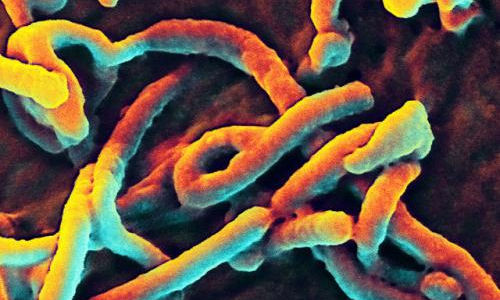
A scanning electron micrograph of Ebola virus budding from a cell (African green monkey kidney epithelial cell line). Photo Credit: NIAID


A prototype drug called FX06, designed to stop haemorrhage, was given to the patient after the doctors got special authorisation from their hospital’s ethics committee, they reported in The Lancet.

“Even though the patient was critically ill, we were able to support him long enough for his body to start antibody production and for the virus to be cleared by his body’s defences,” said Timo Wolf of University Hospital Frankfurt.


The Frankfurt team and the makers of the experimental drug had announced its use in early November.

Publication in The Lancet, a leading peer-reviewed medical journal, validates this announcement.

The unnamed 38-year-old male doctor had been airlifted to Frankfurt in early October, five days after the onset of Ebola symptoms, and admitted to a Biosafety 4 facility, the highest level of medical security, the study said.


Within three days of admission, he was suffering from failure of the lungs, kidneys and gastro-intestinal tract, as well as haemorrhaging blood vessels, a hallmark of Ebola infection.

He was placed on a ventilator and kidney dialysis and administered with antibiotics and a three-day course of FX06.


Called a fibrin-derived peptide, FX06 is designed to seal off the walls of blood vessels, which become permeable when infected by a haemorrhagic virus.


The peptide works by binding to the surface of endothelial cells, which form the inner cell layer of blood vessels. It latches onto the cells via a target called VE-cadherin.

The drug was invented at Vienna General Hospital and is made by a small Austrian firm called MChE-F4Pharma.


It had previously been tested on lab mice infected with the dengue virus and was also put through a trial among 234 European patients to assess its potential for limiting damage to cardiac tissue after a heart attack.


The combination of intensive care and the drug helped the Ugandan patient to stabilise and then recover, the doctors reported.


After a 30-day observation period, no trace of Ebola was found in his blood, and he was released from hospital to return to his family.


But the Lancet study also reported that shortly after this case, another Ebola patient with acute Ebola and haemorrhage was treated with FX06 at a hospital in the eastern Germany city of Leipzig, but died.

Treating Ebola cases in intensive care is a task with “complexity and specific challenges,” it warned.

FX06 is “a potentially valuable therapeutic candidate” for fighting the disease, the authors said, calling for the drug to be assessed in clinical trials.


More than 6,900 people have died in the latest Ebola epidemic, which is centred on the west African states of Guinea, Liberia and Sierra Leone, the World Health Organization (WHO) said on Wednesday.


Story Source:


The above story is based on materials provided by The Lancet.


The post German medics report on drug success for Ebola patient appeared first on BIOENGINEER.ORG.


18 Aralık 2014 Perşembe

Lens-free microscope can detect cancer at the cellular level

from

BIOENGINEER.ORG http://bioengineer.org/lens-free-microscope-can-detect-cancer-cellular-level/


UCLA researchers have developed a lens-free microscope that can be used to detect the presence of cancer or other cell-level abnormalities with the same accuracy as larger and more expensive optical microscopes.


Lens-free microscope can detect cancer at the cellular level


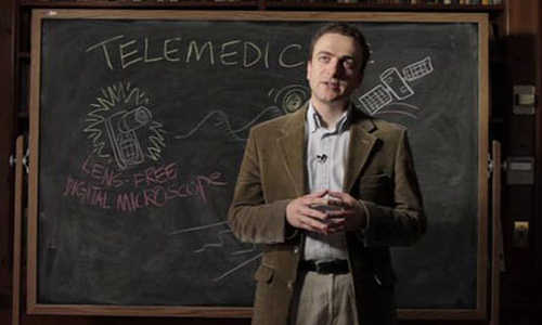
Aydogan Ozcan, the Chancellor’s Professor of Electrical Engineering and Bioengineering at the UCLA Henry Samueli School of Engineering and Applied Science


The invention could lead to less expensive and more portable technology for performing common examinations of tissue, blood and other biomedical specimens. It may prove especially useful in remote areas and in cases where large numbers of samples need to be examined quickly.


The microscope is the latest in a series of computational imaging and diagnostic devices developed in the lab of Aydogan Ozcan, the Chancellor’s Professor of Electrical Engineering and Bioengineering at the UCLA Henry Samueli School of Engineering and Applied Science and a Howard Hughes Medical Institute professor. Ozcan’s lab has previously developed custom-designed smartphone attachments and apps that enable quick analysis of food samples for allergens, water samples for heavy metals and bacteria, cell counts in blood samples, and the use of Google Glass to process the results of medical diagnostic tests.


The latest invention is the first lens-free microscope that can be used for high-throughput 3-D tissue imaging — an important need in the study of disease.


“This is a milestone in the work we’ve been doing,” said Ozcan, who also is the associate director of UCLA’s California NanoSystems Institute. “This is the first time tissue samples have been imaged in 3D using a lens-free on-chip microscope.”


The research is the cover article today in Science Translational Medicine, which is published by the American Association for the Advancement of Science.


The device works by using a laser or light-emitting-diode to illuminate a tissue or blood sample that has been placed on a slide and inserted into the device. A sensor array on a microchip — the same type of chip that is used in digital cameras, including cellphone cameras — captures and records the pattern of shadows created by the sample.


The device processes these patterns as a series of holograms, forming 3-D images of the specimen and giving medical personnel a virtual depth-of-field view. An algorithm color codes the reconstructed images, making the contrasts in the samples more apparent than they would be in the holograms and making any abnormalities easier to detect.


Ozcan’s team tested the device using Pap smears that indicated cervical cancer, tissue specimens containing cancerous breast cells, and blood samples containing sickle cell anemia. In a blind test, a board-certified pathologist analyzed sets of specimen images that had been created by the lens-free technology and by conventional microscopes. The pathologist’s diagnoses using the lens-free microscopic images proved accurate 99 percent of the time.


Another benefit of the lens-free device is that it produces images that are several hundred times larger in area, or field of view, than those captured by conventional bright-field optical microscopes, which makes it possible to process specimens more quickly.


“While mobile health care has expanded rapidly with the growth of consumer electronics — cellphones in particular — pathology is still, by and large, constrained to advanced clinical laboratory settings,” Ozcan said. “Accompanied by advances in its graphical user interface, this platform could scale up for use in clinical, biomedical, scientific, educational and citizen-science applications, among others.”


Story Source:


The above story is based on materials provided by University of California – Los Angeles.


The post Lens-free microscope can detect cancer at the cellular level appeared first on BIOENGINEER.ORG.