31 Aralık 2014 Çarşamba

Using light to produce natural sleep patterns

from

BIOENGINEER.ORG http://bioengineer.org/using-light-to-produce-natural-sleep-patterns/


Getting enough of the right kind of sleep is crucial for keeping both body and mind healthy.

Now a team of researchers at MIT has moved a step closer to being able to produce natural sleep patterns.


sleep_pattern


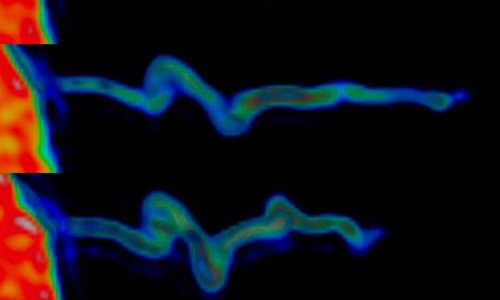
Confocal images show expression of the neurotransmitter, acetylcholine (red), and the light sensitive ion channel, channelrhodopsin (Chr2) (green). The merged images (far right) show neurons expressing both acetylcholine and ChR2 (yellow). The top row displays the entire brainstem; the bottom row shows expression at the cellular level. Photo Credits: Researchers


In a paper published today in the Proceedings of the National Academy of Sciences, the researchers describe how they were able to trigger a period of rapid eye movement (REM), otherwise known as dream sleep, in mice, using a technique that shines light directly on mouse neurons.


Sleep helps the brain recuperate and restore itself, as well as allow it to process memories. It also helps to ensure that the body’s immune and other systems work properly.


The different stages of natural sleep provide different benefits, says the team’s leader, Emery Brown, the Edward Hood Taplin Professor of Medical Engineering at MIT. Studies in rodents have shown that learning occurs during REM sleep, for example, while slow wave sleep, also known as non-REM stage three, is most important for feeling rested and refreshed.


However, these health benefits result only from natural sleep — or alternating 90-minute periods of non-REM and REM sleep — and there are no existing drugs capable of inducing this state.

“What they do is create sedation,” Brown says. “If you are lucky, the sedation allows your natural sleep mechanisms to take over.”


Goal: Creating natural sleep


To develop better approaches to creating natural sleep, researchers need to study the extent to which the various sleep stages can be created first separately, and ultimately together, Brown says.


Previous studies have indicated that neurons called cholinergic cells are active during both wakefulness and REM sleep, says the paper’s lead author, Christa Van Dort, a postdoc in Brown’s group in the Department of Brain and Cognitive Sciences at MIT. “There was a lot of early evidence that cholinergic neurons were involved in this area, but nobody could actually say whether the firing of these specific cells was responsible for the transition to REM sleep,” she says.


To investigate whether cholinergic neurons could induce REM sleep, the team used a technique called optogenetics, in which a head-mounted fiber optic device is used to shine light onto a specific group of neurons.


The neurons are first sensitized to light using a protein found in algae. This protein responds to certain wavelengths of light, allowing the algae to move around, Van Dort says. “In 2005, researchers at Stanford [University] were able to put this algae protein into mammalian cells,” she says. “They realized that if you put the protein into certain types of neurons you could then shine a light on them to activate them, and control the firing of the brain, at a single-cell level.”


Van Dort and her colleagues applied the technique to a mouse known to express this algae protein in cholinergic neurons. “In this way we were able to push the mice into dream sleep,” Van Dort says.

They found that activation of cholinergic neurons during non-REM sleep increased the number of REM sleep episodes the mice experienced. When they analyzed the episodes, they discovered that they closely matched natural periods of REM sleep.


Step toward inducing natural sleep patterns


The technique helps to clarify the mechanism by which REM sleep is controlled, and is a step toward

understanding how to design natural sleep in humans, Van Dort says. Existing drugs used by insomniacs actually repress both REM and the deeper stages of non-REM sleep, and instead put users into a very light sleep state. “Figuring out how each of the components of sleep is controlled can help us design a way of reproducing natural sleep with different drugs in the future,” she says.


In the meantime, triggering more episodes of REM sleep alone could be used to enhance people’s learning and memory, Brown says.


The researchers are now investigating how the cholinergic system connects with other areas of the brain that have also been identified as important in REM sleep production. They are also developing experiments that they hope will lead to the production of better non-REM sleep, Van Dort says.

“The long-term goal is to really understand what controls each phase of non-REM and REM sleep, and then to selectively induce them both, and reproduce the normal cycling of sleep stages,” she says.

Cholinergic control of REM sleep has been an area of longstanding controversy, as previous work has not been able to selectively activate just these neurons, says Robert McCarley, a professor of psychiatry at Harvard Medical School who was not involved in this research. “This pioneering study used optogenetic control of activity of cholinergic neurons to provide evidence that REM induction occurs through activity of cholinergic neurons, those using the neurotransmitter acetylcholine,” McCarley says.


“What is exciting about this study is the use of optogenetics to shed new light on a decades-long conundrum, and it heralds a new era in REM sleep neurophysiology using optogenetics,” he says.


Story Source:


The above story is based on materials provided by MIT News, Helen Knight.


World’s First Human X-Ray 1895 Röntgen’s wife hand

from

BIOENGINEER.ORG http://bioengineer.org/worlds-first-human-x-ray-1895-rontgens-wife-hand/


The discovery of a new and mysterious form of radiation in the late 19th century led to a revolution in medical imaging.


07_11_Foundations01


Wilhelm Rontgen took this radiograph of his wife’s left hand on December 22, 1895, shortly after his discovery of X-rays. Photo Credit: NATIONAL LIBRARY OF MEDICINE


On this day in 1895, physicist Wilhelm Conrad Rontgen (1845-1923) becomes the first person to observe X-rays, a significant scientific advancement that would ultimately benefit a variety of fields, most of all medicine, by making the invisible visible. Rontgen’s discovery occurred accidentally in his Wurzburg, Germany, lab, where he was testing whether cathode rays could pass through glass when he noticed a glow coming from a nearby chemically coated screen. He dubbed the rays that caused this glow X-rays because of their unknown nature.


X-rays are electromagnetic energy waves that act similarly to light rays, but at wavelengths approximately 1,000 times shorter than those of light. Rontgen holed up in his lab and conducted a series of experiments to better understand his discovery. He learned that X-rays penetrate human flesh but not higher-density substances such as bone or lead and that they can be photographed.


Rontgen’s discovery was labeled a medical miracle and X-rays soon became an important diagnostic tool in medicine, allowing doctors to see inside the human body for the first time without surgery. In 1897, X-rays were first used on a military battlefield, during the Balkan War, to find bullets and broken bones inside patients.


Story Source:


The above story is based on materials provided by History.com.


30 Aralık 2014 Salı

Cancer treatment potential discovered in gene repair mechanism

from

BIOENGINEER.ORG http://bioengineer.org/cancer-treatment-potential-discovered-in-gene-repair-mechanism/


Case Western Reserve researchers have identified a two-pronged therapeutic approach that shows great potential for weakening and then defeating cancer cells. The team’s complex mix of genetic and biochemical experiments unearthed a way to increase the presence of a tumor-suppressing protein which, in turn, gives it the strength to direct cancer cells toward a path that leads to their destruction.


case


Photo Credit: Zhang Lab


If the laboratory findings are supported by tests in animal models, the breakthrough could hold the promise of increasing the effectiveness of radiation and chemotherapy in shrinking or even eliminating tumors. The key is to build up a “good” protein – p53-binding protein 1 (53BP1)—so that it weakens the cancer cells, leaving them more susceptible to existing cancer-fighting measures.


The breakthrough detailed appeared in the Nov. 24 online edition of the journal PNAS (Proceedings of the National Academy of Sciences).


“Our discovery one day could lead to a gene therapy where extra amounts of 53BP1 will be generated to make cancer cells more vulnerable to cancer treatment,” said senior author Youwei Zhang, PhD, assistant professor of pharmacology, Case Western Reserve University School of Medicine, and member of the Case Comprehensive Cancer Center. “Alternatively, we could design molecules to increase levels of 53BP1 in cancers with the same cancer-killing end result.”

The cornerstone of the research involves DNA repair – more specifically, double-stand DNA repair. DNA damage is the consequence of an irregular change in the chemical structure of DNA, which in turn damages and even kills cells. The most lethal irregularity to DNA is the DNA double-strand break in the chromosome. DNA double-strand breaks are caused by everything from reactive oxygen components occurring with everyday bodily metabolism to more damaging assaults such as radiation or chemical agents.


The body operates two repair shops, or pathways, to fix these double strand breaks. One provides rapid, but incomplete repair – namely, gluing the DNA strand ends back together. The problem with the glue method is that it leaves the DNA strands unable to transmit enough information for the cell to function properly – leading to a high cell fatality rate.


The second shop, or pathway, uses information from intact, undamaged DNA to instruct damaged cells on how to mend broken double strands. During his study, Zhang and fellow investigators discovered a previously unidentified function of a known gene, UbcH7, in regulating DNA double-strand break repair. Specifically, they found that depleting UbcH7 led to a dramatic increase in the level of the 53BP1 protein.


“What we propose is increasing the level of 53BP1 to force cancer cells into the error-prone pathway where they will die,” Zhang said. “The idea is to suppress deliberately the second accurate repair pathway where cancer cells would prefer to go. It is a strategy that would lead to enhanced effectiveness of cancer therapy drugs.”

The next research step for Zhang and his team will be to test their theory in animal models with cancer. Investigators would study the effects of introducing the protein 53BP1 in lab mice with cancer and then applying chemotherapy and radiotherapy as treatment.


“Each cell in our bodies already contains these UbcH7 proteins that regulate 53BP1,” Zhang said. “In patients with cancer, we want to induce more of 53BP1 proteins within their bodies to make their cancer cells vulnerable to radiation therapy and chemotherapy drugs.”


Story Source:


The above story is based on materials provided by Case Western Reserve University


Molecular network identified underlying autism spectrum disorders

from

BIOENGINEER.ORG http://bioengineer.org/molecular-network-identified-underlying-autism-spectrum-disorders/


Researchers in the United States have identified a molecular network that comprises many of the genes previously shown to contribute to autism spectrum disorders. The findings provide a map of some of the crucial protein interactions that contribute to autism and will help uncover novel candidate genes for the disease. The results are published in Molecular Systems Biology.


otizm


“The study of autism disorders is extremely challenging due to the large number of clinical mutations that occur in hundreds of different human genes associated with autism,” says Michael Snyder, Professor at the Stanford Center for Genomics and Personalized Medicine and the lead author of the study. “We therefore wanted to see to what extent shared molecular pathways are perturbed by the diverse set of mutations linked to autism in the hope of distilling tractable information that would benefit future studies.”


The researchers generated their interactome – the whole set of interactions within a cell – using the BioGrid database of protein and genetic interactions. “We have identified a specific module within this interactome that comprises 119 proteins and which shows a very strong enrichment for autism genes,” remarks Snyder.


Gene expression data and genome sequencing were used to identify the protein interaction module with members strongly enriched for known autism genes. The sequencing of the genomes of 25 patients confirmed the involvement of the module in autism; the candidate genes for autism present in the module were also found in a larger group of more than 500 patients that were analyzed by exome sequencing. The expression of genes in the module was examined using the Allen Human Brain Atlas. The researchers revealed the role of the corpus callosum and oligodendrocyte cells in the brain as important contributors to autism spectrum disorders using genome sequencing, RNA sequencing, antibody staining and functional genomic evidence.


“Much of today’s research on autism is focused on the study of neurons and now our study has also revealed that oligodendrocytes are also implicated in this disease,” says Jingjing Li, Postdoctoral Fellow at the Stanford Center for Genomics and Personalized Medicine who helped to spearhead the work. “In the future, we need to study how the interplay between different types of brain cells or different regions of the brain contribute to this disease.”


“The module we identified which is enriched in autism genes had two distinct components,” says Snyder. “One of these components was expressed throughout different regions of the brain. The second component had enhanced molecular expression in the corpus callosum. Both components of the network interacted extensively with each other.”


The working hypothesis of the scientists, which is consistent with other recent findings, is that disruptions in parts of the corpus callosum interfere with the circuitry that connects the two hemispheres of the brain. This likely gives rise to the different phenotypes of autism that result due to impairment of signaling between the two halves of the brain.


“Our study highlights the importance of building integrative models to study complex human diseases,” says Snyder. “The use of biological networks allowed us to superimpose clinical mutations for autism onto specific disease-related pathways. This helps finding the needles in the haystack worthy of further investigation and provides a framework to uncover functional models for other diseases.”


Story Source:


The above story is based on materials provided by EMBO.


29 Aralık 2014 Pazartesi

Reprogramming stem cells may prevent cancer after radiation

from

BIOENGINEER.ORG http://bioengineer.org/reprogramming-stem-cells-may-prevent-cancer-after-radiation/


The body has evolved ways to get rid of faulty stem cells. A University of Colorado Cancer Center study published today in the journal Stem Cells shows that one of these ways is a “program” that makes stem cells damaged by radiation differentiate into other cells that can no longer survive forever. Radiation makes a stem cell lose its “stemness.” That makes sense: you don’t want damaged stem cells sticking around to crank out damaged cells.


ractive


The study also shows that this same safeguard of “programmed mediocrity” that weeds out stem cells damaged by radiation allows blood cancers to grow in cases when the full body is irradiated. And by reprogramming this safeguard, we may be able to prevent cancer in the aftermath of full body radiation.


“The body didn’t evolve to deal with leaking nuclear reactors and CT scans. It evolved to deal with only a few cells at a time receiving dangerous doses of radiation or other insults to their DNA,” says James DeGregori, PhD, investigator at the CU Cancer Center, professor of Biochemistry and Molecular Genetics at the CU School of Medicine, and the paper’s senior author.


DeGregori, doctoral student Courtney Fleenor, and colleagues explored the effects of full body radiation on the blood stem cells of mice. In this case, radiation increased the probability that cells in the hematopoietic stem cell system would differentiate. Only, while most followed this instruction, a few did not. Stem cells with a very specific mutation were able to disobey the instruction to differentiate and retain their “stemness”. Genetic inhibition of the gene C/EBPA allowed a few stem cells to keep the ability to act as stem cells. With competition from other, healthy stem cells removed, the stem cells with reduced C/EBPA were able to dominate the blood cell production system. In this way, the blood system transitioned from C/EBPA+ cells to primarily C/EBPA- cells.


Mutations and other genetic alterations resulting in inhibition of the C/EBPA gene are associated with acute myeloid leukemia in humans. Thus, it’s not mutations caused by radiation but a blood system reengineered by faulty stem cells that creates cancer risk in people who have experienced radiation.


“It’s about evolution driven by natural selection,” DeGregori says. “In a healthy blood system, healthy stem cells out-compete stem cells that happen to have the C/EBPA mutation. But when radiation reduces the heath and robustness (what we call ‘fitness’) of the stem cell population, the mutated cells that have been there all along are suddenly given the opportunity to take over.”


Think about it in terms of chipmunks and squirrels: reducing an ecosystem’s population of chipmunks may allow squirrels to flourish – especially if the way in which chipmunks are reduced changes the ecosystem to favor squirrels, similar to how radiation changes the body in a way that favors C/EBPA-mutant stem cells).


These studies don’t just tell us why radiation makes hematopoietic stem cells (HSCs) differentiate; they also show that by activating a stem cell maintenance pathway, we can keep it from happening. Even months after irradiation, artificially activating the NOTCH signaling pathway of irradiated HSCs lets them act “stemmy” again – restarting the blood cell assembly line in these HSCs that would have otherwise differentiated in response to radiation.


When DeGregori, Fleenor and colleagues activated NOTCH in previously irradiated HSCs, it kept the population of dangerous, C/EBPA cells at bay. Competition from non-C/EBPA-mutant stem cells, with their fitness restored by NOTCH activation, meant that there was no evolutionary space for C/EBPA-mutant stem cells.


“If I were working in a situation in which I was likely to experience full-body radiation, I would freeze a bunch of my HSCs,” DeGregori says, explaining that an infusion of healthy HSCs after radiation exposure would likely allow the healthy blood system to out-compete the radiation-exposed HSC with their “programmed mediocrity” (increased differentiation) and even HSC with cancer-causing mutations. “But there’s also hope that in the future, we could offer drugs that would restore the fitness of stem cells left over after radiation.”


Story Source:


The above story is based on materials provided by CU Denver.


26 Aralık 2014 Cuma

Noel Tree Decoration in Lab

from

BIOENGINEER.ORG http://bioengineer.org/noel-tree-decoration-in-lab/


Noel Tree Decoration in Lab


1


2


3


4


6


5


Scientists create precursors to human egg and sperm

from

BIOENGINEER.ORG http://bioengineer.org/scientists-create-precursors-to-human-egg-and-sperm/


Scientists at the University of Cambridge working with the Weizmann Institute have created primordial germ cells – cells that will go on to become egg and sperm – using human embryonic stem cells. Although this had already been done using rodent stem cells, the study, published today in the journal Cell, is the first time this has been achieved efficiently using human stem cells.


Scientists create precursors to human egg and sperm


This is an ‘embryoid’ at the start of the appearance of SOX17 positive cells (green cells), which depict birth of the human germ cell lineage. Photo Credit: Walfred Tang, University of Cambridge


When an egg cell is fertilised by a sperm, it begins to divide into a cluster of cells known as a blastocyst, the early stage of the embryo. Within this ball of cells, some cells form the inner cell mass – which will develop into the foetus – and some form the outer wall, which becomes the placenta. Cells in the inner cell mass are ‘reset’ to become stem cells – cells that have the potential to develop into any type of cell within the body. A small number of these cells become primordial germ cells (PGCs) – these have the potential to become germ cells (sperm and egg), which in later life will pass on the offspring’s genetic information to its own offspring.


“The creation of primordial germ cells is one of the earliest events during early mammalian development,” says Dr Naoko Irie, first author of the paper from the Wellcome Trust/Cancer Research UK Gurdon Institute at the University of Cambridge. “It’s a stage we’ve managed to recreate using stem cells from mice and rats, but until now few researches have done this systematically using human stem cells. It has highlighted important differences between embryo development in humans and rodents that may mean findings in mice and rats may not be directly extrapolated to humans.”


Professor Surani at the Gurdon Institute, who led the research, and his colleagues found that a gene known as SOX17 is critical for directing human stem cells to become PGCs (a stage known as ‘specification’). This was a surprise as the mouse equivalent of this gene is not involved in the process, suggesting a key difference between mouse and human development. SOX17 had previously been shown to be involved in directing stem cells to become endodermal cells, which then develop into cells including those for the lung, gut and pancreas, but this is the first time it has been seen in PGC specification.


The group showed that PGCs could also be made from reprogrammed adult cells, such as skin cells, which will allow investigations on patient-specific cells to advance knowledge of the human germline, infertility and germ cell tumours. The research also has potential implications for understanding the process of ‘epigenetic’ inheritance. Scientists have known for some time that our environment – for example, our diet or smoking habits – can affect our genes through a process known as methylation whereby molecules attach themselves to our DNA, acting like dimmer switches to increase or decrease the activity of genes. These methylation patterns can be passed down to the offspring.


Professor Surani and colleagues have shown that during the PGC specification stage, a programme is initiated to erase these methylation patterns, acting as a ‘reset’ switch. However, traces of these patterns might be inherited – it is not yet clear why this might occur.


“Germ cells are ‘immortal’ in the sense that they provide an enduring link between all generations, carrying genetic information from one generation to the next,” adds Professor Surani. “The comprehensive erasure of epigenetic information ensures that most, if not all, epigenetic mutations are erased, which promotes ‘rejuvenation’ of the lineage and allows it to give rise to endless generations. These mechanisms are of wider interest towards understanding age-related diseases, which in part might be due to cumulative epigenetic mutations.”


The research was funded by the Wellcome Trust and BIRAX (the Britain Israel Research and Academic Exchange Partnership).


Story Source:


The above story is based on materials provided by University of Cambridge.


25 Aralık 2014 Perşembe

Christmass and Microbiology

from

BIOENGINEER.ORG http://bioengineer.org/christmass-and-microbiology/


A set of petri dish Christmas tree ornaments


286926638


1


2


3


Story Source:


The above story is based on materials provided by Etsy, Michele Banks.


Immune system may play role in obesity

from

BIOENGINEER.ORG http://bioengineer.org/immune-system-may-play-role-in-obesity/


Scientists had known that the immune cells may help ward off obesity in mice. The new findings are the first to suggest the same is true in humans, researchers report in the Dec. 22 online edition of Nature.


obesity


Photo credit: thinkprogress


The investigators found that the cells, known as ILC2s, were less common in belly fat from obese adults, versus thinner people. What’s more, in experiments with mice, they found that ILC2s seem to spur the development of “beige” fat cells, which boost the body’s calorie burning.


It appears that these (ILC2) cells don’t work properly in obesity, according to senior researcher David Artis, a professor of immunology at Weill Cornell Medical College in New York City.


Exactly why or how that happens is not clear, Artis said, but those are key questions for future research. The ultimate hope, he added, is to develop new approaches to tackling obesity.


It’s only in the past few years that researchers have been gaining an understanding of how the immune system affects metabolism and weight control, according to Artis.


That might sound surprising, since the immune system is best known as the body’s defense against infections. But it makes sense in evolutionary terms, Artis said.


He explained that while the immune system’s immediate job is to fight infection, it’s conceivable that some of its components evolved to have the ability to “communicate” with fat tissue during times of adversity, in order to alter the body’s metabolism.


“You can imagine it basically telling the fat tissue, ‘We’re going to be malnourished for a while. Let’s adapt,'” Artis said.


An obesity researcher who was not involved in the study said the new research adds to evidence that the immune system is a player in weight control.


“It’s really quite intriguing,” said Dr. Charles Billington, an endocrinologist at the University of Minnesota in Minneapolis.


The general idea that immune function and metabolism are connected is not new, according to Billington, who is also a spokesman for the Obesity Society. He noted that when people are injured or have an allergic reaction, the body often goes into “hypermetabolism,” or revved-up calorie burning.


But, Billington said, this study and some other recent work show how the immune system influences metabolism, and possibly longer-term weight control.


He also stressed, however, that there are plenty of unknowns.


“There is some kind of overlap between the immune system and metabolism,” he said, “but we don’t really understand it yet.”


ILC2s are one group of immune cells believed to help fight infections and play a role in allergies. Artis and colleagues wanted to know if these cells might have other jobs, too.


The researchers started with samples of belly fat taken from both obese and normal-weight adults. It turned out that fat from obese people had fewer ILC2s—just like obese lab mice.


Then the researchers tested the effects of injecting lab mice with interleukin-33—an immune system protein that acts like a “chemical messenger” among cells.


The study authors found that the treatment boosted ILC2s in the animals’ white fat, which in turn increased calorie burning.


White fat, Billington explained, is the kind that stores extra calories and shows up as a beer belly or love handles. But there is another fat, called brown fat, which actually takes up little space in the body and burns calories to generate heat.


Scientists have long been interested in finding a way to turn up the dial on brown fat, according to Artis. But in addition to the white and brown varieties, he said, there’s a third type of body fat—so-called beige fat.


Like brown fat, it burns calories and creates heat. What’s more, Artis said, it may play an important role in preventing obesity.


In his team’s experiments, ILC2 cells seemed to boost calorie burning by enhancing the animals’ stores of beige fat.


And what does that mean for humans?


“Obviously, we’re in the infancy of this research, and there’s a lot more work to do,” Artis stressed. But the goal, he said, is to develop new approaches to treating obesity, by better understanding the communication between the immune system and body fat.


That will be a long road, according to Billington. He pointed to one big question: Since immune system cells have multiple jobs, how do you get them to only boost beige fat, without doing things you don’t want—like spur allergic reactions?


And in the bigger picture, obesity research has made one thing clear: Metabolism and weight control are complex. “There’s unlikely to be any ‘magic bullet’ against obesity,” Billington said.


Story Source:


The above story is based on materials provided by HealthDay, Amy Norton, Healthday Reporter.


24 Aralık 2014 Çarşamba

Optogenetics captures neuronal transmission in live mammalian brain

from

BIOENGINEER.ORG http://bioengineer.org/optogenetics-captures-neuronal-transmission-in-live-mammalian-brain/


Swiss scientists have used a cutting-edge method to stimulate neurons with light. They have successfully recorded synaptic transmission between neurons in a live animal for the first time.


Optogenetics captures neuronal transmission in live mammalian brain


Reconstruction of a pair of synaptically connected neurons. Photo Credit: Aurélie Pala/EPFL


Neurons, the cells of the nervous system, communicate by transmitting chemical signals to each other through junctions called synapses. This “synaptic transmission” is critical for the brain and the spinal cord to quickly process the huge amount of incoming stimuli and generate outgoing signals. However, studying synaptic transmission in living animals is very difficult, and researchers have to use artificial conditions that don’t capture the real-life environment of neurons. Now, EPFL scientists have observed and measured synaptic transmission in a live animal for the first time, using a new approach that combines genetics with the physics of light. Their breakthrough work is published in Neuron.


Aurélie Pala and Carl Petersen at EPFL’s Brain Mind Institute used a novel technique, “optogenetics”, that has been making significant inroads in the field of neuroscience in the past ten years. This method uses light to precisely control the activity of specific neurons in living, even moving, animals in real time. Such precision is critical in being able to study the hundreds of different neuron types, and understand higher brain functions such as thought, behavior, language, memory – or even mental disorders.


Activating neurons with light


Optogenetics works by inserting the gene of a light-sensitive protein into live neurons, from a single cell to an entire family of them. The genetically modified neurons then produce the light-sensitive protein, which sits on their outside, the membrane. There, it acts as an electrical channel – something like a gate. When light is shone on the neuron, the channel opens up and allows electrical ions to flow into the cell; a bit like a battery being charged by a solar cell.


The addition of electrical ions changes the voltage balance of the neuron, and if the optogenetic stimulus is sufficiently strong it generates an explosive electrical signal in the neuron. And that is the impact of optogenetics: controlling neuronal activity by switching a light on and off.


Recording neuronal transmissions


Pala used optogenetics to stimulate single neurons of anesthetized mice and see if this approach could be used to record synaptic transmissions. The neurons she targeted were located in a part of the mouse’s brain called the barrel cortex, which processes sensory information from the mouse’s whiskers.


When Pala shone blue light on the neurons that contained the light-sensitive protein, the neurons activated and fired signals. At the same time, she measured electrical signals in neighboring neurons using microelectrodes that can record small voltage changes across a neuron’s membrane.


Using these approaches, the researchers looked at how the light-sensitive neurons connected to some of their neighbors: small, connector neurons called “interneurons”. In the brain, interneurons are usually inhibitory: when they receive a signal, they make the next neuron down the line less likely to continue the transmission.

The researchers recorded and analyzed synaptic transmissions from light-sensitive neurons to interneurons. In addition, they used an advanced imaging technique (two-photon microscopy) that allowed them to look deep into the brain of the live mouse and identify the type of each interneuron they were studying. The data showed that the neuronal transmissions from the light-sensitive neurons differed depending on the type of interneuron on the receiving end.


“This is a proof-of-concept study,” says Aurélie Pala, who received her PhD for this work. “Nonetheless, we think that we can use optogenetics to put together a larger picture of connectivity between other types of neurons in other areas of the brain.”


The scientists are now aiming to explore other neuronal connections in the mouse barrel cortex. They also want to try this technique on awake mice, to see how switching neuronal activity on and off with a light can affect higher brain functions.


Story Source:


The above story is based on materials provided by Ecole Polytechnique Federale de Lausanne.


In Search of the Origin of Our Brain

from

BIOENGINEER.ORG http://bioengineer.org/in-search-of-the-origin-of-our-brain/


Researchers show that nerve cell centralisation does begin in multicellular animals. While searching for the origin of our brain, biologists at Heidelberg University have gained new insights into the evolution of the central nervous system (CNS) and its highly developed biological structures. The researchers analysed neurogenesis at the molecular level in the model organism Nematostella vectensis. Using certain genes and signal factors, the team led by Prof. Dr. Thomas Holstein of the Centre for Organismal Studies demonstrated how the origin of nerve cell centralisation can be traced back to the diffuse nerve net of simple and original lower animals like the sea anemone. The results of their research will be published in the journal “Nature Communications”.


In search of the origin of our brain


Nervous system in Nematostella vectensis embryos with different nerve cell populations, where the different neurons (here in green, blue and magenta) evidence asymmetry. Photo Credit: Hiroshi Watanabe, Thomas Holstein / Nature


Like corals and jellyfish, the sea anemone – Nematostella vectensis – is a member of the Cnidaria family, which is over 700 million years old. It has a simple sack-like body, with no skeleton and just one body orifice. The nervous system of this original multicellular animal is organised in an elementary nerve net that is already capable of simple behaviour patterns. Researchers previously assumed that this net did not evidence centralisation, that is, no local concentration of nerve cells. In the course of their research, however, the scientists discovered that the nerve net of the embryonic sea anemone is formed by a set of neuronal genes and signal factors that are also found in vertebrates.

According to Prof. Holstein, the origin of the first nerve cells depends on the Wnt signal pathway, named for its signal protein, Wnt. It plays a pivotal role in the orderly evolution of different types of animal cells. The Heidelberg researchers also uncovered an initial indication that another signal path is active in the neurogenesis of sea anemones – the BMP pathway, which is instrumental for the centralisation of nerve cells in vertebrates.


Named after the BMP signal protein, this pathway controls the evolution of various cell types depending on the protein concentration, similar to the Wnt pathway, but in a different direction. The BMP pathway runs at a right angle to the Wnt pathway, thereby creating an asymmetrical pattern of neuronal cell types in the widely diffuse neuronal net of the sea anemone. “This can be considered as the birth of centralisation of the neuronal network on the path to the complex brains of vertebrates,” underscores Prof. Holstein.


While the Wnt signal path triggers the formation of the primary body axis of all animals, from sponges to vertebrates, the BMP signal pathway is also involved in the formation of the secondary body axis (back and abdomen) in advanced vertebrates. “Our research results indicate that the origin of a central nervous system is closely linked to the evolution of the body axes,” explains Prof. Holstein.


Story Source:


The above story is based on materials provided by Heidelberg, Universität.


Researchers shed light on how ‘microbial dark matter’ might cause disease

from

BIOENGINEER.ORG http://bioengineer.org/researchers-shed-light-on-how-microbial-dark-matter-might-cause-disease/


One of the great recent discoveries in modern biology was that the human body contains 10 times more bacterial cells than human cells. But much of that bacteria is still a puzzle to scientists. It is estimated by scientists that roughly half of bacteria living in human bodies is difficult to replicate for scientific research — which is why biologists call it “microbial dark matter.” Scientists, however, have long been determined to learn more about these uncultivable bacteria, because they may contribute to the development of certain debilitating and chronic diseases.


Researchers shed light on how 'microbial dark matter' might cause disease


The left image shows the tight physical association between TM7x cells and XH001. The right image shows TM7x cells (red) attach to the surface of XH001 (white). Photo Credit: Batbileg Bor/UCLA and Ryan Hunter/U of Minnesota


For decades, one bacteria group that has posed a particular challenge for researchers is the Candidate Phylum TM7, which has been thought to cause inflammatory mucosal diseases because it is so prevalent in people with periodontitis, an infection of the gums.


Now, a landmark discovery by scientists at the UCLA School of Dentistry, the J. Craig Venter Institute and the University of Washington School of Dentistry has revealed insights into TM7’s resistance to scientific study and to its role in the progression of periodontitis and other diseases. Their findings shed new light on the biological, ecological and medical importance of TM7, and could lead to better understanding of other elusive bacteria.


The team’s findings are published online in the December issue of the Proceedings of the National Academy of Sciences.


“I consider this the most exciting discovery in my 30-year career,” said Dr. Wenyuan Shi, a UCLA professor of oral biology. “This study provides the roadmap for us to make every uncultivable bacterium cultivable.”


The researchers cultivated a specific type of TM7 called TM7x, a version of TM7 found in people’s mouths, and found the first known proof of a signaling interaction between the bacterium and an infectious agent called Actinomyces odontolyticus, or XH001, which causes mucosal inflammation.


“Once the team grew and sequenced TM7x, we could finally piece together how it makes a living in the human body,” said Dr. Jeff McLean, acting associate professor at the University of Washington School of Dentistry. “This may be the first example of a parasitic long-term attachment between two different bacteria — where one species lives on the surface of another species gaining essential nutrients and then decides to thank its host by attacking it.”


To prove that TM7x needs XH001 to grow and survive, the team attempted to mix isolated TM7x cells with other strains of bacteria. Only XH001 was able to establish a physical association with TM7x, which led researchers to believe that TM7x and XH001 might have evolved together during their establishment in the mouth.


What makes TM7x even more intriguing are its potential roles in chronic inflammation of the digestive tract, vaginal diseases and periodontitis. The co-cultures collected in this study allowed researchers to examine, for the first time ever, the degree to which TM7x helps cause these conditions.


“Uncultivable bacteria presents a fascinating ‘final frontier’ for dental microbiologists and are a high priority for the NIDCR research portfolio,” said Dr. R. Dwayne Lunsford, director of the National Institute of Dental and Craniofacial Research’s microbiology program. “This study provides a near-perfect case of how co-cultivation strategies and a thorough appreciation for interspecies signaling can facilitate the recovery of these elusive organisms. Although culture-independent studies can give us a snapshot of microbial diversity at a particular site, in order to truly understand physiology and virulence of an isolate, we must ultimately be able to grow and manipulate these bacteria in the lab.”


It was previously known that XH001 induces inflammation. But by infecting bone marrow cells with XH001 alone and then with the TM7x/XH001 co-culture, the researchers also found that inflammation was greatly reduced when TM7x was physically attached to XH001. This is the only known study that has provided evidence of this relationship between TM7 and XH001.


The researchers plan to further study the unique relationship between TM7X and XH001 and how they jointly cause mucosal disease. Their findings could have implications for potential treatment and therapeutics.


Story Source:


The above story is based on materials provided by UCLA News Room.


Cells ‘feel’ their surroundings using finger-like structures

from

BIOENGINEER.ORG http://bioengineer.org/cells-feel-their-surroundings-using-finger-like-structures/


Cells have finger-like projections that they use to feel their surroundings. They can detect the chemical environment and they can ‘feel’ their physical surroundings using ultrasensitive sensors. New research from the Niels Bohr Institute shows how the finger-like structures, called filopodia can extend themselves, contract and bend in dynamic movements. The results are published in the scientific journal, Proceedings of the National Academy of Sciences, PNAS.


Cells 'feel' their surroundings using finger-like structures


The image shows how the actin within the filopodia contracts while it forms a spiral-shaped structure. The actin inside the filopodium exhibits a marked rotational movement and when it contracts, it spiral-shaped folds form – just like when you twist an elastic band, holding tight to one end and pulling the other. Photo Credit: Niels Bohr Institute


In many biological processes, cell interaction and communication with their environment are critical to their functioning. To feel their surroundings, the cells use finger-like structures that are actually tube-like protrusions from the cell membrane. These tubes are called filopodia and they can bring messages back to the cell about both the chemical environment and the physical surroundings. Fore example, the cells use the filopodia structures for correct development of the embryo, for growing nerve cells and when cells (like macrophages) need to migrate towards pathogenic bacteria in order to remove them.


“The filopodia structures are very dynamic and can both contract and elongate and bend actively in all directions. But what is it that allows them to move, how do they control their movements and what forces do they use? This is what we wanted to find out,” explains Poul Martin Bendix, Associate Professor in the research group BioComplexity at the Niels Bohr Institute, University of Copenhagen.


The researchers Natascha Leijnse, Lene Oddershede and Poul Martin Bendix studied the physical properties of filopodia using an optical trap, which is a microscope where you can hold onto and influence individual living cells using a highly focused laser while you observe, measure and follow their movements.


In order to follow the movements better, the researchers placed a small plastic ball on the tip of the filopodia structure and by performing ultrasensitive force measurements, they could measure the dynamic activity in the individual filopodia. In addition to the force measurements, the internal ‘skeleton’ of the filopodia, called actin, which is responsible for the movement of the filopodia, was marked with fluorescent markers in order to monitor the movements in the microscope.


Discovered a new mechanism


“In the experiment we grasped the ball sitting on the end of the filopodia antenna and pulled it with the ultrasensitive force microscope for up to 20 minutes. We could measure that the cells pulled back with a force of 1-100 piconewtons – the equivalent of the gravity on a single red blood cell. Furthermore, the study revealed a new mechanism that the filopodia use to move. We observed that the actin inside the filopodia exhibited a marked twisting motion and when it drew back, spiral folds were formed – just like when you twist an elastic band, holding tight to one end and pulling the other,” explains Poul Martin Bendix.


These spiral folds were filmed using fluorescence microscopy, all while measuring the contraction. The rotational mechanism that formed the spiral in the actin structure is important for making it possible for the filopodia to explore their environment by means of the rotary movement.


“These new results show a surprising new mechanism where rotation is converted into a mechanical feature that makes it possible for the cell to interact with neighbouring cells,” says Poul Martin Bendix.


He explains that spiral-shaped structures are found everywhere in nature, for example, it the twisted DNA strand, as well as the hair-like cilia and flagella, which are a kind of rotating helix that give some bacteria and sperm cells the ability to swim.


Spiral-shaped filopodia were previously predicted in theory, but the predictions were based on a different mechanism for spiral formation. However, these spiral-shaped folds could also be proven theoretically by modelling a rotating actin structure inside a membrane tube.


“Our results show that experiments and theoretical calculations work well together when studying biological mechanisms”, Poul Martin Bendix says.


Story Source:


The above story is based on materials provided by University of Copenhagen.


Bioengineering stem cells shows promise in HIV resistance

from

BIOENGINEER.ORG http://bioengineer.org/bioengineering-stem-cells-shows-promise-in-hiv-resistance/


Using modified human stem cells, a team of UC Davis scientists has developed an improved gene therapy strategy that in animal models shows promise as a functional cure for the human immunodeficiency virus (HIV) that causes AIDS. The achievement, which involves an improved technique to purify populations of HIV-resistant stem cells, opens the door for human clinical trials that were recently approved by the U.S. Food and Drug Administration.


hiv


This scanning electron micrograph shows HIV particles infecting a human T cell. Photo Credit: National Institutes of Health


“We have devised a gene therapy strategy to generate an HIV-resistant immune system in patients,” said Joseph Anderson, principal investigator of the study and assistant professor of internal medicine. “We are now poised to evaluate the effectiveness of this therapy in human clinical trials.”


Anderson and his colleagues modified human stem cells with genes that resist HIV infection and then transplanted a near-purified population of these cells into immunodeficient mice. The mice subsequently resisted HIV infection, maintaining signs of a healthy immune system.


The findings are now online in a paper titled “Safety and efficacy of a tCD25 pre-selective combination anti-HIV lentiviral vector in human hematopoietic stem and progenitor cells,” and will be published in the journal Stem Cells.


Using a viral vector, the researchers inserted three different genes that confer HIV resistance into the genome of human hematopoietic stem cells – cells destined to develop into immune cells in the body. The vector also contains a gene which tags the surface of the HIV-resistant stem cells. This allows the gene-modified stem cells to be purified so that only the ones resistant to HIV infection are transplanted. The stem cells were then delivered into the animal models, with the genetically engineered human stem cells generating an HIV-resistant immune system in the mice.


The three HIV-resistant genes act on different aspects of HIV infection – one prevents HIV from exposing its genetic material when inside a human cell; another prevents HIV from attaching to target cells; and the third eliminates the function of a viral protein critical for HIV gene expression. In combination, the genes protect against different HIV strains and provide defense against HIV as it mutates.


After exposure to HIV infection, the mice given the bioengineered cells avoided two important hallmarks of HIV infection: a drop in human CD4+ cell levels and a rise in HIV virus in the blood. CD4+ is a glycoprotein found on the surface of white blood cells, which are an important part of the normal immune system. CD4+ cells in patients with HIV infection are carefully monitored by physicians so that therapies can be adjusted to keep them at normal level: If levels are too low, patients become susceptible to opportunistic infections characteristic of AIDS. In the experiments, mice that received the genetically engineered stem cells and infected with two different strains of HIV were still able to maintain normal CD4+ levels. The mice also showed no evidence of HIV virus in their blood.


Although other HIV investigators had previously bioengineered stem cells to be resistant to HIV and conducted clinical trials in human patients, efforts were stymied by technical problems in developing a pure population of the modified cells to be transplanted into patients. During the process of genetic engineering, a significant percentage of stem cells remain unmodified, leading to poor resistance when the entire population of modified cells is transplanted into humans or animal models. In the current investigation, the UC Davis team introduced a “handle” onto the surface of the bioengineered cells so that the cells could be recognized and selected. This development achieved a population of HIV-resistant stem cells that was greater than 94 percent pure.


“Developing a technique to purify the population of HIV-resistant stem cells is the most important breakthrough of this research,” said Anderson, whose laboratory is based at the UC Davis Institute for Regenerative Cures. “We now have a strategy that shows great promise for offering a functional cure for the disease.”


A “functional” cure of HIV means that the virus is no longer detectable in the blood and the patient has no signs or symptoms of the disease. Viruses may still be hiding in cells in the body, but it is believed they are no longer causing harm. This line of research is inspired by the so-called “Berlin patient,” a man who was HIV-positive and developed acute myeloid leukemia, requiring a stem cell transplant involving complete replacement of his immune system. The donor supplying the stem cells had a mutation known to resist HIV infection. Since undergoing the transplant, the Berlin patient has been free of disease despite being off antiretroviral therapy.


The new cellular therapy method was also found to be safe: at six months, no signs of toxicity or tumor formation were found, and the animal models provided with the genetically modified cells appeared to generate a normal immune system. For the upcoming human clinical trials, the stem cells will be autologous – taken from the bone marrow of a patient’s own body, which avoids the common transplantation risk of rejection.


Story Source:


The above story is based on materials provided by UC Davis Health System.


23 Aralık 2014 Salı

Researchers map paths to cancer drug resistance

from

BIOENGINEER.ORG http://bioengineer.org/researchers-map-paths-to-cancer-drug-resistance/


A team of researchers led by Duke Cancer Institute has identified key events that prompt certain cancer cells to develop resistance to otherwise lethal therapies.


wood lab


By mapping the specific steps that cells of melanoma, breast cancer and a blood cancer called myelofibrosis use to become resistant to drugs, the researchers now have much better targets for blocking those pathways and keeping current therapies effective.


The findings are published in two papers Dec. 23, 2014, in the journal Science Signaling.


“Clinical resistance to anticancer therapies is a major problem,” said lead author Kris Wood, Ph.D., assistant professor of Pharmacology and Cancer Biology at Duke. “The most logical way to solve the problem is to understand why tumor cells become resistant to drugs, and develop strategies to thwart these processes.

“In our studies, we developed a screening technology that allows us to quickly identify the routes cells can use to become resistant, and using that information, we were able to show that these mechanisms seen in the laboratory are actually also occurring in patients’ tumors,” Wood said.

Wood and colleagues conducted a broad survey of the known cell-signaling pathways that, when activated, have the potential to trigger resistance to drugs. Using this screening technology, they were able to corroborate the results of earlier drug-resistance studies, while also finding new pathways that had not previously been described.

The new mechanisms they identified in the laboratory were also clinically relevant, appearing in tumor cells from patients who had grown resistant to therapies.


“Interestingly, the mechanisms are quite similar among all three of the cancer types,” Wood said. “In breast cancer and melanoma, our findings suggest the same Notch-1 pathway as a potential driver of resistance to a wide array of targeted therapies—a role that has not been widely acknowledged previously.”


Wood said that in myelofibrosis, the researchers tracked a pair of separate signaling pathways downstream of an important signaling molecule called RAS. When activated, these pathways promote resistance to current standard-of-care targeted drugs by suppressing cell death. In the second Science Signaling paper, the researchers suggest that targeting the pathways downstream of RAS may sustain the potency of current therapies.


“Together, these findings improve our ability to stratify patients into groups more and less likely to respond to therapy and design drug combinations that work together to block or delay resistance,” Wood said.


Story Source:


The above story is based on materials provided by Duke University.


Neuroscientists identify brain mechanisms that predict generosity in children

from

BIOENGINEER.ORG http://bioengineer.org/neuroscientists-identify-brain-mechanisms-that-predict-generosity-in-children/


University of Chicago developmental neuroscientists have found specific brain markers that predict generosity in children. Those neural markers appear to be linked to both social and moral evaluation processes.


Neuroscientists identify brain mechanisms that predict generosity


Children were monitored with EEGs while watching animated characters perform prosocial and antisocial behaviors, and later participated in a task measuring generosity. Photo Credit: Jean Decety/University of Chicago


There are many sorts of prosocial behaviors. Although young children are natural helpers, their perspective on sharing resources tends to be selfish. Jean Decety, the Irving B. Harris Professor of Psychology and Psychiatry, and Jason Cowell, a postdoctoral scholar in Decety’s Child NeuroSuite lab, wanted to find out how young children’s brains evaluate whether to share something with others out of generosity. In this study, generosity was used as a proxy for moral behavior. The paper is published online by Current Biology and will appear in the Jan. 5, 2015 issue.


“We know that generosity in children increases as they get older,” said Decety. He added that neuroscientists have not yet examined the mechanisms that guide the increase in generosity. “The results of this study demonstrate that children exhibit both distinct early automatic and later more controlled patterns of neural responses when viewing scenarios showing helping and harmful behaviors. It’s that later more controlled neural response that is predictive of generosity.”


The study included recording brain waves by EEG and eye tracking of 57 children, ages three to five, while they viewed short animations depicting prosocial and antisocial behaviors of cartoon-like characters helping or hurting each other. Following that testing, the children played a modified version of a scenario called the “dictator game.” The children were given ten stickers and were told that the stickers were theirs to keep. They were then asked if they wanted to share any of their stickers with an anonymous child who was to come to the lab later that day.


The children had two boxes, one for themselves and one for the anonymous child. In an effort to prevent bias, the experimenter turned around while the child decided whether or how much to share. On average, the children shared fewer than two stickers (1.78 out of 10) with the anonymous child. There was no significant difference in sharing behavior by gender or age. The authors also found that the nature of the animations the children watched at the outset could influence the children’s likelihood of behaving in a generous way.


The study shows how young children’s brains process moral situations presented in these scenarios and the direct link to actual prosocial behavior in the act of generosity by sharing the stickers. “The results shed light on the theory of moral development by documenting the respective contribution of automatic and cognitive neural processes underpinning moral behavior in children,” Decety concluded in the paper.


The developmental scientists found evidence from the EEG that the children exhibited early automatic responses to morally laden stimuli (the scenarios) and then reappraised the same stimuli in a more controlled manner, building to produce implicit moral evaluations.


“This is the first neuro-developmental study of moral sensitivity that directly links implicit moral evaluations and actual moral behavior, and identifies the specific neuro markers of each,” said Decety. “These findings provide an interesting idea that by encouraging children to reflect upon the moral behavior of others, we may be able to foster sharing and generosity in them.” Decety added that these findings show that, contrary to several predominant theories of morality, while gut reactions to the behavior of others do exist, they are not associated with one’s own moral behavior, as in how generous the children were with their stickers.


Story Source:


The above story is based on materials provided by Technischen Universität München.


22 Aralık 2014 Pazartesi

New species found in the deepest trench on Earth

from

BIOENGINEER.ORG http://bioengineer.org/new-species-found-in-the-deepest-trench-on-earth/


The Mariana Trench – located in the Western Pacific near Guam – has been the focus of high-profile voyages to conquer Challenger Deep, the deepest place on Earth. This recent expedition to the Trench onboard Research Vessel Falkor targeted multiple depths and found active thriving communities of animals. The expedition set many new records, including the deepest rock samples ever collected and the discovery of new fish species at the greatest depths ever recorded.


New species found in the deepest trench on Earth


David Barclay, preparing one of the Deep Sound instrument housings for a dive. Photo Credit: Cynthia Matzke


This Hadal Ecosystem Studies (HADES) expedition departed from other deep-sea trench research by sampling a broad spectrum of environments using five deep-sea vehicle systems called landers at specifically targeted depths from 5000 to 10,600 meters (16,404 to 34,777 feet). Rather than solely focusing on the deepest point in the Mariana Trench, a concerted effort was made to gain a better understanding of the interplay between life and geologic processes across the entire hadal zone.


Dr. Jeff Drazen, co-chief scientist, expressed the drive behind this method: “Many studies have rushed to the bottom of the trench, but from an ecological view that is very limiting. It’s like trying to understand a mountain ecosystem by only looking at its summit.”


The findings from this research will help to answer important questions about Earth’s largest and least explored habitat, including what organisms live there and how life adapts to these extreme conditions, as well as, how much carbon in the atmosphere reaches the deep sea and if it affects the food chains there.


New species were discovered on this expedition that will provide insight into the physiological adaptations of animals to this high-pressure environment. This research is being conducted in the lab of Whitman College’s Professor of Biology Paul Yancey. In the past, Yancey and his students, working on animals from moderate depths, discovered certain organic molecules that protect the cells of deep-sea animals from the effects of high pressure, which distorts proteins such as enzymes. These kinds of protective molecules are also being tested to treat human diseases that are caused by malformed proteins, such as cystic fibrosis. Additionally, his work on protective molecules in fishes predicted that fish would not be able to live below about 8,200 meters (27,060 feet). Prior to this expedition, the deepest documented fish was from 7,700 meters (25,410 feet).


“In this new research, my students Chloe Weinstock ’17 and Anna Downing ’16 and I want to see if such molecules help animals at the greatest ocean depths – about 35,000 feet in the Mariana Trench,” said Yancey. “In a preliminary analysis of amphipods we got from the Kermadec Trench (33,000 feet deep) last spring, Gemma Wallace ‘14 and I discovered high levels of a potentially protective molecule, scyllo-inositol, that is coincidentally being tested by medical researchers to treat malformed proteins thought to cause Alzheimer’s Disease.”


The expedition also broke several records for the deepest living fish either caught or seen on video. Setting the record at 8,143 meters, (26,872 feet) was a completely unknown variety of snailfish, which stunned scientists when it was filmed several times during seafloor experiments. The white translucent fish had broad wing-like fins and an eel-like tail, and slowly glided over the bottom.


Additionally, the deepest rock samples ever obtained from the inner slope of the Trench represent some of the earliest volcanic eruptions of the Mariana Island arc. These rocks can provide significant information on the geology of the trench system.


Wendy Schmidt, co-founder and vice president of Schmidt Ocean Institute, was delighted with the success of the expedition. “Rarely do we get a full perspective of the ocean’s unique deep environments. The questions that the scientists will be able to answer following this cruise will pave the way for a better understanding of the deep sea, which is not exempt from human impact.”


Story Source:


The above story is based on materials provided by Whitman College.


New technology makes tissues, someday maybe organs

from

BIOENGINEER.ORG http://bioengineer.org/new-technology-makes-tissues-someday-maybe-organs/


A new device for building large tissues from living components of three-dimensional microtissues borrows on ideas from electronics manufacturing. The Bio-Pick, Place, and Perfuse (BioP3) is a step toward someday making whole organs. A new grant from the National Science Foundation will allow for major improvements including automation.


Video Credit: Mike Cohea/Brown University


A new instrument could someday build replacement human organs the way electronics are assembled today: with precise picking and placing of parts.


In this case, the parts are not resistors and capacitors, but 3-D microtissues containing thousands to millions of living cells that need a constant stream of fluid to bring them nutrients and to remove waste. The new device is called “BioP3” for pick, place, and perfuse. A team of researchers led by Jeffrey Morgan, a Brown University bioengineer, and Dr. Andrew Blakely, a surgery fellow at Rhode Island Hospital and the Warren Alpert Medical School, introduces BioP3 in a new paper in the journal Tissue Engineering Part C.


Because it allows assembly of larger structures from small living microtissue components, Morgan said, future versions of BioP3 may finally make possible the manufacture of whole organs such as livers, pancreases, or kidneys.


“For us it’s exciting because it’s a new approach to building tissues, potentially organs, layer by layer with large, complex living parts,” said Morgan, professor of molecular pharmacology, physiology and bBiotechnology. “In contrast to 3-D bioprinting that prints one small drop at a time, our approach is much faster because it uses pre-assembled living building parts with functional shapes and a thousand times more cells per part.”


New technology makes tissues, someday maybe organs


Bio building blocks – Honeycombs of bioengineered tissue, top, can be stacked and arranged to build larger living structures. Photo Credit: Brown University


Morgan’s research has long focused on making individual microtissues in various shapes such as spheres, long rods, donut rings and honeycomb slabs. He uses a novel micromolding technique to direct the cells to self-assemble and form these complex shapes. He is a founder of the Providence startup company MicroTissues Inc., which sells such culture-making technology.


Now, the new paper shows, there is a device to build even bigger tissues by combining those living components.


“This project was particularly interesting to me since it is a novel approach to large-scale tissue engineering that hasn’t been previously described,” Blakely said.


The BioP3 prototype


The BioP3, made mostly from parts available at Home Depot for less than $200, seems at first glance to be a small, clear plastic box with two chambers: one side for storing the living building parts and one side where a larger structure can be built with them. It’s what rests just above the box that really matters: a nozzle connected to some tubes and a microscope-like stage that allows an operator using knobs to precisely move it up, down, left, right, out and in.


The plumbing in those tubes allows a peristaltic pump to create fluid suction through the nozzle’s finely perforated membrane. That suction allows the nozzle to pick up, carry and release the living microtissues without doing any damage to them, as shown in the paper.


Once a living component has been picked, the operator can then move the head from the picking side to the placing side to deposit it precisely. In the paper, the team shows several different structures Blakely made including a stack of 16 donut rings and a stack of four honeycombs. Because these are living components, the stacked microtissues naturally fuse with each other to form a cohesive whole after a short time.


Because each honeycomb slab had about 250,000 cells, the stack of four achieved a proof-of-concept, million-cell structure more than 2 millimeters thick.


That’s not nearly enough cells to make an organ such as a liver (an adult’s has about 100 billion cells), Morgan said, but the stack did have a density of cells consistent with that of human organs. In 2011, Morgan’s lab reported that it could make honeycomb slabs 2 centimeters wide, with 6 million cells each. Complex stacks with many more cells are certainly attainable, Morgan said.


If properly nurtured, stacks of these larger structures could hypothetically continue to grow, Morgan said. That’s why the BioP3 keeps a steady flow of nutrient fluid through the holes of the honeycomb slabs to perfuse nutrients and remove waste. So far, the researchers have shown that stacks survive for days.


In the paper the team made structures with a variety of cell types including H35 liver cells, KGN ovarian cells, and even MCF-7 breast cancer cells (building large tumors could have applications for testing of chemotherapeutic drugs or radiation treatments). Different cell types can also be combined in the microtissue building parts. In 2010, for example, Morgan collaborated on the creation of an artificial human ovary unifying three cell types into a single tissue.


Improvements underway


Because version 1.0 of the BioP3 is manually operated, it took Blakely about 60 minutes to stack the 16 donut rings around a thin post, but he and Morgan have no intention of keeping it that way.


In September, Morgan received a $1.4-million, three-year grant from the National Science Foundation in part to make major improvements, including automating the movement of the nozzle to speed up production.


“Since we now have the NSF grant, the Bio-P3 will be able to be automated and updated into a complete, independent system to precisely assemble large-scale, high-density tissues,” Blakely said.


In addition, the grant will fund more research into living building parts — how large they can be made and how they will behave in the device over longer periods of time. Those studies include how their shape will evolve and how they function as a stack.


“We are just at the beginning of understanding what kinds of living parts we can make and how they can be used to design vascular networks within the structures,” Morgan said. “Building an organ is a grand challenge of biomedical engineering. This is a significant step in that direction.”


Story Source:


The above story is based on materials provided by Brown University.


Newest computer neural networks can identify visual objects as well as the primate brain

from

BIOENGINEER.ORG http://bioengineer.org/newest-computer-neural-networks-can-identify-visual-objects-as-well-as-the-primate-brain/


For decades, neuroscientists have been trying to design computer networks that can mimic visual skills such as recognizing objects, which the human brain does very accurately and quickly.


Newest computer neural networks can identify visual objects as well as the primate brain


A team of MIT neuroscientists has found that some computer programs can identify the objects in these images just as well as the primate brain.


Until now, no computer model has been able to match the primate brain at visual object recognition during a brief glance. However, a new study from MIT neuroscientists has found that one of the latest generation of these so-called “deep neural networks” matches the primate brain.


Because these networks are based on neuroscientists’ current understanding of how the brain performs object recognition, the success of the latest networks suggest that neuroscientists have a fairly accurate grasp of how object recognition works, says James DiCarlo, a professor of neuroscience and head of MIT’s Department of Brain and Cognitive Sciences and the senior author of a paper describing the study in the Dec. 18 issue of the journal PLoS Computational Biology.


“The fact that the models predict the neural responses and the distances of objects in neural population space shows that these models encapsulate our current best understanding as to what is going on in this previously mysterious portion of the brain,” says DiCarlo, who is also a member of MIT’s McGovern Institute for Brain Research.


This improved understanding of how the primate brain works could lead to better artificial intelligence and, someday, new ways to repair visual dysfunction, adds Charles Cadieu, a postdoc at the McGovern Institute and the paper’s lead author.


Other authors are graduate students Ha Hong and Diego Ardila, research scientist Daniel Yamins, former MIT graduate student Nicolas Pinto, former MIT undergraduate Ethan Solomon, and research affiliate Najib Majaj.


Inspired by the brain


Scientists began building neural networks in the 1970s in hopes of mimicking the brain’s ability to process visual information, recognize speech, and understand language.


For vision-based neural networks, scientists were inspired by the hierarchical representation of visual information in the brain. As visual input flows from the retina into primary visual cortex and then inferotemporal (IT) cortex, it is processed at each level and becomes more specific until objects can be identified.


To mimic this, neural network designers create several layers of computation in their models. Each level performs a mathematical operation, such as a linear dot product. At each level, the representations of the visual object become more and more complex, and unneeded information, such as an object’s location or movement, is cast aside.


“Each individual element is typically a very simple mathematical expression,” Cadieu says. “But when you combine thousands and millions of these things together, you get very complicated transformations from the raw signals into representations that are very good for object recognition.”


For this study, the researchers first measured the brain’s object recognition ability. Led by Hong and Majaj, they implanted arrays of electrodes in the IT cortex as well as in area V4, a part of the visual system that feeds into the IT cortex. This allowed them to see the neural representation — the population of neurons that respond — for every object that the animals looked at.


The researchers could then compare this with representations created by the deep neural networks, which consist of a matrix of numbers produced by each computational element in the system. Each image produces a different array of numbers. The accuracy of the model is determined by whether it groups similar objects into similar clusters within the representation.


“Through each of these computational transformations, through each of these layers of networks, certain objects or images get closer together, while others get further apart,” Cadieu says.


The best network was one that was developed by researchers at New York University, which classified objects as well as the macaque brain.


More processing power


Two major factors account for the recent success of this type of neural network, Cadieu says. One is a significant leap in the availability of computational processing power. Researchers have been taking advantage of graphical processing units (GPUs), which are small chips designed for high performance in processing the huge amount of visual content needed for video games. “That is allowing people to push the envelope in terms of computation by buying these relatively inexpensive graphics cards,” Cadieu says.

The second factor is that researchers now have access to large datasets to feed the algorithms to “train” them. These datasets contain millions of images, and each one is annotated by humans with different levels of identification. For example, a photo of a dog would be labeled as animal, canine, domesticated dog, and the breed of dog.


At first, neural networks are not good at identifying these images, but as they see more and more images, and find out when they were wrong, they refine their calculations until they become much more accurate at identifying objects.


Cadieu says that researchers don’t know much about what exactly allows these networks to distinguish different objects.


“That’s a pro and a con,” he says. “It’s very good in that we don’t have to really know what the things are that distinguish those objects. But the big con is that it’s very hard to inspect those networks, to look inside and see what they really did. Now that people can see that these things are working well, they’ll work more to understand what’s happening inside of them.”


The high performance of the latest computer models “is exciting not just as an engineering feat, but it also gives us better computational tools for modeling how biological brains work, including the human brain,” says Nikolaus Kriegeskorte, a principal investigator in the United Kingdom’s Medical Research Council Cognition and Brain Sciences Unit, who was not part of the research team. “Along with two other recent studies, this work suggests that the deep learning models solve the complex task of visual recognition in ways somewhat similar to biological brains.”


DiCarlo’s lab now plans to try to generate models that can mimic other aspects of visual processing, including tracking motion and recognizing three-dimensional forms. They also hope to create models that include the feedback projections seen in the human visual system. Current networks only model the “feedforward” projections from the retina to the IT cortex, but there are 10 times as many connections that go from IT cortex back to the rest of the system.


Story Source:


The above story is based on materials provided by MIT News, Anne Trafton.


The computational framework for standardizing neuroscience data worldwide

from

BIOENGINEER.ORG http://bioengineer.org/the-computational-framework-for-standardizing-neuroscience-data-worldwide/


Thanks to standardized image file formats—like JPEG, PNG or TIFF—which store information every time you take a digital photo, you can easily share selfies and other pictures with anybody connected to a computer, mobile phone or the Internet. Nobody needs to download any special software to see your picture.


brain


Photo credit: Unknown


But in many science fields—like neuroscience—sharing data isn’t that simple because no standard data format exists. So in November 2014, the Neurodata without Borders initiative—which is supported by the Kavli Foundation, GE, Janelia Farm, Allen Institute for Brain Science and the International Neuroinformatics Coordinating Facility (INCF)—hosted a hackathon to consolidate ideas for designing and implementing a standard neuroscience file format. And BrainFormat, a neuroscience data standardization framework developed at the Lawrence Berkeley National Laboratory (Berkeley Lab), is among the candidates selected for further investigation. It is now a strong contender to contribute to and develop a community-wide data format and storage standard for the neuroscience research community. BrainFormat is free to use, and can be downloaded here.


“This issue of standardizing data formats and sharing files isn’t unique to neuroscience. Many science areas, including the global climate community, have grappled with this,” says Oliver Ruebel, Berkeley Lab Computational Scientist who developed BrainFormat. “Sharing data allows researchers to do larger, more comprehensive studies. This in-turn increases confidence in scientific results and ultimately leads to breakthroughs.”


In conjunction with this work, Berkeley Lab’s National Energy Research Scientific Computing Center (NERSC) is also working with Jeff Teeters and Fritz Sommer of the Redwood Center for Theoretical Neuroscience at UC Berkeley on the Collaborative Research Computational Neuroscience (CRCNS) data-sharing portal, which will allow neuroscience researchers worldwide to easily share files without having to download any special software.


Both BrainFormat and CRCNS are being developed as part of a tri-institutional partnership between Berkeley Lab, UC Berkeley and UC San Francisco (UCSF). The computational tools could also help facilitate the White House’s Brain Research through Advancing Innovative Neurotechnologies (BRAIN) Initiative.


In 2013, President Barack Obama challenged the neuroscience community to gain fundamental insights into how the mind develops and functions, and discover new ways to address brain diseases and trauma. He called this the BRAIN Initiative.


This work is expected to generate a deluge of data for the neuroscience community. After all, measuring activity from a fraction of neurons in the brain of a single mouse could generate almost as much data as the Hadron Collider, which is 17-miles in circumference. So before researchers can even begin taking measurements, they must first develop a standard format for labeling and organizing data, sharing files, and scaling up analytical and visualization methods and software to handle massive amounts of information.


“Neuroscience is currently a field of individual principle investigators, doing individual experiments, and analyzing that data on customized software. This means that data is stored in many different formats and described in different ways, which hinders community access to data,” says Kristofer Bouchard, a neuroscientist at Berkeley Lab. “As data volumes grow, we are going to need more people to look at the same data in different ways.”


Berkeley Lab is actively seeking ways to expand its contribution to the BRAIN Initiative, and as a scientist in the Computational Research Division (CRD) Ruebel is familiar with helping scientists from a variety of disciplines organize, store, access, analyze, share and massive complex datasets.

To come up with a convention for labeling, organizing, storing and accessing neuroscience data, Ruebel worked closely with Bouchard for applications from UCSF neurosurgeon Edward Chang and Berkeley Lab physicist Peter Denes to design BrainFormat using open source Hierarchical Data Format (HDF) technologies. Over the last 15 years, HDF has helped a variety of scientific disciplines organize and share their data. One prominent user of HDF is NASA’s Earth Observing System, the primary data repository for understanding global climate change.


In addition to data format standardization, HDF is also optimized to run on supercomputers. So by building BrainFormat on this technology, neuroscientists will be able to use supercomputers to process and analyze their massive datasets.


“This work really highlights the unique strength of a Berkeley Lab, UC Berkeley and UCSF partnership,” says Denes. “UCSF is renowned for its clinical and experimental neuroscience experience with in vivo cortical electrophysiology; UC Berkeley contributes world-class expertise in theoretical neuroscience, statistical learning and data analysis; and Berkeley Lab brings supercomputing and applied mathematics expertise together with electronics and micro- and nano-fabrication.”


Denes heads Berkeley Lab’s contingent of the tri-institutional partnership to develop instrumentation and computational methods for recording neuroscience data. In addition to developing tools to deal with the data deluge, the BRAIN Initiative is also going to require new hardware to collect more data at higher-resolution, and process it in real-time. Researchers will also need novel algorithms for analyzing data. The tri-institutional partnership is also leveraging tools and expertise from different areas of science to tackle these challenges as well.


“Berkeley Lab’s strength has always been in science of scale,” says Prabhat, Berkeley Lab computational scientist. “Over the years, many science areas have struggled with issues of file format standardization, as well as managing and sharing massive datasets, and our staff built similar infrastructures for them. This isn’t a new problem, with BrainFormat and the CRCNS portal we’ve just extended these solutions to the field of neuroscience.”


Story Source:


The above story is based on materials provided by Lawrence Berkeley National Laboratory.


Researchers Model the Mechanics of Cells’ Long-range Communication

from

BIOENGINEER.ORG http://bioengineer.org/researchers-model-the-mechanics-of-cells-long-range-communication/


Interdisciplinary research at the University of Pennsylvania is showing how cells interact over long distances within fibrous tissue, like that associated with many diseases of the liver, lungs and other organs.


A fibrosis progresses, bridges of extracellular matrix appear between cells


A fibrosis progresses, “bridges” of extracellular matrix appear between cells


By developing mathematical models of how the collagen matrix that connects cells in tissue stiffens, the researchers are providing insights into the pathology of fibrosis, cirrhosis of the liver and certain cancers.


Tissue stiffness has long been know to be clinically relevant in these diseases, but the underlying changes that alter the mechanics of tissues are poorly understood. Consisting of a complex network of fibers, tissues have proven difficult to simulate and model beyond local, neighbor-to-neighbor interactions.


Fiber Reorientation


The team’s simulations showed that these bridges formed as collegen fibers alligned in the direction the tissue was stretched.


Developing a better understanding of the large-scale mechanical changes that occur over longer distances, specifically the process by which the extracellular matrix is pulled into compact, highly-aligned “bridges,” could eventually form the basis of treatments for related diseases.


Vivek Shenoy, professor in the Department of Materials Science and Engineering in Penn’s School of Engineering and Applied Science, has led an interdisciplinary research team to tackle this problem, authoring a pair of papers that were published in Biophysical Journal.


One, “Remodeling of Fibrous Extracellular Matrices by Contractile Cells: Predictions from Discrete Fiber Network Simulations” involved developing simulations that extrapolated the overall remodeling of the extracellular matrix based on the behavior of neighboring pairs of cells. The other, “Long Range Force Transmission in Fibrous Matrices Enabled by Tension-Driven Alignment of Fibers,” took a more mathematical approach, producing a coarse-grained model of this remodeling that could be more broadly applied to fibrotic tissue.


“We’re trying to understand how force is transmitted in tissues,” Shenoy said. “Cells are the ones that generate force, and it has to be transmitted through what surrounds the cell, the extracellular matrix, or ECM. But imagine trying to model the ECM by trying to keep track of each collagen fibril in your liver; there are tens of millions of those. So we’re taking what we learn from simulating those networks to turn it into a model that captures the main features with only a few parameters.


“The key here is the mechanics,” he said. “In particular, how does ECM, as a fibrous material, differ from solids, gels and other materials that are better studied.”


Rebecca Wells, an associate professor in Penn’s Perelman School of Medicine and a co-author on the latter paper, provided insight into the clinical relevance of the mechanics that characterize ECM-related disorders.


“Fibrosis occurs when you have an injury and the tissue responds by depositing ECM, forming scar tissue,” Wells said. “In liver fibrosis, the liver can stiffen by up to an order of magnitude, so measuring stiffness is a common diagnostic test for the disease. Increased stiffness also occurs in cancer, where tumors are typically stiffer than the surrounding tissue.”


Existing experimental evidence showed that mechanical forces were at play in the changes in both fibrosis and cancer and that these forces were important to their development and progression but could not explain the long-ranging changes cells were able to produce to change their environments. When put in tissue-simulating gels, cells can deform their immediate surroundings but are unable to pull on more distant cells. In real, ECM-linked tissue, however, cells’ range of influence can be up to 20 times their own diameter.


“If you look at a normal tissue,” Shenoy said, “you see the cells are more rounded, and the network of ECM fibers is more random. But as cancer progresses, you see more elliptical cells, more ECM, and you see that the ECM fibers are more aligned. The cells are the ones generating force, so they’re contracting and pulling the fibers, stretching them out into bridges.”


“That’s also the pathology of cirrhosis,” Wells said. “My group had been looking at the early mechanical changes associated with liver fibrosis, which progresses to cirrhosis, but then, by collaborating with Vivek, we started to wonder if these large scale changes in the architecture of the liver could have a mechanical basis and if something similar to what is seen in gels might be occurring in the liver. This is a new way of approaching the problem, which has largely been thought of as biochemical in origin. And there are other tissues where it is probably the same thing, the lung, for example.”


The researchers found that the critical difference between the existing models and ECM’s long-range behavior was rooted in its elastic properties. Materials with linear elasticity cannot transmit force over the distances observed, but the team’s simulations showed that nonlinear elasticity could arise from the ECM’s fibrous structure.


“In our model, every component is linearly elastic,” Shenoy said, “but the collective behavior is nonlinear; it emerges because of the connectivity. When you deform the network, it’s easy to bend the ‘sticks’ that represent collagen fibers but hard to stretch them. When you deform it to a small extent, it’s all the bending of the fibers, but, as you deform further, it can’t accommodate bending any more and moves over to stretching, forming the bridges we see in the tissue.”


Such simulations can’t predict which fibers will end up in which bridge, necessitating the coarser-grained model the researchers described in their second paper. By showing the point at which linear elasticity gives way to its nonlinear counterpart, the team produced a more complete picture of how the alignment of collagen bridges under tension transmit force between distant cells.


Further studies are needed to elucidate the feedback loops between ECM stiffening and cell contraction strength. The team is conducting physical experiments to confirm and refine their in silico findings.


“Right now,” Wells said,” we’re hypothesizing that the mechanical interactions modeled by the Shenoy lab explain aspects of cancer and fibrosis, and we’re developing the experimental systems to confirm it with real cells.”


Story Source:


The above story is based on materials provided by Penn News, Evan Lerner.