31 Mayıs 2014 Cumartesi

Scientists control rapid re-wiring of brain circuits

from

Bioengineer.org http://bioengineer.org/scientists-control-rapid-re-wiring-brain-circuits/


In a new study, published in this week’s issue of the journal Science, researchers show for the first time how the brain re-wires and fine-tunes its connections differently depending on the relative timing of sensory stimuli. In most neuroscience textbooks today, there is a widely held model that explains how nerve circuits might refine their connectivity based on patterned firing of brain cells, but it has not previously been directly observed in real time. This “Hebbian Theory”, named after the McGill University psychologist Donald Olding Hebb who first proposed it in 1949 has been summarized as:


nerves in action in transparent xenopus tadpoles


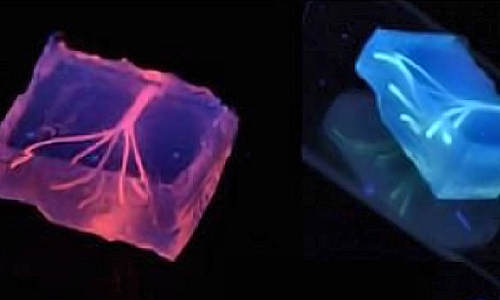
Nerves in action in transparent xenopus tadpoles.


“Cells that fire together, wire together. Cells that fire out of sync, lose their link”


In other words, a nerve cell that fires at the same time as its nerve cell neighbors will cooperatively form strong, stable connections onto its partner cells. On the other hand, a nerve cell that fires out of synchrony with its neighbours, will end up destabilizing and withdrawing its connections. “For the first time, we have direct, real-time evidence from watching brain cells in an intact animal to support Hebb’s model, but, we also provide surprising, new details, fundamentally updating the model for the 21st century,” says Dr. Edward Ruthazer, senior investigator on the study at the Montreal Neurological Institute and Hospital –The Neuro at McGill University and the McGill University Health Centre.


The study, which used multiphoton laser-scanning microscopy to observe cells in the brains of intact animals, discovered that asynchronous firing, or “firing out of sync” not only caused brain cells to lose their ability to make other cells fire, but unexpectedly, also caused them to dramatically increase their elaboration of new branches in search of better matched partners. “The surprising and entirely unexpected finding is that even though nerve circuit remodeling from asynchronous stimulation actively weakens connections, there is a 60% increase in axon branches that are exploring the environment but these exploratory branches are not long-lived,” said Dr. Ruthazer.


Dr. Ruthazer’s lab charts the formation of brain circuitry during development in the hopes of better understanding the rules that control healthy brain wiring and of advancing treatments for injuries to the nervous system and therapies for neurodevelopmental disorders such as autism and schizophrenia. Astoundingly, nearly one out of every 100 Canadians suffers from one of these disorders, estimated to cost the Canadian economy over $10 billion annually in addition to inflicting a devastating impact on patients and their families.


In the developing brain, initially imprecise connections between nerve cells are gradually pruned away, leaving connections that are stronger and more specific. This refinement occurs in response to patterned stimulation from the environment. “The way we perceive the world as adults is directly impacted by what we saw when we were younger,” says Dr. Ruthazer.


Dr. Ruthazer’s team studies brain development in Xenopus tadpoles, which have the distinct advantage of being transparent, enabling the team to clearly see the nervous system inside. They have developed a model that allows them to watch nerve cell remodeling in vivo, in real time, and to measure the efficacy of connections between cells. Optic fibers were used to stimulate the eyes of the tadpoles with different light patterns, while imaging and recording nerve cell branch formation. Asynchronous stimulation involved light flashes presented to each eye at different times, while synchronous stimulation involved simultaneous stimulation of both eyes.


Importantly, Dr. Ruthazer’s group also has begun to identify the molecular mechanisms underlying these changes in the nervous system. They show that the stabilization of the retinal nerve cell branches caused by synchronous firing involves signaling downstream of the synaptic activation of a neurotransmitter receptor called the N-methyl-D-aspartate receptor. In contrast, the enhanced exploratory growth that occurs with asynchronous activity does not appear to require the activation of this receptor.


Story Source:


The above story is based on materials provided by McGill University.


The post Scientists control rapid re-wiring of brain circuits appeared first on Bioengineer.org.


Skin grafts from genetically modified pigs

from

Bioengineer.org http://bioengineer.org/skin-grafts-genetically-modified-pigs/


A specially-bred strain of miniature swine lacking the molecule responsible for the rapid rejection of pig-to-primate organ transplants may provide a new source of skin grafts to treat seriously burned patients. A team of investigators from Massachusetts General Hospital (MGH) report that skin grafts from pigs lacking the Gal sugar molecule were as effective in covering burn-like injuries on the backs of baboons as skin taken from other baboons, a finding that could double the length of time burns can be protected while healing. The report in the journal Transplantation has been published online.


Skin grafts from genetically modified pigs


“This exciting work suggests that these GalT-knockout porcine skin grafts would be a useful addition to the burn-management armamentarium,” says Curtis Cetrulo, MD, of the MGH Transplantation Biology Research Center (TBRC) and the Division of Plastic and Reconstructive Surgery, corresponding author of the Transplantation paper. “We are actively exploring options for establishing clinical-grade production of these grafts and hope to begin a clinical trial in due course.”


A key component in the treatment of major burns, particularly those involving more than 30 percent of the body surface, is removing the damaged skin and covering the injury, preferably with a graft of a patient’s own tissue. When insufficient undamaged skin is available for grafting, tissue from deceased donors is used as a temporary covering. But deceased-donor skin grafts are in short supply and expensive – disadvantages also applying to artificial skin grafts – must be carefully tested for pathogens and are eventually rejected by a patient’s immune system. Once a deceased-donor graft has been rejected, a patient’s immune system will reject any subsequent deceased-donor grafts almost immediately.


The current study was designed to investigate whether a resource already available at the MGH might help expand options for protecting burned areas following removal of damaged skin. For more than 30 years David H. Sachs, MD, founder and scientific director of the TBRC, has been investigating ways to allow the human body to accept organ and tissue transplants from animals. Sachs and his team developed a strain of inbred miniature swine with organs that are close in size to those of adult humans. Since pig organs implanted into primates are rapidly rejected due to the presence of the Gal (alpha-1,3-galactose) molecule, Sachs and his collaborators used the strain that he developed to generate miniature swine in which both copies of the gene encoding GalT (galactosyltransferase), the enzyme responsible for placing the Gal molecule on the cell surface, were knocked out.


When Cetrulo’s team used skin from these Gal-free pigs to provide grafts covering burn-like injuries on the backs of baboons – injuries made while the animals were under anesthesia – the grafts adhered and developed a vascular system within 4 days of implantation. Signs of rejection began to appear on day 10, and rejection was complete by day 12 – a time frame similar to what is seen with deceased-donor grafts and identical to that observed when the team used skin grafts from other baboons. As with the use of second deceased-donor grafts to treat burned patients, a second pig-to-baboon graft was rapidly rejected. But if a pit-to baboon was followed by a graft using baboon skin, the second graft adhered to the wound and remained in place for around 12 days before rejection. The researchers also showed that acceptance of a second graft was similar no matter whether a pig xenograft or a baboon skin graft was used first.


“These results raise the possibility not only of providing an alternative to deceased-donor skin for many patients but also that, in patients whose burns are particularly extensive and require prolonged coverage, sequential use of GalT-knockout and deceased-donor skin could provide extended, high-quality wound coverage,” says co-author David Leonard, MBChB, of the TBRC and Division of Plastic and Reconstructive Surgery. “A high-quality alternative to deceased-donor skin that could be produced from a specially maintained, pathogen-free herd of GalT-knockout miniature swine would be an important resource for burn management in both civilian and military settings.”


Story Source:


The above story is based on materials provided by Massachusetts General Hospital.


The post Skin grafts from genetically modified pigs appeared first on Bioengineer.org.


3-D bioprinting builds a better blood vessel

from

Bioengineer.org http://bioengineer.org/3-d-bioprinting-builds-better-blood-vessel/


The tangled highway of blood vessels that twists and turns inside our bodies, delivering essential nutrients and disposing of hazardous waste to keep our organs working properly has been a conundrum for scientists trying to make artificial vessels from scratch. Now a team from Brigham and Women’s Hospital (BWH) has made headway in fabricating blood vessels using a three-dimensional (3D) bioprinting technique.


3D bioprinting builds a better blood vessel


Artificial blood vessels are created using hydrogel constructs that combine advances in 3-D bioprinting technology and biomaterials. Photo Credit: Image courtesy of Khademhosseini Lab


The study is published online this month in Lab on a Chip.


“Engineers have made incredible strides in making complex artificial tissues such as those of the heart, liver and lungs,” said senior study author, Ali Khademhosseini, PhD, biomedical engineer, and director of the BWH Biomaterials Innovation Research Center. “However, creating artificial blood vessels remains a critical challenge in tissue engineering. We’ve attempted to address this challenge by offering a unique strategy for vascularization of hydrogel constructs that combine advances in 3D bioprinting technology and biomaterials.”


The researchers first used a 3D bioprinter to make an agarose (naturally derived sugar-based molecule) fiber template to serve as the mold for the blood vessels. They then covered the mold with a gelatin-like substance called hydrogel, forming a cast over the mold which was then reinforced via photocrosslinks.


“Our approach involves the printing of agarose fibers that become the blood vessel channels. But what is unique about our approach is that the fiber templates we printed are strong enough that we can physically remove them to make the channels,” said Khademhosseini. “This prevents having to dissolve these template layers, which may not be so good for the cells that are entrapped in the surrounding gel.”


Khademhosseini and his team were able to construct microchannel networks exhibiting various architectural features. They were also able to successfully embed these functional and perfusable microchannels inside a wide range of commonly used hydrogels, such as methacrylated gelatin or poly(ethylene glycol)-based hydrogels at different concentrations.


Methacrylated gelatin laden with cells, in particular, was used to show how their fabricated vascular networks functioned to improve mass transport, cellular viability and cellular differentiation. Moreover, successful formation of endothelial monolayers within the fabricated channels was achieved.

“In the future, 3D printing technology may be used to develop transplantable tissues customized to each patient’s needs or be used outside the body to develop drugs that are safe and effective,” said Khademhosseini.


Story Source:


The above story is based on materials provided by Brigham and Women’s Hospital.


The post 3-D bioprinting builds a better blood vessel appeared first on Bioengineer.org.


Uncovering Clues to the Genetic Cause of Schizophrenia

from

Bioengineer.org http://bioengineer.org/uncovering-clues-to-the-genetic-cause-of-schizophrenia/


The overall number and nature of mutations—rather than the presence of any single mutation—influences an individual’s risk of developing schizophrenia, as well as its severity, according to a discovery by Columbia University Medical Center researchers published in the latest issue of Neuron. The findings could have important implications for the early detection and treatment of schizophrenia.


Uncovering Clues to the Genetic Cause of Schizophrenia


Taking a closer look at severe loss-of-function mutations revealed the contribution of both inherited (yellow pieces) and new (red piece) mutations to the puzzling genetic architecture of schizophrenia. Photo Credit: Lab of Maria Karayiorgou, M.D., and Joseph Gogos, M.D.


Maria Karayiorgou, MD, professor of psychiatry and Joseph Gogos, MD, PhD, professor of physiology and cellular biophysics and of neuroscience, and their team sequenced the “exome”—the region of the human genome that codes for proteins—of 231 schizophrenia patients and their unaffected parents. Using this data, they demonstrated that schizophrenia arises from collective damage across several genes.


“This study helps define a specific genetic mechanism that explains some of schizophrenia’s heritability and clinical manifestation,” said Dr. Karayiorgou, who is acting chief of the Division of Psychiatric and Medical Genetics at the New York State Psychiatric Institute. “Accumulation of damaged genes inherited from healthy parents leads to higher risk not only to develop schizophrenia but also to develop more severe forms of the disease.”


Schizophrenia is a severe psychiatric disorder in which patients experience hallucination, delusion, apathy and cognitive difficulties. The disorder is relatively common, affecting around 1 in every 100 people, and the risk of developing schizophrenia is strongly increased if a family member has the disease. Previous research has focused on the search for individual genes that might trigger schizophrenia. The availability of new high-throughput DNA sequencing technology has contributed to a more holistic approach to the disease.


The researchers compared sequencing data to look for genetic differences and identify new loss-of-function mutations—which are rarer, but have a more severe effect on ordinary gene function—in cases of schizophrenia that had not been inherited from the patients’ parents. They found an excess of such mutations in a variety of genes across different chromosomes.


Using the same sequencing data, the researchers also looked at what types of mutations are commonly passed on to schizophrenia patients from their parents. It turns out that many of these are “loss-of-function” types. These mutations were also found to occur more frequently in genes with a low tolerance for genetic variation.


“These mutations are important signposts toward identifying the genes involved in schizophrenia,” said Dr. Karayiorgou.


The researchers then looked more deeply into the sequencing data to try to determine the biological functions of the disrupted genes involved in schizophrenia. They were able to verify two key damaging mutations in a gene called SETD1A, suggesting that this gene contributes significantly to the disease.

SETD1A is involved in a process called chromatin modification. Chromatin is the molecular apparatus that packages DNA into a smaller volume so it can fit into the cell and physically regulates how genes are expressed. Chromatin modification is therefore a crucial cellular activity.


The finding fits with accumulating evidence that damage to chromatin regulatory genes is a common feature of various psychiatric and neurodevelopmental disorders. By combining the mutational data from this and related studies on schizophrenia, the authors found that “chromatin regulation” was the most common description for genes that had damaging mutations.


“A clinical implication of this finding is the possibility of using the number and severity of mutations involved in chromatin regulation as a way to identify children at risk of developing schizophrenia and other neurodevelopmental disorders,” said Dr. Gogos. “Exploring ways to reverse alterations in chromatic modification and restore gene expression may be an effective path toward treatment.”


In further sequencing studies, the researchers hope to identify and characterize more genes that might play a role in schizophrenia and to elucidate common biological functions of the genes.


Story Source:


The above story is based on materials provided by Columbia University Medical Center.


The post Uncovering Clues to the Genetic Cause of Schizophrenia appeared first on Bioengineer.org.


New genetic sequencing methods mean quicker, cheaper, and accurate embryo screening

from

Bioengineer.org http://bioengineer.org/new-genetic-sequencing-methods-mean-quicker-cheaper-accurate-embryo-screening/


Results from the first study of the clinical application of next generation DNA sequencing (NGS) in screening embryos for genetic disease prior to implantation in patients undergoing in-vitro fertilisation treatments show that it is an effective reliable method of selecting the best embryos to transfer, the annual conference of the European Society of Human Genetics will hear tomorrow (Sunday). Dr Francesco Fiorentino, from the GENOMA Molecular Genetics Laboratory, Rome, Italy, will say that his team’s research has shown that NGS, a high throughput sequencing method, has the potential to revolutionise pre-implantation genetic screening (PGS). The technique can result in reduced cost, faster results, and accurate identification of good embryos resulting in more ongoing pregnancies, he will say.


Professor Brunhilde Wirth, Head of the Institute of Human Genetics


Professor Brunhilde Wirth, Head of the Institute of Human Genetics


The researchers undertook a prospective, double blind trial using two methods of embryo screening, NGS, and the older method array-comparative genomic hybridisation (Array-CGH) of 192 blastocysts, or early embryos, obtained from 55 consecutive clinical pre-implantation genetic screening (PGS) cycles. Array-CGH was the first technology to be widely available for the accurate analysis of chromosomal abnormalities in the embryo and is used extensively across the world for this purpose.


Fifty five patients with an average age of 40 years were enrolled; in 45 cases they were undertaking IVF because of advanced age and in ten because of repeated IVF failures. Two different teams of researchers carried out biopsies and analysed the genetic make-up of the embryos at between five and six/seven days, depending on the speed of growth, and then measured the consistency of the diagnosis by comparing results from the two sequencing methods.


This comparison showed concordant results for 191 of the 192 embryos analysed. One embryo showed a false positive for three copies of chromosome 22 (trisomy 22) using the NGS technique. But analysis of this embryo also showed concordance between the two methods in detecting several other chromosomal abnormalities, and it would therefore have been ruled for transfer in any event. There were no other false negative diagnoses for chromosome abnormalities, and no inaccurate predictions of gender. NGS also showed itself to be as capable of identifying small, difficult to detect abnormalities.


“We found that results from the NGS and array-CGH diagnostic tests were highly concordant,” Dr Fiorentino will say. “NGS allowed us to detect a number of different abnormalities in 4608 chromosomes with a very high degree of accuracy, and following the transfer of 50 healthy embryos in 46 women, 30 pregnancies continued.”


These pregnancies were confirmed by the presence of a foetal sac and a heartbeat, and all have now completed at least 20 weeks of gestation.


PGS has been the subject of controversy over recent years. Initially hailed as an opportunity to improve clinical outcome in sub-fertile patients undergoing IVF, a number of studies later appeared to show that it might not help to identify and select chromosomally normal embryos for transfer based on its lack of benefit with respect to improving life birth rates.


“However, these studies used an older screening technique, fluorescent in-situ hybridisation (FISH),” says Dr Fiorentino, “and we hypothesised that NGS might come up with more accurate results. The results of our study have proved this to be the case, and that NGS can improve clinical outcomes. We expect that the use of NGS technologies will increase as evidence of their utility becomes better-known.


“A further advantage of the technique is that it is quicker and cheaper, while remaining just as sensitive as other methods of screening. Our next step will be to participate in a large randomised controlled trial, the results of which will be critical for the acceptance of NGS-based pre-implantation embryo assessment into wider clinical practice.”


Story Source:


The above story is based on materials provided by European Society of Human Genetics, Mary Rice.


The post New genetic sequencing methods mean quicker, cheaper, and accurate embryo screening appeared first on Bioengineer.org.


For the first time in the lab, researchers see stem cells take initial step toward development

from

Bioengineer.org http://bioengineer.org/first-time-lab-researchers-see-stem-cells-take-initial-step-toward-development/


The gap between stem cell research and regenerative medicine just became a lot narrower, thanks to a new technique that coaxes stem cells, with potential to become any tissue type, to take the first step to specialization. It is the first time this critical step has been demonstrated in a laboratory.


For the first time in the lab, researchers see stem cells take initial step toward development


Professor Ning Wang led a team that found the precise combination of mechanical forces, chemistry and timing to help stem cells differentiate into three germ layers, the first step toward developing specialized tissues and organs.


University of Illinois researchers, in collaboration with scientists at Notre Dame University and the Huazhong University of Science and Technology in China, published their results in the journal Nature Communications.


“Everybody knows that for an embryo to form, somehow a single cell has a way to self-organize into multiple cells, but the in vivo microenvironment is not well understood,” said study leader Ning Wang, a professor of mechanical science and engineering at the U. of I. “We want to know how they develop into organized structures and organs. It doesn’t happen by random chance. There are biological rules that we don’t yet understand.”


During fetal development, all the specialized tissues and organs of the body form out of a small ball of stem cells. First, the ball of generalized cells separates into three different cell lines, called germ layers, which will become different systems of the body. This crucial first step has eluded researchers in the lab. No one has yet been able to induce the cells to form the three distinct germ layers, in the correct order – endoderm on the inside, mesoderm in the middle and ectoderm on the outside. This represents a major hurdle in the application of stem cells to regenerative medicine, since researchers need to understand how tissues develop before they can reliably recreate the process.


“It’s very hard to generate tissues or organs, and the reason is that we don’t know how they form in vivo,” Wang said. “The problem, fundamentally, is that the biological process is not clear. What is the biological environment that controls this, so they can become more organized and specialized?”


Wang’s team demonstrated that not only is it possible for mouse embryonic stem cells to form three distinct germ layers in the lab, but also that achieving the separation requires a careful combination of correct timing, chemical factors and mechanical environment. The team uses cell lines that fluoresce in different colors when they become part of a germ layer, which allows the researchers to monitor the process dynamically.


The researchers deposited the stem cells in a very soft gel matrix, attempting to recreate the properties of the womb. They found that several mechanical forces played a role in how the cells organized and differentiated – the stiffness of the gel, the forces each cell exerts on its neighbors, and the matrix of proteins that the cells themselves deposit as a scaffolding to give the developing embryo structure.


By adjusting the mechanical environment, the researchers were able to observe how the forces affected the developing cells, and found the particular combination that yielded the three germ layers. They also found that they could direct layer development by changing the mechanics, even creating an environment that caused the layers to form in reverse order.


Now, Wang’s group is working to improve their technique for greater efficiency. He hopes that other researchers will be able to use the technique to bridge the gap between stem cells and tissue engineering.


“It’s the first time we’ve had the correct three-germ-layer organization in mammalian cells,” Wang said. “The potential is huge. Now we can push it even further and generate specific organs and tissues. It opens the door for regenerative medicine.”


Story Source:


The above story is based on materials provided by University of Illinois at Urbana-Champaign, Liz Ahlberg.


The post For the first time in the lab, researchers see stem cells take initial step toward development appeared first on Bioengineer.org.


Research details how developing neurons sense a chemical cue

from

Bioengineer.org http://bioengineer.org/research-details-developing-neurons-sense-chemical-cue/


Symmetry is an inherent part of development. As an embryo, an organism’s brain and spinal cord, like the rest of its body, organize themselves into left and right halves as they grow. But a certain set of nerve cells do something unusual: they cross from one side to the other. New research in mice delves into the details of the molecular interactions that help guide these neurons toward this anatomical boundary.


Research details how developing neurons sense a chemical cue


Chemical cues help developing neurons make the right connections by telling them where to extend their branches, or axons. New structural studies of the interactions between one such cue and receptor molecules on the axons reveal how Netrin-1 molecules (blue and green) bind to two neogenin molecules (magenta and orange).


In an embryo, a neuron’s branches, or axons, have special structures on their tips that sense chemical cues telling them where to grow. The new findings, by researchers at Memorial Sloan Kettering Cancer Center and The Rockefeller University, reveal the structural details of how one such cue, Netrin-1, interacts with two sensing molecules on the axons, DCC and a previously less well characterized player known as neogenin, as a part of this process.


“Our work provides the first high-resolution view of the molecular complexes that form on the surface of a developing axon and tell it to move in one direction or another,” says Dimitar Nikolov, a structural biologist at Memorial Sloan Kettering. “This detailed understanding of these assemblies helps us better understand neural wiring, and may one day be useful in the development of drugs to treat spinal cord or brain injuries.”


In a developing nervous system, the signaling molecule, Netrin-1, identified by Rockefeller University Professor Marc Tessier-Lavigne and colleagues, can guide neurons by attracting or repulsing them. In the case of axons that cross from one side to the other, extended by so-called commissural neurons, Netrin-1 attracts them toward the middle.


With a technique that uses X-rays to visualize the structure of crystalized proteins, research scientist Kai Xu and colleagues in Nikolov’s laboratory revealed that Netrin-1 has two separate binding sites on opposite ends, enabling it to simultaneously bind to different receptors. This may explain how Netrin-1, which is an important axon-guiding molecule, can affect in different ways neurons that express different combinations of receptors, Nikolov says.


For some time, scientists have known commissural neurons used the receptor molecule DCC to detect Netrin-1. Neogenin has a structure similar to DCC, and this research, described today in Science, confirms neogenin too acts as a sensing molecule for commissural neurons in mammals.


In experiments that complemented the structural work, conducted by Nicolas Renier and Zhuhao Wu in Tessier-Lavigne’s lab, the researchers confirmed that, like DCC, neogenin senses Netrin-1 for the growing commissural neurons in mice.


These neurons are part of the system by which one side of the brain controls movement on the opposite side of the body. As a result, a mutation in the gene responsible for DCC interferes with this coordination, causing congenital mirror movement disorder. People with this disorder cannot move one side of the body in isolation; for example, a right-handed wave is mirrored by a similar gesture by the left hand.


The work also has implications for understanding why DCC, neogenin and other cell-surface receptors come in slightly different forms, called splice isoforms. The structural research revealed these isoforms bind differently to Netrin-1. However, it is not yet clear what this means for neuron wiring, Nikolov says.


“With this structural knowledge, and with the identification of an additional receptor involved in axon guidance in the spinal cord, we are gaining deeper insight into the mechanisms through which neurons make connections that produce a functioning nervous system, as well as the dysfunction that arises from miswiring of connections” says Tessier-Lavigne.


Story Source:


The above story is based on materials provided by The Rockefeller University, Zach Veilleux.


The post Research details how developing neurons sense a chemical cue appeared first on Bioengineer.org.


28 Mayıs 2014 Çarşamba

Artificial lung the size of a sugar cube

from

Bioengineer.org http://bioengineer.org/artificial-lung-size-sugar-cube/


What medications can be used to treat lung cancer, and how effective are they? Until now, drug companies have had to rely on animal testing to find out. But in the future, a new 3D model lung is set to achieve more precise results and ultimately minimize – or even completely replace – animal testing. From June 23-26, researchers will be presenting their new model at the BIO International Convention in San Diego, California (Germany Pavilion, Booth 4513-03).


Artificial lung the size of a sugar cube


Lung cancer is a serious condition. Once patients are diagnosed with it, chemotherapy is often their only hope. But nobody can accurately predict whether or not this treatment will help. To start with, not all patients respond to a course of chemotherapy in exactly the same way. And then there’s the fact that the systems drug companies use to test new medications leave a lot to be desired. “Animal models may be the best we have at the moment, but all the same, 75 percent of the drugs deemed beneficial when tested on animals fail when used to treat humans,” explains Prof. Dr. Heike Walles, head of the Würzburg-based “Regenerative Technologies for Oncology” project group belonging to the Fraunhofer Institute for Interfacial Engineering and Biotechnology IGB.


These tests are set to achieve better results in the future: “We’ve developed an innovative 3D test system that allows us to superbly simulate what happens in the human body. Our plan is for this system to replace animal tests in the future,” says Walles. Essentially what the researchers have done is to recreate the human lung in miniature – with a volume of half a cubic centimeter, each model is no bigger than a sugar cube. In a parallel effort, scientists at the Department of Bioinformatics at the University of Würzburg are working up computer simulation models for different patient groups. These are necessary because patients may have genetic variations that inhibit therapies from having the desired effect. Comparing the theoretical and biological models allows each research group to optimize their results.


The biological model is based human lung cancer cells growing on tissue. Thus an artificial lung is created. A bioreactor is used to make it breathe and to pump a nutrient medium through its blood vessels in the same way our bodies supply our lungs with blood. The reactor also makes it possible to regulate factors such as how fast and deeply the model lung breathes.


With the scientists having managed to construct the lung tissue, Walles is delighted to report that “treatments that generate resistance in clinics do the same in our model.” Researchers are now planning to explore the extent to which their artificial lung can be used to test new therapeutic agents. Should resistance crop up during testing, doctors can opt to treat the patient with a combination therapy from the outset and thus side-step the problem. Thinking long-term, there is even the possibility of creating an individual model lung for each patient. This would make it possible to accurately predict which of the various treatment options will work. The required lung cells are collected as part of the biopsy performed to allow doctors to analyze the patient’s tumor.


On the trail of metastases


Testing new medications is by no means the only thing the model lung can be used for. It is also designed to help researchers to better understand the formation of metastases; it is these that often make a cancer fatal. “As metastases can’t be examined in animals – or in 2D models where cells grow only on a flat surface – we’ve only ever had a rough understanding of how they form. Now for the first time, our 3D lung tissue makes it possible to perform metastases analysis,” explains Walles. “In the long term, this may enable us to protect patients from metastases altogether.” In order to travel through the body, tumor cells alter their surface markers – in other words, the molecules that bind them to a particular area of the body. Cancer cells are then free to spread throughout the body via the body’s circulatory system before taking up residence somewhere else by expressing their original surface markers. The scientists plan to use their model lung’s artificial circulatory system to research exactly how this transformation occurs. And in doing so, they may someday succeed in developing medication that will stop metastases from forming in the first place.


Story Source:


The above story is based on materials provided by Fraunhofer.


The post Artificial lung the size of a sugar cube appeared first on Bioengineer.org.


Researchers use light to coax stem cells to regrow teeth

from

Bioengineer.org http://bioengineer.org/researchers-use-light-coax-stem-cells-regrow-teeth/


A Harvard-led team is the first to demonstrate the ability to use low-power light to trigger stem cells inside the body to regenerate tissue, an advance they reported in Science Translational Medicine. The research, led by David J. Mooney, Robert P. Pinkas Family Professor of Bioengineering at the Harvard School of Engineering and Applied Sciences (SEAS), lays the foundation for a host of clinical applications in restorative dentistry and regenerative medicine more broadly, such as wound healing, bone regeneration, and more.


Researchers use light to coax stem cells to regrow teeth


The team used a low-power laser to trigger human dental stem cells to form dentin, the hard tissue that is similar to bone and makes up the bulk of teeth. What’s more, they outlined the precise molecular mechanism involved, and demonstrated its prowess using multiple laboratory and animal models.


A number of biologically active molecules, such as regulatory proteins called growth factors, can trigger stem cells to differentiate into different cell types. Current regeneration efforts require scientists to isolate stem cells from the body, manipulate them in a laboratory, and return them to the body—efforts that face a host of regulatory and technical hurdles to their clinical translation. But Mooney’s approach is different and, he hopes, easier to get into the hands of practicing clinicians.


“Our treatment modality does not introduce anything new to the body, and lasers are routinely used in medicine and dentistry, so the barriers to clinical translation are low,” said Mooney, who is also a Core Faculty Member at the Wyss Institute for Biologically Inspired Engineering at Harvard. “It would be a substantial advance in the field if we can regenerate teeth rather than replace them.”


The team first turned to lead author and dentist Praveen Arany, Ph.D. ’11, who is now an Assistant Clinical Investigator at the National Institutes of Health (NIH). At the time of the research, he was a Harvard graduate student and then postdoctoral fellow affiliated with SEAS and the Wyss Institute.

Arany took rodents to the laboratory version of a dentist’s office to drill holes in their molars, treat the tooth pulp that contains adult dental stem cells with low-dose laser treatments, applied temporary caps, and kept the animals comfortable and healthy. After about 12 weeks, high-resolution x-ray imaging and microscopy confirmed that the laser treatments triggered the enhanced dentin formation.


“It was definitely my first time doing rodent dentistry,” said Arany, who faced several technical challenges in performing oral surgery on such a small scale. The dentin was strikingly similar in composition to normal dentin, but did have slightly different morphological organization. Moreover, the typical reparative dentin bridge seen in human teeth was not as readily apparent in the minute rodent teeth, owing to the technical challenges with the procedure.


“This is one of those rare cases where it would be easier to do this work on a human,” Mooney said.

Next the team performed a series of culture-based experiments to unveil the precise molecular mechanism responsible for the regenerative effects of the laser treatment. It turns out that a ubiquitous regulatory cell protein called transforming growth factor beta-1 (TGF-β1) played a pivotal role in triggering the dental stem cells to grow into dentin. TGF-β1 exists in latent form until activated by any number of molecules.


Here is the chemical domino effect the team confirmed: In a dose-dependent manner, the laser first induced reactive oxygen species (ROS), which are chemically active molecules containing oxygen that play an important role in cellular function. The ROS activated the latent TGF-β1complex which, in turn, differentiated the stem cells into dentin.


Nailing down the mechanism was key because it places on firm scientific footing the decades-old pile of anecdotes about low-level light therapy (LLLT), also known as Photobiomodulation (PBM).


Since the dawn of medical laser use in the late 1960s, doctors have been accumulating anecdotal evidence that low-level light therapy can stimulate all kind of biological processes including rejuvenating skin and stimulating hair growth, among others. But interestingly enough, the same laser can be also be used to ablate skin and remove hair—depending on the way the clinician uses the laser. The clinical effects of low-power lasers have been subtle and largely inconsistent. The new work marks the first time that scientists have gotten to the nub of how low-level laser treatments work on a molecular level, and lays the foundation for controlled treatment protocols.


“The scientific community is actively exploring a host of approaches to using stem cells for tissue regeneration efforts,” said Wyss Institute Founding Director Don Ingber, “and Dave and his team have added an innovative, noninvasive and remarkably simple but powerful tool to the toolbox.”


Next Arany aims to take this work to human clinical trials. He is currently working with his colleagues at the National Institute of Dental and Craniofacial Research (NIDCR), which is one of the National Institutes of Health (NIH), to outline the requisite safety and efficacy parameters. “We are also excited about expanding these observations to other regenerative applications with other types of stem cells,” he said.


Story Source:


The above story is based on materials provided by Wyss Institute, Kristen Kusek.


The post Researchers use light to coax stem cells to regrow teeth appeared first on Bioengineer.org.


Sperm cells are extremely efficient at swimming against a current

from

Bioengineer.org http://bioengineer.org/sperm-cells-extremely-efficient-swimming-current/


Like salmon traveling upstream to spawn, sperm cells are extremely efficient at swimming against the current, according to research to be published this week.


sperm


Superimposed photographs of a human sperm cell swimming upstream along the wall of a microfluidic channel, with overlaid virtual tracer particles indicating the flow direction. Photo Credit: Vasily Kantsler


The discovery, to be published in the journal eLife by researchers at MIT and Cambridge University, may help us to understand how some sperm travel such long distances, through difficult terrain, to reach and fertilize an egg.


Of the hundreds of millions of sperm cells that begin the journey up the oviducts, only a few hardy travelers will ever reach their destination. Not only do the cells have to swim in the right direction over distances that are around 1,000 times their own length, but they are exposed to different chemicals and currents along the way.


While we know that sperm cells can “smell” chemicals given off by the egg once they get very close to it, this does not explain how they navigate for the majority of their journey, says Jörn Dunkel, an assistant professor of mathematics at MIT, and a member of the research team.


“We wanted to know which physical mechanisms could be responsible for navigation,” says Dunkel, who carried out the research alongside Vasily Kantsler of the Skolkovo Institute of Science and Technology and the University of Warwick (and currently visiting at MIT); Raymond E. Goldstein of Cambridge; and Martyn Blayney of the Bourn Hall Clinic in the U.K. “If you think of salmon, for example, they can swim against the stream, and the question was whether something similar could really be confirmed for human sperm cells.”


Microchannels in lieu of oviducts


However, observing sperm cells swimming within the human body itself is no easy task. So in a bid to understand what the cells are capable of, the researchers instead built a series of artificial microchannels of different sizes and shapes, into which they inserted the sperm. They were then able to modify the flow of fluid through the tubes, to investigate how the cells responded to different current speeds.


They discovered that at certain flow speeds, the sperm cells were able to swim very efficiently upstream. “We found that if you create the right flow velocities, you can observe them swimming upstream for several minutes,” Dunkel says. “The mechanism is very robust.”


What’s more, the researchers were also surprised to observe that the sperm were not swimming in a straight line upstream, but in a spiraling motion, along the walls of the channel. The sperm cells react to the difference in the speed of current near the walls of the chamber — where the fluid is attracted to the surface, and is therefore at its slowest — and the free-flowing center of the tube, Dunkel says.


If biologists are able to observe similar fluid-flow speeds within the oviduct, it could help confirm whether sperm cells are indeed using this mechanism to navigate through the body, he says.


Possible advances in artificial insemination


Not only would this improve our understanding of human reproduction, but it could also one day allow us to design new diagnostic tools and more efficient artificial-insemination techniques, the researchers claim. Reproduction specialists could take sperm samples and artificially recreate the conditions within the body to identify the cells that are the best swimmers, in a bid to preselect those most likely to succeed, Dunkel says.


The researchers can also experiment with different fluid viscosities within the microchannels, to determine which result in the strongest upstream swimming effect, he says. “So the idea would be to fine-tune the properties of the fluid medium that the sperm cells are contained in, before you insert it into the body, so that you know the cells can achieve optimal upstream swimming.”


Jackson Kirkman-Brown, honorary reader in reproductive science at the University of Birmingham and science lead for the Birmingham Women’s Fertility Centre, both in the U.K., says the research gives us an important new insight into a mechanism that sperm may be using to navigate inside the human body.

“We really know effectively nothing about how sperm cells navigate, so this gives us more information about a potential mechanism that may be important,” he says. “It’s telling us that human sperm seem to move differently to other things that propel themselves with a tail.”


However, much more work will be needed to determine if sperm cells behave in the same way in the much more complex terrain inside the oviduct itself. “This is a huge step in understanding what might influence sperm in that environment, but it’s far from explaining what does influence them,” Kirkman-Brown says. “People will certainly try to find this mechanism happening [inside the body], but it’s going to be a complicated chase.”


In the meantime, the researchers plan to begin investigating whether sperm cells can work together to reach the egg. “It is a commonly held belief that there is competition between sperm cells, with the fittest reaching the egg first,” Dunkel says. “But recent studies by our team and others show that sperm practically always accumulate at the surface of a tube, and you can end up with a high local concentration of sperm cells, so there could actually be cooperation among these cells that allows them to swim faster collectively.”


The research was supported by the European Research Council.


Story Source:


The above story is based on materials provided by MIT News, Helen Knight.


The post Sperm cells are extremely efficient at swimming against a current appeared first on Bioengineer.org.


How DNA is ‘edited’ to correct genetic diseases

from

Bioengineer.org http://bioengineer.org/dna-edited-correct-genetic-diseases/


An international team of scientists has made a major step forward in our understanding of how enzymes ‘edit’ genes, paving the way for correcting genetic diseases in patients.


How DNA is 'edited' to correct genetic diseases


An illustration of DNA attached to magnetic beads, as used in the single molecule microscope. Photo Credit: Professor Mark Szczelkun


Researchers at the Universities of Bristol, Münster and the Lithuanian Institute of Biotechnology have observed the process by which a class of enzymes called CRISPR – pronounced ‘crisper’ – bind and alter the structure of DNA.


The results, published in the Proceedings of the National Academy of Sciences (PNAS) today [26 May], provide a vital piece of the puzzle if these genome editing tools are ultimately going to be used to correct genetic diseases in humans.


CRISPR enzymes were first discovered in bacteria in the 1980s as an immune defence used by bacteria against invading viruses. Scientists have more recently shown that one type of CRISPR enzyme – Cas9 – can be used to edit the human genome – the complete set of genetic information for humans.


These enzymes have been tailored to accurately target a single combination of letters within the three billion base pairs of the DNA molecule. This is the equivalent of correcting a single misspelt word in a 23-volume encyclopaedia.


To find this needle in a haystack, CRISPR enzymes use a molecule of RNA – a nucleic acid similar in structure to DNA. The targeting process requires the CRISPR enzymes to pull apart the DNA strands and insert the RNA to form a sequence-specific structure called an ‘R-loop’.


The global team tested the R-loop model using specially modified microscopes in which single DNA molecules are stretched in a magnetic field. By altering the twisting force on the DNA, the researchers could directly monitor R-loop formation events by individual CRISPR enzymes.


This allowed them to reveal previously hidden steps in the process and to probe the influence of the sequence of DNA bases.


Professor Mark Szczelkun, from Bristol University’s School of Biochemistry, said: “An important challenge in exploiting these exciting genome editing tools is ensuring that only one specific location in a genome is targeted.


“Our single molecule assays have led to a greater understanding of the influence of DNA sequence on R-loop formation. In the future this will help in the rational re-engineering of CRISPR enzymes to increase their accuracy and minimise off-target effects. This will be vital if we are to ultimately apply these tools to correct genetic diseases in patients. ”


Story Source:


The above story is based on materials provided by University of Bristol.


The post How DNA is ‘edited’ to correct genetic diseases appeared first on Bioengineer.org.


Hybrid system mimics photosynthesis

from

Bioengineer.org http://bioengineer.org/hybrid-energy-transfer-system-mimics-photosynthesis/


Scientists from the University of Southampton, in collaboration with the Universities of Sheffield and Crete, have developed a new hybrid energy transfer system, which mimics the processes responsible for photosynthesis.


Hybrid energy transfer system mimics photosynthesis


This is the device trapping photons between two mirrors in which two different organic molecules reside. Photo Credit: University of Southampton


From photosynthesis to respiration, the processes of light absorption and its transfer into energy represent elementary and essential reactions that occur in any biological living system.


This energy transfer is known as Forster Resonance Energy Transfer (FRET), a radiationless transmission of energy that occurs on the nanometer scale from a donor molecule to an acceptor molecule. The donor molecule is the dye or chromophore that initially absorbs the energy and the acceptor is the chromophore to which the energy is subsequently transferred without any molecular collision. However, FRET is a strongly distance dependent process which occurs over a scale of typically 1 to 10 nm.


In a new study, published in the journal Nature Materials, the researchers demonstrate an alternate non-radiative, intermolecular energy transfer that exploits the intermediating role of light confined in an optical cavity. The advantage of this new technique which exploits the formation of quantum states admixture of light and matter, is the length over which the interaction takes places, that is in fact, considerably longer than conventional FRET-type processes.


Dr Niccolo Somaschi, from the University of Southampton’s Hybrid Photonics group (which is led by Prof Lagoudakis) and co-author of the paper, says: “The possibility to transfer energy over distances comparable to the wavelength of light has the potential to be of both fundamental and applied interest. Our deep understanding of energy transfer elucidates the basic mechanisms behind the process of photosynthesis in biological systems and therefore gets us closer to the reproduction of fully synthetic systems which mimic biological functionalities. At the fundamental level, the present work suggests that the coherent coupling of molecules may be directly involved in the energy transfer process which occurs in the photosynthesis.


“On the applied perspective instead, organic semiconductors continue to receive significant interest for application in optoelectronic devices, for example light-emitting or photovoltaic devices, in which performance is dependent on our ability to control the formation and transport of carriers in molecular systems.”


The new device consists of an optical cavity made by two metallic mirrors which trap the photons in a confined environment where two different organic molecules reside in. By engineering the spacing between the mirrors based on the optical properties of the organic materials, it is possible to create a new quantum state that is a combination of the trapped photons and the excited states in the molecules. The photon essentially “glues” together these quantum mechanical states, forming a new half-light half-matter particle, called polariton, which is responsible for the efficient transfer of energy from one material to the other.


Story Source:


The above story is based on materials provided by University of Southampton.


The post Hybrid system mimics photosynthesis appeared first on Bioengineer.org.


Learning early in life may help keep brain cells alive

from

Bioengineer.org http://bioengineer.org/learning-early-life-may-help-keep-brain-cells-alive/


Using your brain – particularly during adolescence – may help brain cells survive and could impact how the brain functions after puberty.


brain


Photo Credits: Doggygraph/Shutterstock via mnn


According to a recently published study in Frontiers in Neuroscience, Rutgers behavioral and systems neuroscientist Tracey Shors, who co-authored the study, found that the newborn brain cells in young rats that were successful at learning survived while the same brain cells in animals that didn’t master the task died quickly.

“In those that didn’t learn, three weeks after the new brain cells were made, nearly one-half of them were no longer there,” said Shors, professor in the Department of Psychology and Center for Collaborative Neuroscience at Rutgers. “But in those that learned, it was hard to count. There were so many that were still alive.”


The study is important, Shors says, because it suggests that the massive proliferation of new brain cells most likely helps young animals leave the protectiveness of their mothers and face dangers, challenges and opportunities of adulthood.


Scientists have known for years that the neurons in adult rats, which are significant but fewer in numbers than during puberty, could be saved with learning, but they did not know if this would be the case for young rats that produce two to four times more neurons than adult animals.


By examining the hippocampus – a portion of the brain associated with the process of learning – after the rats learned to associate a sound with a motor response, scientists found that the new brain cells injected with dye a few weeks earlier were still alive in those that had learned the task while the cells in those who had failed did not survive.


“It’s not that learning makes more cells,” says Shors. “It’s that the process of learning keeps new cells alive that are already present at the time of the learning experience.”


Since the process of producing new brain cells on a cellular level is similar in animals, including humans, Shors says ensuring that adolescent children learn at optimal levels is critical.

“What it has shown me, especially as an educator, is how difficult it is to achieve optimal learning for our students. You don’t want the material to be too easy to learn and yet still have it too difficult where the student doesn’t learn and gives up,” Shors says.


So, what does this mean for the 12-year-old adolescent boy or girl?


While scientists can’t measure individual brain cells in humans, Shors says this study, on the cellular level, provides a look at what is happening in the adolescent brain and provides a window into the amazing ability the brain has to reorganize itself and form new neural connections at such a transformational time in our lives.


“Adolescents are trying to figure out who they are now, who they want to be when they grow up and are at school in a learning environment all day long,” says Shors. “The brain has to have a lot of strength to respond to all those experiences.”


Story Source:


The above story is based on materials provided by Rutgers University, Robin Lally.


The post Learning early in life may help keep brain cells alive appeared first on Bioengineer.org.


27 Mayıs 2014 Salı

Using Thoughts to Control Airplanes

from

Bioengineer.org http://bioengineer.org/using-thoughts-control-airplanes/


Pilots of the future could be able to control their aircraft by merely thinking commands. Scientists of the Technische Universität München and the TU Berlin have now demonstrated the feasibility of flying via brain control – with astonishing accuracy.


Using Thoughts to Control Airplanes


Simulating brain controlled flying at the Institute for Flight System Dynamics. Photo Credit: A. Heddergott/TU München


The pilot is wearing a white cap with myriad attached cables. His gaze is concentrated on the runway ahead of him. All of a sudden the control stick starts to move, as if by magic. The airplane banks and then approaches straight on towards the runway. The position of the plane is corrected time and again until the landing gear gently touches down. During the entire maneuver the pilot touches neither pedals nor controls.


This is not a scene from a science fiction movie, but rather the rendition of a test at the Institute for Flight System Dynamics of the Technische Universität München (TUM). Scientists working for Professor Florian Holzapfel are researching ways in which brain controlled flight might work in the EU-funded project “Brainflight.” “A long-term vision of the project is to make flying accessible to more people,” explains aerospace engineer Tim Fricke, who heads the project at TUM. “With brain control, flying, in itself, could become easier. This would reduce the work load of pilots and thereby increase safety. In addition, pilots would have more freedom of movement to manage other manual tasks in the cockpit.”


Surprising accuracy


The scientists have logged their first breakthrough: They succeeded in demonstrating that brain-controlled flight is indeed possible – with amazing precision. Seven subjects took part in the flight simulator tests. They had varying levels of flight experience, including one person without any practical cockpit experience whatsoever. The accuracy with which the test subjects stayed on course by merely thinking commands would have sufficed, in part, to fulfill the requirements of a flying license test. “One of the subjects was able to follow eight out of ten target headings with a deviation of only 10 degrees,” reports Fricke. Several of the subjects also managed the landing approach under poor visibility. One test pilot even landed within only few meters of the centerline.


The TU München scientists are now focusing in particular on the question of how the requirements for the control system and flight dynamics need to be altered to accommodate the new control method. Normally, pilots feel resistance in steering and must exert significant force when the loads induced on the aircraft become too large. This feedback is missing when using brain control. The researchers are thus looking for alternative methods of feedback to signal when the envelope is pushed too hard, for example.


Electrical potentials are converted into control commands


In order for humans and machines to communicate, brain waves of the pilots are measured using electroencephalography (EEG) electrodes connected to a cap. An algorithm developed by scientists from Team PhyPA (Physiological Parameters for Adaptation) of the Technische Universität Berlin allows the program to decipher electrical potentials and convert them into useful control commands.


Only the very clearly defined electrical brain impulses required for control are recognized by the brain-computer interface. “This is pure signal processing,” emphasizes Fricke. Mind reading is not possible.


Story Source:

The above story is based on materials provided by Technische Universitaet Muenchen.


The post Using Thoughts to Control Airplanes appeared first on Bioengineer.org.


HIV can cut and paste in the human genome

from

Bioengineer.org http://bioengineer.org/hiv-can-cut-paste-human-genome/


For the first time researchers have succeeded in altering HIV virus particles so that they can simultaneously, as it were, ‘cut and paste’ in our genome via biological processes. Developed at the Department of Biomedicine at Aarhus University, the technology makes it possible to repair genomes in a new way.


HIV can cut and paste in the human genome


A technology developed at Aarhus University makes it possible to repair the human genome in a new way with the help of manipulated virus particles.


It also offers good perspectives for individual treatment of both hereditary diseases and certain viral infections:


“Now we can simultaneously cut out the part of the genome that is broken in sick cells, and patch the gap that arises in the genetic information which we have removed from the genome. The new aspect here is that we can bring the scissors and the patch together in the HIV particles in a fashion that no one else has done before,” says associate professor in genetics Jacob Giehm Mikkelsen from Aarhus University.


‘Hit-and-run’ technique leaves no traces


At the same time, the team of researchers from Aarhus have developed a technique that increases the safety of the cutting process, the so-called “gene editing”:


“In the past, the gene for the scissors has been transferred to the cells, which is dangerous because the cell keeps on producing scissors which can start cutting uncontrollably. But because we make the scissors in the form of a protein, they only cut for a few hours, after which they are broken down. And we ensure that the virus particle also brings along a small piece of genetic material to patch the hole,” says Jacob Giehm Mikkelsen.


“We call this a ‘hit-and-run’ technique because the process is fast and leaves no traces”.


Viruses become nanoparticles


The researchers have benefited from many years of intense research into HIV as this has e.g. shown that HIV particles can be converted into transporters of genetic information. But when they also become transporters of proteins that are not normally found in the cells, as is the case now, the particles are altered. Virus particles are converted into nanoparticles which carry the substances that can have a direct effect on the treated cells.


HIV infection is one of the areas where the researchers want to make use of the technique, and here the goal is to stop a specific gene from functioning – something that the protein scissors can do.


“By altering relevant cells in the immune system (T cells) we can make them resistant to HIV infection and perhaps even at the same time also equip them with genes that help fight HIV.

“So in this way HIV can in time become a tool in the fight against HIV,” says postdoc and PhD Yujia Cai of the research team.


The results have recently been published in the scientific journal eLIFE.


Story Source:


The above story is based on materials provided by Aarhus University.


The post HIV can cut and paste in the human genome appeared first on Bioengineer.org.


25 Mayıs 2014 Pazar

Nature inspires drones of the future

from

Bioengineer.org http://bioengineer.org/nature-inspires-drones-future/


Based on the mechanisms adopted by birds, bats, insects and snakes, 14 distinguished research teams have developed solutions to some of the common problems that drones could be faced with when navigating through an urban environment and performing novel tasks for the benefit of society.


Nature inspires drones of the future


Biologically-inspired flapping-wing robots are shown. Photo Credit: Pakpong Chirarattananon et al. 2014


Whether this is avoiding obstacles, picking up and delivering items or improving the take-off and landing on tricky surfaces, it is hoped the solutions can lead to the deployment of drones in complex urban environments in a number of different ways, from military surveillance and search and rescue efforts to flying camera phones and reliable courier services. For this, drones need exquisite flight control.


The research teams have presented their work today, 23 May, in a special issue of IOP Publishing’s journal Bioinspiration and Biomimetics, devoted to bio-inspired flight control.


The first small drones have already been used in search and rescue operations to investigate difficult-to-reach and hazardous areas, such as in Fukushima, Japan. A research team from Hungary believe these efforts could be improved if robots are able to work in tandem, and have developed an algorithm that allows a number of drones to fly together like a flock of birds.


The effectiveness of the algorithm was demonstrated by using it to direct the movements of a flock of nine individual quadcopters whilst they followed a moving car.


While this collective movement may be helpful when searching vast expanses of land, a group of researchers from Harvard University have developed a millimetre-sized drone with a view to using it to explore extremely cramped and tight spaces.


The microrobot they designed, which was the size of a one cent coin, could take off and land and hover in the air for sustained periods of time. In their new paper, the researchers have demonstrated the first simple, fly-like manoeuvres. In the future, millimetre-sized drones could also be used in assisted agriculture pollination and reconnaissance, and could aid future studies of insect flight.


Once deployed into the real world, drones will be faced with the extremely tricky task of dealing with the elements, which could be extreme heat, the freezing cold, torrential rain or thunderstorms.


The most challenging problem for airborne robots will be strong winds and whirlwinds, which a research team, from the University of North Caroline at Chapel Hill, University of California and The Johns Hopkins University, have begun to tackle by studying the hawk moth.


In their study, the researchers flew hawk moths through a number of different whirlwind conditions in a vortex chamber, carefully examining the mechanisms that the hawk moths used to successfully regain flight control.


Researchers must also find a way of reducing the amount of power that is required to operate drones, which a team from the Université de Sherbrooke and Stanford University have achieved by creating a “jumpglider”.


Inspired by vertebrates like the flying squirrel, the flying fish and the flying snake, which use their aerodynamic bodies to extend their jumping range to avoid predators, the “jumpglider” combines an aeroplane-shaped body with a spring-based mechanical foot that propels the robot into the air.


The researchers believe the “jumpglider” can be used in search and rescue efforts, operating at low power and offering a significant advantage over land-based robots by being able to navigate around obstacles and over rough terrain.


In his opening editorial, Guest Editor of the special issue, Dr David Lentink, from Stanford University, writes: “Flying animals can be found everywhere in our cities. From scavenging pigeons to alcohol-sniffing fruit flies that make precision landings on our wine glasses, these animals have quickly learnt how to control their flight through urban environments to exploit our resources.


“To enable our drones to fly equally well in wind and clutter, we need to solve several flight control challenges during all flight phases: take-off, cruising, and landing.


“This special issue provides a unique integration between biological studies of animals and bio-inspired engineering solutions. Each of the 14 papers presented in this special issue offer a unique perspective on bio-mimetic flight, providing insights and solutions to the take-off, obstacle avoidance, in-flight grasping, swarming, and landing capabilities that urban drones need to succeed.”


Story Source:


The above story is based on materials provided by Institute of Physics.


The post Nature inspires drones of the future appeared first on Bioengineer.org.


Study Identifies How Signals Trigger Cancer Cells to Spread

from

Bioengineer.org http://bioengineer.org/study-identifies-signals-trigger-cancer-cells-spread/


Researchers at Albert Einstein College of Medicine of Yeshiva University have discovered a signaling pathway in cancer cells that controls their ability to invade nearby tissues in a finely orchestrated manner. The findings offer insights into the early molecular events involved in metastasis, the deadly spread of cancer cells from primary tumor to other parts of the body. The study was published today in the online edition of Nature Cell Biology.


Study Identifies How Signals Trigger Cancer Cells to Spread


This electron micrograph shows a cancer cell (upper darker area) that has formed three invadopodia that are penetrating the adjacent extracellular matrix (lower lighter area).


To migrate from a primary tumor, a cancer cell must first break through surrounding connective tissue known as the extracellular matrix (ECM). The cancer cell does so by forming short-lived invadopodia—foot-like protrusions these cells use to invade. Invadopodia release enzymes that degrade the ECM, while other protrusions pull the cancer cell along, much like a locomotive pulls a train. The invading cancer cell relies on the cycle of invadopodium formation/disappearance to successfully travel from the tumor and enter nearby blood vessels to be carried to distant parts of the body.


“We’ve known for some time that invadopodia are driven by protein filaments called actin,” said study leader Louis Hodgson, Ph.D., assistant professor of anatomy and structural biology at Einstein. “But exactly what was regulating the actin in invadopodia was not clear.”


Previous studies had suggested that a protein called Rac1 played a role in cancer-cell invasion. When Rac1 levels are elevated, cancer cells display more invasive characteristics. But this suspected Rac1 activity in invadopodia had never been directly observed, only indirectly inferred.


To surmount this hurdle, Dr. Hodgson and his colleagues in the Gruss Lipper Biophotonics Center at Einstein devised a new fluorescent protein biosensor that, combined with live-cell imaging, revealed exactly when and where Rac1 is activated inside cancer cells.


Using this biosensor in highly invasive breast cancer cells taken from rodents and humans, the Einstein team discovered that when an individual invadopodium forms and is actively degrading the ECM, its Rac1 levels are low; on the other hand, elevated Rac1 levels coincide with the invadopodium’s disappearance. “So high levels of Rac1 induce the disappearance of ECM-degrading invadopodia, while low levels allow them to stay—which is the complete opposite of what Rac1 was thought to be doing in invadopodia,” said Dr. Hodgson.


To confirm this observation, the researchers used siRNAs (molecules that silence gene expression) to turn off the RAC1 gene, which synthesizes Rac1 protein. When the gene was silenced, ECM degradation increased. Conversely, when Rac1 activity was enhanced—using light to activate a form of the Rac1 protein—the invadopodia disappeared.


In subsequent experiments, the Einstein team deciphered other parts of the Rac1 signaling cascade during invasion and showed that this signaling mechanism is regulated differently in normal breast epithelial cells. “Rac1 levels in invadopodia of invasive tumor cells appear to surge and ebb at precisely timed intervals in order to maximize the cells’ invasive capabilities,” said Dr. Hodgson.


Most of the 580,000 U.S. cancer deaths each year are caused by complications from the spread of cancer to distant tissues and organs, rather than from the primary tumor itself. So throwing a monkey wrench into the inner workings of invasive tumor cells—perhaps with a drug that prevents them from locally activating or inhibiting Rac1—could be extremely useful.


“Rac1 inhibitors have been developed,” Dr. Hodgson said, “but it wouldn’t be safe to use them indiscriminately. Rac1 is an important molecule in healthy cells, including immune cells. So we’d need to find a way to shut off this signaling pathway specifically in cancer cells.”


The paper is titled “A Trio-Rac1-PAK1 signaling axis driving invadopodia disassembly.” The other contributors were: John Condeelis, Ph.D., Jose Javier Bravo-Cordero, Ph.D., and Ph.D. students Yasmin Moshfegh and Veronika Miskolci, all at Einstein.


Story Source:


The above story is based on materials provided by Einstain University.


The post Study Identifies How Signals Trigger Cancer Cells to Spread appeared first on Bioengineer.org.


22 Mayıs 2014 Perşembe

Biomedical engineers offer much-needed update for blood-sampling process

from

Bioengineer.org http://bioengineer.org/biomedical-engineers-offer-much-needed-update-for-blood-sampling-process/


Dr. Erwin Berthier studied biomedical engineering because he wanted to change the world. Since he co-founded Tasso, Inc., his dream of making a difference in people’s lives is one step closer to reality.


Biomedical engineers offer much-needed update for blood-sampling process


In Berthier’s work in the UW-Madison lab of Associate Chair of Research and Faculty Development and Professor of Biomedical Engineering David Beebe, he made note of technological innovations that allow researchers to study blood samples with incredible precision and accuracy. But while sample analysis techniques have improved the ability to diagnose and even predict certain diseases, how blood samples are collected – using needles to draw blood – has not changed for decades. Berthier and his co-founders at Tasso have developed an alternative to traditional blood draws, and they may just change the world in the process.


According to Berthier, patients with medical conditions requiring frequent blood testing have to take time off work, find parking, and deal with overbooked clinicians and painful needles. All of these factors make it “really miserable for people.”


And the complicated logistics associated with clinical visits are more than an annoyance – they can limit a patient’s access to the most effective treatments, particularly in cases where more frequent monitoring of a disease is required. HIV, for instance, needs to be frequently analyzed to determine how much of the virus exists in the blood. Due to the complications of sample collection, most HIV patients get their blood work screened only three times a year, limiting their access to the most timely and efficacious treatments.


With its innovative HemoLink™ device, Tasso offers a state-of-the-art method to get blood samples from the patient to the lab. The HemoLink™ is a patch a patient applies to the shoulder, waiting two minutes while it painlessly draws blood from the skin. Once the sample is collected, the patient removes the patch, places it in an envelope, and ships it to the lab by mail.


“It’s just like returning a Netflix DVD,” Berthier quipped.


Like most startups, Tasso started out humbly in Berthier’s living room, with each co-founder investing $50 in seed money. The team now works in offices in downtown Madison with a Capitol view and have acquired $2.3 million in federal SBIR/STTR funding with the assistance of the Center for Technology Commercialization (CTC).


This success, however, was not without its share of hurdles. The three co-founders began as student entrepreneurs, and quickly learned there was a lot they didn’t know about starting a business. Berthier stresses the importance of talking with others to gain practical knowledge and insight.


This, he said, is where CTC staff proved to be incredibly helpful. “[CTC Director] Cheryl Vickroy was one of the first people we talked to, even before Tasso existed. Besides providing a micro-grant and working with the team and a CTC Service Provider to obtain their $2.3-million DARPA [Defense Advanced Research Projects Agency] grant, the staff at the Center directed us to additional resources such as accountants, and patent lawyers. The introductions and connections provided were very useful.”


Backed by knowledge and connections gained from CTC staff, the Wisconsin Entrepreneurial Bootcamp at UW-Madison, and conversations with many experts and mentors, Tasso now is working to develop relationships with larger corporate partners, including diagnostic companies and distributors. The team hopes to see the HemoLink™ widely available to patients who depend on frequent analysis of their blood to remain healthy.


Berthier notes, “This is a very profound change in how we interface with health care. From your home computer, you and your doctor can get results without the complicated logistics of the clinic. Ultimately this leads to the patient being able to plan ahead and identify issues before they become major health emergencies.”


For someone who wants to make the world a better place, it seems like Berthier is on the right track in commercializing Tasso’s technology.


Story Source:


The above story is based on materials provided by University of Wisconsin System.


The post Biomedical engineers offer much-needed update for blood-sampling process appeared first on Bioengineer.org.


21 Mayıs 2014 Çarşamba

Neuroscience’s grand question

from

Bioengineer.org http://bioengineer.org/neurosciences-grand-question/


Neurons live for many years but their components, the proteins and molecules that make up the cell are continually being replaced. How this continuous rebuilding takes place without affecting our ability to think, remember, learn or otherwise experience the world is one of neuroscience’s biggest questions.


Neuroscience's grand question


Eve Marder Professor of Neuroscience Photo credit: Mike Lovett.


And it’s one that has long intrigued Eve Marder, the Victor and Gwendolyn Beinfield Professor of Neuroscience. As reported in Neuron on May 21, Marder’s lab has built a new theoretical model to understand how cells monitor and self-regulate their properties in the face of continual turnover of cellular components.


Ion channels, the molecular gates on the surface of cells, determine neuronal properties needed to regulate everything from the size and speed of limb movement to how sensory information is processed. Different combinations of types of ion channels are found in each kind of neuron. Receptors are the molecular ‘microphones’ that enable neurons to communicate with each other.


Receptors and ion channels are constantly turning over, so cells need to regulate the rate at which they are replaced in a way that avoids disrupting normal nervous system function. Scientists have considered the idea of a ‘factory’ or ‘default’ setting for the numbers of ion channels and receptors in each neuron. But this idea seems implausible because there is so much change in a neuron’s environment over the course of its life.


If there is no factory setting, then neurons need an internal gauge to monitor electrical activity and adjust ion channel expression accordingly, the team asserts. Because a single neuron is always part of a larger circuit, it also needs to do this while maintaining homeostasis across the nervous system.


The Marder lab built a new theoretical model of ion channel regulation based on the concept of an internal monitoring system. The team, comprised of postdoctoral fellow Timothy O’Leary, lab technician Alex Williams, Alessio Franci, of the University of Liege in Belgium, and Marder, discovered that cells don’t need to measure every detail of activity to keep the system functioning. In fact, too much detail can derail the process.


“Certain target properties can contradict each other,” O’Leary says. “You would not set your air conditioning to 64 degrees and your heat to 77 degrees. One might win over the other but they would be continually fighting each other and you would end up paying a big energy bill.”


The team also learned that cells can have similar properties but different ion channel expression rates — like cellular homophones, they sound alike but look very different.


The model showed that the very internal monitoring system designed to control runaway electrical activity can actually lead to neuronal hyperexcitability, the basis of seizures. Even if set points are maintained in single neurons, overall homeostasis in the system can be lost.


The study represents an important advance in understanding the most complex machinery ever built — the human brain. And it may lead to entirely different therapeutic strategies for treating diseases, O’Leary says. “To understand and cure some diseases, we need to pick apart and understand how biological systems control their internal properties when they are in a normal healthy state, and this model could help researchers do that.”


Story Source:


The above story is based on materials provided by Brandeis University, Leah Burrows.


The post Neuroscience’s grand question appeared first on Bioengineer.org.


Functional nerve cells from skin cells

from

Bioengineer.org http://bioengineer.org/functional-nerve-cells-skin-cells/


A new method of generating mature nerve cells from skin cells could greatly enhance understanding of neurodegenerative diseases, and could accelerate the development of new drugs and stem cell-based regenerative medicine.


Functional nerve cells from skin cells


These are mature nerve cells generated from human cells using enhanced transcription factors. Photo Credit: Fahad Ali


The nerve cells generated by this new method show the same functional characteristics as the mature cells found in the body, making them much better models for the study of age-related diseases such as Parkinson’s and Alzheimer’s, and for the testing of new drugs.


Eventually, the technique could also be used to generate mature nerve cells for transplantation into patients with a range of neurodegenerative diseases.


By studying how nerves form in developing tadpoles, researchers from the University of Cambridge were able to identify ways to speed up the cellular processes by which human nerve cells mature. The findings are reported in the May 27th edition of the journal Development.


Stem cells are our master cells, which can develop into almost any cell type within the body. Within a stem cell, there are mechanisms that tell it when to divide, and when to stop dividing and transform into another cell type, a process known as cell differentiation. Several years ago, researchers determined that a group of proteins known as transcription factors, which are found in many tissues throughout the body, regulate both mechanisms.


More recently, it was found that by adding these proteins to skin cells, they can be reprogrammed to form other cell types, including nerve cells. These cells are known as induced neurons, or iN cells. However, this method generates a low number of cells, and those that are produced are not fully functional, which is a requirement in order to be useful models of disease: for example, cortical neurons for stroke, or motor neurons for motor neuron disease.


In addition, for age-related diseases such as Parkinson’s and Alzheimer’s, both of which affect millions worldwide, mature nerve cells which show the same characteristics as those found in the body are crucial in order to enhance understanding of the disease and ultimately determine the best way to treat it.


“When you reprogramme cells, you’re essentially converting them from one form to another but often the cells you end up with look like they come from embryos rather than looking and acting like more mature adult cells,” said Dr Anna Philpott of the Department of Oncology, who led the research. “In order to increase our understanding of diseases like Alzheimer’s, we need to be able to work with cells that look and behave like those you would see in older individuals who have developed the disease, so producing more ‘adult’ cells after reprogramming is really important.”


By manipulating the signals which transcription factors send to the cells, Dr Philpott and her collaborators were able to promote cell differentiation and maturation, even in the presence of conflicting signals that were directing the cell to continue dividing.


When cells are dividing, transcription factors are modified by the addition of phosphate molecules, a process known as phosphorylation, but this can limit how well cells can convert to mature nerves. However, by engineering proteins which cannot be modified by phosphate and adding them to human cells, the researchers found they could produce nerve cells that were significantly more mature, and therefore more useful as models for disease such as Alzheimer’s.


Additionally, very similar protein control mechanisms are at work to mature important cells in other tissues such as pancreatic islets, the cell type that fails to function effectively in type 2 diabetes. As well as making more mature nerves, Dr Philpott’s lab is now using similar methods to improve the function of insulin-producing pancreas cells for future therapeutic applications.


“We’ve found that not only do you have to think about how you start the process of cell differentiation in stem cells, but you also have to think about what you need to do to make differentiation complete – we can learn a lot from how cells in developing embryos manage this,” said Dr Philpott.


Story Source:


The above story is based on materials provided by University of Cambridge, Sarah Collins.


The post Functional nerve cells from skin cells appeared first on Bioengineer.org.