26 Ekim 2015 Pazartesi

Earth’s first bacteria made their own sunscreen

from
BIOENGINEER.ORG http://bioengineer.org/earths-first-bacteria-made-their-own-sunscreen/

Earth in the days when life was just beginning had no protective ozone layer, so light-dependent, iron-oxidizing bacteria formed iron minerals around themselves to protect them from damaging ultraviolet rays. In this way, living beings were able to survive in the rough environment of 3-4 billion years ago. This is the conclusion reached by Tübingen geomicrobiologists Tina Gauger and Professor Andreas Kappler following a series of laboratory experiments in collaboration with Professor Kurt Konhauser of the University of Alberta in Edmonton, Canada. The results of this research have been published in the latest issue of Geology.

firts bacteria

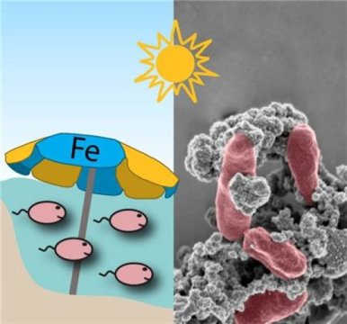
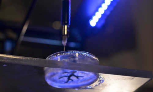
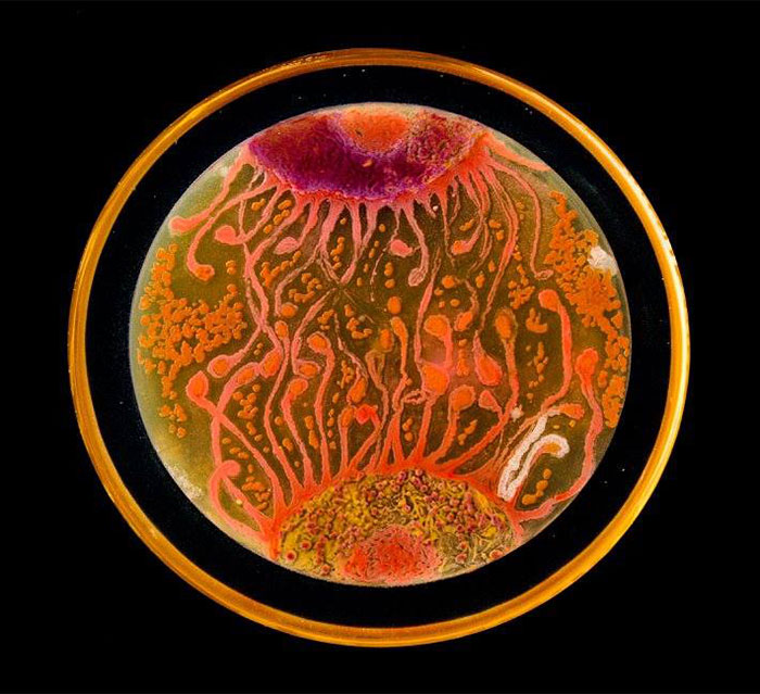
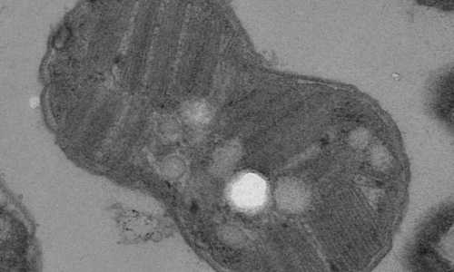
A sunshield for iron-oxidizing bacteria: These tiny organisms build their own sun umbrella by forming iron minerals or rust around their cells; this protects them from harmful UV rays. Photo Credit: Kappler/Gauger/University of Tübingen

The atmosphere we breathe today is composed of about 20 percent oxygen, which is not just essential to many organisms — it also provides protection from the sun’s more dangerous rays. In the presence of sunlight, oxygen molecules in the atmosphere react to form ozone. Up in the stratosphere, the ozone layer absorbs harmful UV radiation coming from space — protecting humans, animals and plants from the damage UV does. Three to four billion years ago, the atmosphere contained little oxygen and there was no ozone layer. “The earth’s surface — and areas of shallow water — were subject to high levels of ultra-violet radiation,” Andreas Kappler explains. “And yet, microbial life came to be. We wondered how that was possible.”

Certain bacteria which need light are able to eat dissolved iron (Fe2+) and to carry out photosynthesis in the presence of sunlight. But unlike today’s green plants, they did not release oxygen in the process. The process produces rust and other iron minerals as waste products. The iron minerals have special qualities — They absorb harmful ultraviolet radiation, but the part of the sunlight needed for photosynthesis can still be used by organisms. “The iron needed to form the minerals was available in much greater amounts in the oceans than it is today,” says Kappler. There are many indications that the photosynthesizing bacteria lived in these early oceans and oxidized iron. “We can still see the results of this today in the form of enormous iron-bearing rocks known as banded iron formations. They are the biggest deposits of iron we have.”

In their experiments, the geomicrobiologists subjected the bacteria to high doses of ultraviolet radiation — either in the presence or the absence of the iron minerals the bacteria themselves produce. “In the presence of their own rust, considerably more bacteria survived and were active,” says Tina Gauger. “We also saw that the bacterial cells’ DNA suffered less damage. In our experiments, more bacteria survived with mineral sunscreen than without.” The new findings are helping the researchers to understand how very early organisms survived despite the high level of radiation, and how life was even able to develop in shallow seas with sufficient sunlight.

Story Source:

The above post is reprinted from materials provided by Universitaet Tübingen.

Researchers hack off-the-shelf 3-D printer towards rebuilding the heart

from
BIOENGINEER.ORG http://bioengineer.org/researchers-hack-off-the-shelf-3-d-printer-towards-rebuilding-the-heart/

As of this month, over 4,000 Americans are on the waiting list to receive a heart transplant. With failing hearts, these patients have no other options; heart tissue, unlike other parts of the body, is unable to heal itself once it is damaged. Fortunately, recent work by a group at Carnegie Mellon could one day lead to a world in which transplants are no longer necessary to repair damaged organs.

bioprinting

“We’ve been able to take MRI images of coronary arteries and 3-D images of embryonic hearts and 3-D bioprint them with unprecedented resolution and quality out of very soft materials like collagens, alginates and fibrins,” said Adam Feinberg, an associate professor of Materials Science and Engineering and Biomedical Engineering at Carnegie Mellon University. Feinberg leads the Regenerative Biomaterials and Therapeutics Group, and the group’s study was published in the October 23 issue of the journal Science Advances.

“As excellently demonstrated by Professor Feinberg’s work in bioprinting, our CMU researchers continue to develop novel solutions like this for problems that can have a transformational effect on society,” said Jim Garrett, Dean of Carnegie Mellon’s College of Engineering. “We should expect to see 3-D bioprinting continue to grow as an important tool for a large number of medical applications.”

Traditional 3-D printers build hard objects typically made of plastic or metal, and they work by depositing material onto a surface layer-by-layer to create the 3-D object. Printing each layer requires sturdy support from the layers below, so printing with soft materials like gels has been limited.

“3-D printing of various materials has been a common trend in tissue engineering in the last decade, but until now, no one had developed a method for assembling common tissue engineering gels like collagen or fibrin,” said TJ Hinton, a graduate student in biomedical engineering at Carnegie Mellon and lead author of the study.

“The challenge with soft materials — think about something like Jello that we eat — is that they collapse under their own weight when 3-D printed in air,” explained Feinberg. “So we developed a method of printing these soft materials inside a support bath material. Essentially, we print one gel inside of another gel, which allows us to accurately position the soft material as it’s being printed, layer-by-layer.”

One of the major advances of this technique, termed FRESH, or “Freeform Reversible Embedding of Suspended Hydrogels,” is that the support gel can be easily melted away and removed by heating to body temperature, which does not damage the delicate biological molecules or living cells that were bioprinted. As a next step, the group is working towards incorporating real heart cells into these 3-D printed tissue structures, providing a scaffold to help form contractile muscle.

Bioprinting is a growing field, but to date, most 3-D bioprinters have cost over $100,000 and/or require specialized expertise to operate, limiting wider-spread adoption. Feinberg’s group, however, has been able to implement their technique on a range of consumer-level 3-D printers, which cost less than $1,000 by utilizing open-source hardware and software.

“Not only is the cost low, but by using open-source software, we have access to fine-tune the print parameters, optimize what we’re doing and maximize the quality of what we’re printing,” Feinberg said. “It has really enabled us to accelerate development of new materials and innovate in this space. And we are also contributing back by releasing our 3-D printer designs under an open-source license.”

Video: https://www.youtube.com/watch?v=Zfl_tFdt2D4&feature=youtu.be

Story Source:

The above post is reprinted from materials provided by Carnegie Mellon University.

25 Ekim 2015 Pazar

Microbiologists Create ‘Starry Night’ And Other Art With Bacteria

from
BIOENGINEER.ORG http://bioengineer.org/microbiologists-create-starry-night-and-other-art-with-bacteria/

The American Society for Microbiologists recently hosted its first international ‘Agar Art’ challenge in which microbiologists from around the world used various microbes and germs to create beautiful works of art in petri dishes. The submissions included recognizable paintings like Van Gogh’s ‘Starry Night’ as well as original microbe paintings.

The scientists used nutritious agar jelly as a “canvas” for their colorful microbes. While they do add an element of randomness as they grow, they can also do things that paint cannot – some of them emit bioflourescent light under certain conditions, while others, guided by the scientists, grew into perfect tree-branch patterns or jelly-fish tentacles.

For more about the process behind art like this, read about the work of Tasha Sturm, a microbiologist who used an agar dish to capture the germs on an eight-year-old boy’s hand.

First place: Neurons by Mehmet Berkmen and artist Maria Pernil, Nesterenkonia, Deinociccus, Sphingomonas

microbe-art-petri-dish-agar-contest-van-gogh-starry-night-american-society-microbiologists-44

Second place: NYC Biome Map by Christine Marizzi, Escherichia coli K12

microbe-art-petri-dish-agar-contest-van-gogh-starry-night-american-society-microbiologists-49

Third place: Harvest season by Maria Eugenia Inda, Saccharomyces cerevisiae

microbe-art-petri-dish-agar-contest-van-gogh-starry-night-american-society-microbiologists-41

People’s choice: Cell to Cell by Mehmet Berkmen and artist Maria Pernil, Nesterenkonia, Deinociccus, Sphingomonas

microbe-art-petri-dish-agar-contest-van-gogh-starry-night-american-society-microbiologists-47

Van Gogh’s “Starry Night,” Proteus mirabilis, Acinetobacter baumanii, Enterococcus faecalis, Klebsiella pneumonia

microbe-art-petri-dish-agar-contest-van-gogh-starry-night-american-society-microbiologists-43

Flowering Sunshine, Shigella, Salmonella

microbe-art-petri-dish-agar-contest-van-gogh-starry-night-american-society-microbiologists-40

The Great Wave of Candida by Cristina Marcos, Candida albicans, Candida glabrata, Candida parapsilosis

microbe-art-petri-dish-agar-contest-van-gogh-starry-night-american-society-microbiologists-42

The Streptomyces Sky, Streptomyces coelicolor

microbe-art-petri-dish-agar-contest-van-gogh-starry-night-american-society-microbiologists-3635

Mushrooms, Nesterenkonia, Deinociccus, Sphingomonas

microbe-art-petri-dish-agar-contest-van-gogh-starry-night-american-society-microbiologists-37

Yeast Go Viral, S. cerevisiae, L-A virus

microbe-art-petri-dish-agar-contest-van-gogh-starry-night-american-society-microbiologists-45

Cells, Nesterenkonia, Deinociccus, Sphingomonas

microbe-art-petri-dish-agar-contest-van-gogh-starry-night-american-society-microbiologists-46

Blocking enzymes in hair follicles promotes hair growth

from
BIOENGINEER.ORG http://bioengineer.org/blocking-enzymes-in-hair-follicles-promotes-hair-growth/

Inhibiting a family of enzymes inside hair follicles that are suspended in a resting state restores hair growth, a new study from researchers at Columbia University Medical Center has found. The research was published today in the online edition of Science Advances.

mice-hair-growth

Within 3 weeks, mice that received topical ruxolitinib or tofacitinib had regrown nearly all their hair (right photo; drug was applied only to the right side of the mouse). Little to no hair growth occurred in control mice during the same timeframe (left photo). Photo Credit: From S. Harel et al., Sci. Adv. 1, e1500973 (2015). Distributed under a Creative Commons Attribution Non Commercial License 4.0 (CC BY-NC). 10.1126/sciadv.1500973

In experiments with mouse and human hair follicles, Angela M. Christiano, PhD, and colleagues found that drugs that inhibit the Janus kinase (JAK) family of enzymes promote rapid and robust hair growth when directly applied to the skin.

The study raises the possibility that drugs known as JAK inhibitors could be used to restore hair growth in multiple forms of hair loss such as that induced by male pattern baldness, and additional types that occur when hair follicles are trapped in a resting state. Two JAK inhibitors have been approved by the U.S. Food and Drug Administration. One is approved for treatment of blood diseases (ruxolitinib) and the other for rheumatoid arthritis (tofacitinib). Both are being tested in clinical trials for the treatment of plaque psoriasis and alopecia areata, an autoimmune disease that causes hair loss.

“What we’ve found is promising, though we haven’t yet shown it is effective for male pattern baldness,” said Dr. Christiano. “More work needs to be done to test formulations of JAK inhibitors specially made for the scalp to determine whether they can induce hair growth in humans.”

Christiano and her colleagues serendipitously discovered the effect of JAK inhibitors on hair follicles when they were studying a type of hair loss known as alopecia areata, caused by an autoimmune attack on the hair follicles. Christiano and colleagues reported last year that JAK inhibitors shut off the signal that provokes the autoimmune attack, and that oral forms of the drug restore hair growth in some people with the disorder.

In the course those experiments, Dr. Christiano noticed that mice grew more hair when the drug was applied topically to the skin than when given internally. This suggested JAK inhibitors might have a direct effect on the hair follicles in addition to inhibiting the immune attack.

When the researchers looked more closely at normal mouse hair follicles, they found that JAK inhibitors rapidly awakened resting follicles out of dormancy. Hair follicles do not produce hair constantly but rather by cycling between resting and growing phases.

JAK inhibitors trigger the follicles’ normal reawakening process, the researchers found. Mice treated for five days with one of two JAK inhibitors sprouted new hair within 10 days, greatly accelerating the hair follicle growth phase. No hair grew on untreated control mice in the same time period.

“There are very few compounds that can push hair follicles into their growth cycle so quickly,” said Dr. Christiano. “Some topical agents induce tufts of hair here and there after a few weeks, but very few have such a potent and rapid-acting effect.” The drugs also produce longer hair from human hair follicles grown in culture and on skin grafted onto mice.

It’s likely that the drugs that are so effective in enhancing hair growth in the mice could affect the same pathways in human follicles, suggesting they could induce new hair growth and extend the growth of existing hairs in humans.

It remains to be seen if JAK inhibitors can reawaken hair follicles that have been suspended in a resting state because of androgenetic alopecia (which causes male and female pattern baldness) or other forms of hair loss. So far, all the experiments have been conducted in normal mice and human follicles. Experiments to address hair follicles affected by hair loss disorders are under way.

The title of the paper is: Pharmacologic inhibition of JAK-STAT signaling promotes hair growth. Other CUMC authors: Sivan Harel, Claire Higgins (now at Imperial College London) Jane E. Cerise, Zhenpeng Dai, James C. Chen, and Raphael Clynes (now at Bristol-Myers Squib).

The work was supported in part by the NIH (grants R01AR056016, P30AR044535, T32GM082771 and T32GM007088), Locks of Love Foundation, Alopecia Areata Initiative, and the Dermatology Foundation (Career Development Award).

Story Source:

The above post is reprinted from materials provided by Columbia University Medical Center.

22 Ekim 2015 Perşembe

What you didn’t know about naked mole-rats

from
BIOENGINEER.ORG http://bioengineer.org/what-you-didnt-know-about-naked-mole-rats/

The naked mole-rat is a particularly ugly or cute animal, depending on your definition. It is tubular in shape, like the tunnels it creates, hairless and wrinkled, for wiggling through those tunnels, and has long, chisel-like front teeth. It looks somewhat like a walrus in miniature. And these rodents can chew through concrete!

mole

Do a Google search of the naked mole-rat, or read through a number of biology textbooks, and you will find numerous references to this African mammal as being “inbred” and “eusocial,” meaning — similar to some insects — it has a fertile “queen” at the head of the colony, helpers who tend to her and may mate with her, and female “workers” who are sterile, expending their energy building tunnels and finding food.

This social system is rare among animals, and almost unheard-of among mammals, so evolutionary biologists have long taken particular interest in the unusual eusocial mating system of the naked mole-rat and its essentially homogeneous genetics. Why would this rodent have evolved to socialize and mate so differently from other mammals? From a natural selection standpoint — where advantageous traits are passed down to succeeding generations — what is gained by limiting genetic diversity by limiting the breeding pool?

Evolutionary biologists have puzzled over and debated this for decades. For this reason, the naked mole-rat has been an interesting oddball study model.

Well, it turns out the long-held conventional wisdom about the naked mole-rat being inbred is wrong, according to a University of Virginia-led study published recently in the journal Molecular Ecology.

UVA biologist Colleen Ingram and a team of researchers from several U.S. universities and the American Museum of Natural History conducted genetics studies of different mole-rat populations from Africa, and compared them to the genetics of a long-studied mole-rat population. They found that the populations of mole-rats studied for decades are “inbred” only because they originally came from a small, genetically isolated population of naked mole-rats from south of Kenya’s Athi River. The researchers discovered that larger wild populations from north of that river and from the Tana River region are genetically variable, like other mammals — meaning they are not inbred, despite their unusual eusocial mating behavior.

“We now know, from looking at the big picture from a much larger geographic area than previously studied, that the naked mole-rat is not inbred at all,” Ingram said. “What we thought we knew was based on early genetics studies of a small inbred sample from an otherwise genetically variable species. This shows that long-held assumptions, even from heavily studied model species, can and should always be questioned and further studied.”

It also suggests, she said, that laboratory animals, isolated and repeatedly re-bred for studies, might over time represent behaviors and genetics that are different from the diverse wild populations from which they originally came.

The study also means that some biology websites and textbooks need updating.

Story Source:

The above post is reprinted from materials provided by University of Virginia.

Cancer-causing parasite may accelerate wound healing

from
BIOENGINEER.ORG http://bioengineer.org/cancer-causing-parasite-may-accelerate-wound-healing/

It’s short, ugly and deadly. But James Cook University scientists have found a cancer-causing, parasitic worm could help patients recover from their wounds.

cancer-causing

JCU scientists at the Australian Institute of Tropical Health and Medicine (AITHM) have discovered that the parasitic worm that kills tens of thousands of people every year may also supercharge recovery from wounds.

The oriental liver fluke, Opisthorchis viverrini is caught by eating raw fish. It infects millions of people in south-east Asia and kills 26,000 people each year due to a parasite-induced bile duct cancer it causes, known as cholangiocarcinoma (CCA).

JCU scientists, Dr Michael Smout and Professor Alex Loukas found that a growth factor secreted by the one centimetre-long worm drives wound healing and blood vessel growth. However an unfortunate consequence of this accelerated wound repair over many years is an increased risk of developing liver cancer.

Dr Smout said the discovery means it’s possible the growth factor could be used to accelerate the healing of chronic wounds such as diabetic ulcers and to develop a vaccine against the worm-induced cancer.

He said the vaccine would obviously benefit the people directly at risk of cancer, but the growth factor would also benefit the developed world as a possible wound healing agent.

“Diabetes is a big problem as we live longer and get heavier,” he said. “There are increasing numbers of inflammatory diseases such as diabetes and associated non-healing wounds. A powerful wound healing agent designed by millennia of host-parasite co-evolution may accelerate the impaired healing processes that plague diabetic and elderly patients”

Dr Smout said the parasite could live for decades in the human body before CCA developed and it had an incentive to keep its host healthy while chewing away at its cells.

He said scientists are still learning how this growth factor controls healing, and ultimate development of the discovery as a healing agent or vaccine was still a number of years away.

Story Source:

The above post is reprinted from materials provided by James Cook University.

Surprising source for ancient life biomarker

from
BIOENGINEER.ORG http://bioengineer.org/surprising-source-for-ancient-life-biomarker/

Stanford scientists have discovered a surprising source for an organic molecule used as an indicator for life on early Earth.

bacteria

Tetrahymanol is a fatty molecule, or lipid, found in the membranes enclosing eukaryotic cells, the class of cells that carry their genetic material in compartments called nuclei. Eukaryotes can be single-celled or multicellular; humans and plants are eukaryotes, as are plants.

It was thought that tetrahymanol was produced primarily by eukaryotes, but a new study suggests many bacteria might also produce the lipid. The finding, published in this week’s issue of the journal Proceedings of the National Academy of Sciences, could mean scientists will have to reevaluate their views about ancient organisms and ecosystems.

Evidence of tetrahymanol, and thus eukaryotic life, have been found in rocks dating back to 1.6 billion years ago. “Because they are so well preserved, these lipids allow us to go really deep into the rock record and learn about what life was like back then,” said geobiologist Paula Welander, an assistant professor of Earth system science at Stanford School of Earth, Energy & Environmental Sciences.

Unlike most other organic molecules, cyclic lipids — the class of lipids that tetrahymanol belongs to — are quite durable and can linger in the environment. So even after a cell has died and its other biomolecules such as DNA have degraded, the tetrahymanol that helped make up its cell membrane can remain. Over time, the lipid can become part of the rock itself.

Geobiologists use tetrahymanol not only as an indicator of ancient life, but also as a gauge of the environmental conditions that existed when the organisms that made the lipid lived. For example, modern marine eukaryotes ramp up the production of lipids such as tetrahymanol when stressed by a lack of oxygen. From this, scientists infer that ancient eukaryotes — which are also thought to have lived primarily in oceans — produced tetrahymanol when oxygen levels dropped, such as can occur in aquatic zones made up of water layers with varying oxygen concentrations.

“Tetrahymanol is a valuable indicator of water column stratification on the early Earth,” Welander said.

Prior to the new study, scientists knew of only two bacterial species that produced tetrahymanol in small amounts. “The conventional wisdom was that these organisms produced tetrahymanol accidentally,” Welander said.

But recently Amy Banta, a postdoctoral researcher in Welander’s group, and Jeremy Wei, a lab manager at Stanford, found evidence that the bacteria Methylomicrobium alcaliphilum produces lots of tetrahymanol. Using genetic manipulation techniques, the group showed that M. alcaliphilum was not making the lipid by accident.

“We could change the amount of tetrahymanol in the bacteria by tweaking its growth conditions. To us, that means it’s somehow controlling the production of this lipid,” Welander said.

By comparing the genomes of various bacteria, the team was also able to identify and delete the gene in M. alcaliphilum that produces the lipid — an important first step for determining what function tetrahymanol plays in bacteria.

Welander says her team’s finding that eukaryotes are not unique in producing and using tetrahymanol means that geobiologists will have to consider alternative explanations for its presence in ancient records.

“Scientists will have to take on a much more nuanced interpretation of what this molecule is telling us about life on early Earth,” she said.

Story Source:

The above post is reprinted from materials provided by Stanford’s School of Earth, Energy & Environmental Sciences.

21 Ekim 2015 Çarşamba

Biologists Discover Bacteria Communicate Like Neurons in the Brain

from
BIOENGINEER.ORG http://bioengineer.org/biologists-discover-bacteria-communicate-like-neurons-in-the-brain/

Biologists at UC San Diego have discovered that bacteria–often viewed as lowly, solitary creatures–are actually quite sophisticated in their social interactions and communicate with one another through similar electrical signaling mechanisms as neurons in the human brain.

In a study published in this week’s advance online publication of Nature, the scientists detail the manner by which bacteria living in communities communicate with one another electrically through proteins called “ion channels.”

“Our discovery not only changes the way we think about bacteria, but also how we think about our brain,” said Gürol Süel, an associate professor of molecular biology at UC San Diego who headed the research project. “All of our senses, behavior and intelligence emerge from electrical communications among neurons in the brain mediated by ion channels. Now we find that bacteria use similar ion channels to communicate and resolve metabolic stress. Our discovery suggests that neurological disorders that are triggered by metabolic stress may have ancient bacterial origins, and could thus provide a new perspective on how to treat such conditions.”

“Much of our understanding of electrical signaling in our brains is based on structural studies of bacterial ion channels” said Süel. But how bacteria use those ion channels remained a mystery until Süel and his colleagues embarked on an effort to examine long-range communication within biofilms–organized communities containing millions of densely packed bacterial cells. These communities of bacteria can form thin structures on surfaces–such as the tartar that develops on teeth–that are highly resistant to chemicals and antibiotics.

The scientists’ interest in studying long-range signals grew out of a previous study, published in July in Nature, which found that biofilms are able to resolve social conflicts within their community of bacterial cells just like human societies.

When a biofilm composed of hundreds of thousands of Bacillus subtilis bacterial cells grows to a certain size, the researchers discovered, the protective outer edge of cells, with unrestricted access to nutrients, periodically stopped growing to allow nutrients–specifically glutamate, to flow to the sheltered center of the biofilm. In this way, the protected bacteria in the colony center were kept alive and could survive attacks by chemicals and antibiotics.

Realizing that oscillations in biofilm growth required long-range coordination between bacteria at the periphery and interior of the biofilm, together with the fact that bacteria were competing for glutamate, an electrically charged molecule, prompted the researchers to speculate that the metabolic coordination among distant cells within biofilms might involve a form of electrochemical communication. The scientists noted that glutamate is also known to drive about half of all human brain activity.

So they designed an experiment to test their hypothesis. The object was to carefully measure changes in bacterial cell membrane potential during metabolic oscillations.

The researchers observed oscillations in membrane potential that matched the oscillations in biofilm growth and found that ion channels were responsible for these changes in membrane potential. Further experiments revealed that oscillations conducted long-range electrical signals within the biofilms through spatially propagating waves of potassium, a charged ion. As these waves of charged ions propagate through the biofilm, they coordinated the metabolic activity of bacteria in the inner and outer regions of the biofilm. When the ion channel that allows potassium to flow in and out of cells was deleted from the bacteria, the biofilm was no longer able to conduct these electrical signals.

“Just like the neurons in our brain, we found that bacteria use ion channels to communicate with each other through electrical signals,” said Süel. “In this way, the community of bacteria within biofilms appears to function much like a ‘microbial brain’.”

Süel added that the specific mechanism by which the bacteria communicate with one another is surprisingly similar to a process in the human brain known as “cortical spreading depression,” which is thought to be involved in migraines and seizures.

“What’s interesting is that both migraines and the electrical signaling in bacteria we discovered are triggered by metabolic stress,” he said. “This suggests that many drugs originally developed for epilepsy and migraines may also be effective in attacking bacterial biofilms, which have become a growing health problem around the world because of their resistance to antibiotics.”

Story Source:

The above post is reprinted from materials provided by University of California – San Diego.

18 Ekim 2015 Pazar

Sixth sense: How do we sense electric fields?

from
BIOENGINEER.ORG http://bioengineer.org/sixth-sense-how-do-we-sense-electric-fields/

A variety of animals are able to sense and react to electric fields, and living human cells will move along an electric field, for example in wound healing. Now a team lead by Min Zhao at the UC Davis Institute for Regenerative Cures has found the first actual “sensor mechanism” that allows a living cell detect an electric field. The work is published Oct. 9 in the journal Nature Communications.

sixth sense

“We believe there are several types of sensing mechanisms, and none of them are known. We now provide experimental evidence to suggest one which has not been even hypothesized before, a two-molecule sensing mechanism,” Zhao said.

Zhao and colleagues have been studying these “electric senses” in cells from both larger animals (fish skin cells, human cell lines) and in the soil-dwelling amoeba Dictyostelium. By knocking out some genes in Dictyostelium, they previously identified some of the genes and proteins that allow the amoeba to move in a certain direction when exposed to an electric field.

In the new work, carried out in a human cell line, they found that two elements, a protein called Kir4.2 (made by gene KCNJ15) and molecules within the cell called polyamines, were needed for signaling to occur. Kir4.2 is a potassium channel — it forms a pore through the cell membrane that allows potassium ions to enter the cell. Such ion channels are often involved in transmitting signals into cells. Polyamines are molecules within the cell that carry a positive charge.

Zhao and colleagues found that when the cells were in an electric field, the positively-charged polyamines tend to accumulate at the side of the cell near the negative electrode. The polyamines bind to the Kir4.2 potassium channel, and regulate its activity.

He cautioned that they do not yet have definitive evidence of how “switching” of the potassium channel by polyamines translates into directional movement by the cell.

Story Source:

The above post is reprinted from materials provided by University of California – Davis.

Chemical Microdroplet Computers

from
BIOENGINEER.ORG http://bioengineer.org/chemical-microdroplet-computers/

Scientists from the Institute of Physical Chemistry of the Polish Academy of Sciences in Warsaw in cooperation with the Institute of Physics of the PAS and the University of Jena have developed the concept of a simple chemical computer made of microdroplets capable of searching databases. Computer simulations, carried out on databases of malignant tumours, have confirmed the validity of the adopted new design strategy, which opens the door to the popularisation of chemical methods of processing information.

comp

These are different spatio-temporal structures that appear in chemical systems can be used for information coding and processing. Photo Credit: IPC PAS, Grzegorz Krzyzewski

Under the appropriate conditions, oscillating chemical reactions can occur inside a droplet. If there is more than one droplet and they are in contact with each other, the resulting chemical waves are able to penetrate into neighbouring droplets and disperse throughout the whole complex. This phenomenon is well-known, and attempts are being made to use it, among other things, for chemical data processing. Propagation of information throught many droplet system depends on their geometrical arrangement. Up to now, not much was known about how to design the shape of the microdroplet complexes for them to execute specific tasks. So, at the Institute of Physical Chemistry of the Polish Academy of Sciences (IPC PAS) in Warsaw, Poland, a novel strategy has been proposed. Instead of laboriously designing complex systems of microdroplets for a particular purpose, it is better to first produce a system, and then try to teach it something useful.

“We adopted a strategy that nature uses with great efficiency. Let’s just look at ourselves. After all, our brains don’t evolve to, for instance, recognise letters! First the brain comes into existence, and only then does it learn to read and write. Why not approach complex microdroplet systems in a similar manner, since we know that they also process information? Our proposal is therefore as follows: first let’s make a system of interacting chemically microdroplets, and then let’s check what it can learn to do,” says Prof. Jerzy Górecki (IPC PAS).

Research on the chemical processing of information by microdroplet systems, funded by the Polish Ministry of Science and Higher Education, the Foundation for Polish Science and the European Union, was carried out using the Belousov-Zhabotinsky oscillating reaction. When the conditions of this reaction are suitably selected, a chemical front wandering in space appears. Oscillation reactions are common in living organisms. In humans, at the stage of embryonic development, they form the beginnings of the spinal vertebrae; in adults they are responsible, among others, for the contractions of heart muscle.

“In the Belousov-Zhabotinsky reaction the passing of a chemical front is accompanied by changes in ion concentrations leading to a change in the colour of the solution. When the reaction occurs inside the droplet, clear pulses radiating in all directions can be seen within it under the microscope. The bigger the drop, the more often it pulsates,” explains PhD student Konrad Gi?y?ski (IPC PAS).

Chemical pulses in complexes of adjoining droplets spread much like electrical stimulation in nerve fibres. Researchers from IPC PAS used pulse frequencies in individual drops to encode information: a high frequency corresponded to TRUE, a low frequency to FALSE. In order to control the pulses, and thus, among other things, to input data, the sensitivity of the reactions taking place in the droplets to blue light was used: in droplets illuminated by this the reactions die off completely.

Computer simulations were used to examine the calculating possibilities of a planar array of adjoining microdroplets arranged in a 5×5 square. Within the array droplets for inputting data and droplets for processing information were distinguished. Data was entered by simulating appropriately long exposure of the input droplets. Learning took place by the selective interruption of reactions taking place in the drops (in a real system the interruption would also be performed by light). Researchers took the droplet whose oscillations were the best match with the correct answer as the droplet giving the answer. The aim of the learning process was to select the light exposure time of all the droplets in the system in such a way as to obtain the highest number of correct answers for all the records in the database.

The simulated array of oscillating microdroplets classified tumours that were in the CANCER database. This database is composed of 699 records, of which 66% correspond to benign tumour cells. This means that on seeing the next entry if we randomly say “Don’t worry, your tumour is not malignant” we have a 66% chance of giving the right answer.

“Our little chemical computer answered correctly in more than 90% of cases. This is a very good result and proves the effectiveness of the strategy we adopted. It is not completely unequivocal, but even the classic computer does not have to give the right response to cases outside the database. In any case, we humans also don’t always make the right decisions,” says Prof. Gorecki.

Microdroplet information processing systems can be built using microfluidic devices. These are usually small plates made of transparent plastic, in which a carrier liquid flows through a system of appropriately designed channels, carrying droplets of other liquids immiscible in the carrier. In such systems, it is relatively easy to produce drops of different sizes, substrate concentrations, or even substrates themselves.

“We are able, in a controlled and repeatable manner, to arrange the microdroplets in space, for example, enclosing many droplets of one liquid within a droplet of another liquid — and in such a way that the selected droplet always has the same neighbours. What is more, we also have techniques that allow us to influence the rate of chemical exchange through the membranes of the adjoining droplets,” describes Prof. Piotr Garstecki (IPC PAS) and gives the example of an arrangement of nine microdroplets enclosed within another droplet, recently constructed at his laboratory.

Systems processing information chemically cannot replace consumer electronics — they are too slow. Their important advantages, however, include their capability of parallel processing of information, and the potential possibility of working in extreme environments, e.g. at significant pressures and/or high temperatures, which is where modern electronics fails. An interesting perspective is intelligent medicines, responding to many factors within the body and which are activated only under specific, strictly defined, circumstances. But chemical computers can offer even more: theoretically they could arise using the phenomenon of self-organization. This possibility lets us think about, among others, futuristic space probes, capable of independently building key components from materials available on other planets.

See chemical pulses dispersing in a system of adjoining microdroplets. After only a dozen-or-so pulses, the biggest droplet starts to dominate over the others at https://www.youtube.com/watch?v=I0sBISsZX-w. (Source: IPC PAS)

Story Source:

The above post is reprinted from materials provided by Institute of Physical Chemistry of the Polish Academy of Sciences.

Structure of Cellular Memory Mechanism Uncovered

from
BIOENGINEER.ORG http://bioengineer.org/structure-of-cellular-memory-mechanism-uncovered/

Calcium is a crucial element in the body that controls thought, movement and other bodily functions. These events are directed by specialized proteins called ion channels that allow the flow of calcium ions in and out of cells and among cell compartments. For years, scientists have been unsure how calcium ion channels function.

cell memory

UTHealth biochemists studying the structures of molecular mechanisms from the left are Irina Serysheva, Ph.D., Mariah R. Baker, Ph.D., and Guizhen Fan, Ph.D. Photo Credit: The University of Texas Health Science Center at Houston (UTHealth)

New atomic scale images of the structure of calcium’s gatekeeper, IP3R, could go a long way toward solving this mystery and lead to treatments for the many diseases tied to channel malfunctions.

The IP3R channel was imaged by scientists in the Department of Biochemisty and Molecular Biology at The University of Texas Health Science Center at Houston (UTHealth). Their findings appear in the journal Nature.

“We now know the structure of the gating machinery of IP3R,” said Irina Serysheva, Ph.D., the study’s senior author and an associate professor of biochemistry and molecular biology at UTHealth Medical School. “This work will fuel many functional and translational studies and allow for new drug design venues.”

When the IP3R calcium channel receives signals, it creates a pathway for calcium ions to move across cell membranes. While it works flawlessly most of the time, serious health issues occur when everything does not go to plan.

“Those health issues include Alzheimer’s disease, Parkinson’s disease, Huntington’s disease, cardiac hypertrophy, heart failure, cancer and stroke,” she said.

The study involved IP3R channel proteins that were purified from rat brain. The researchers used electron cryomicroscopy and computer reconstruction techniques to visualize the channel at near-atomic resolution. They then built a model representing the protein’s atomic 3-D arrangement as it may exist in the cell.

“Knowledge of the 3-D structure of these channels is required to understand the molecular basis of channel opening and closing, and how this process is controlled by a wide variety of endogenous molecules and pharmacological modifiers,” said Rodney Kellems, Ph.D., professor and chair of the Department of Biochemistry and Molecular Biology at UTHealth Medical School.

“We can’t explore function if we don’t know structure,” Serysheva said.

Story Source:

The above post is reprinted from materials provided by University of Texas Health Science Center at Houston.

17 Ekim 2015 Cumartesi

Dreams turned off and on with a neural switch

from
BIOENGINEER.ORG http://bioengineer.org/dreams-turned-off-and-on-with-a-neural-switch/

At the flip of a switch, University of California, Berkeley, neuroscientists can send a sleeping mouse into dreamland.

dr

When a laser triggers an optogenetic switch in neurons in the medulla of a sleeping mouse, the animal goes from non-REM sleep (NREM) into REM or dream sleep. The axons of these neurons (green) reach into distant parts of the primitive brain, such as the hypothalamus, broadly affecting brain function. Photo Credit: Franz Weber/UC Berkeley

The researchers inserted an optogenetic switch into a group of nerve cells located in the ancient part of the brain called the medulla, allowing them to activate or inactivate the neurons with laser light.

When the neurons were activated, sleeping mice entered REM sleep within seconds. REM sleep, characterized by rapid eye movements, is the dream state in mammals accompanied by activation of the cortex and total paralysis of the skeletal muscles, presumably so that we don’t act out the dreams flashing through our mind.

Inactivating the neurons reduced or even eliminated a mouse’s ability to enter REM sleep.

“People used to think that this region of the medulla was only involved in the paralysis of skeletal muscles during REM sleep,” said lead author Yang Dan, a UC Berkeley professor of molecular and cell biology and a Howard Hughes Medical Institute Investigator. “What we showed is that these neurons triggered all aspects of REM sleep, including muscle paralysis and the typical cortical activation that makes the brain look more awake than in non-REM sleep.”

While other types of neurons in the brainstem and hypothalamus have been shown to influence REM sleep, Dan said, “Because of the strong induction of REM sleep — in 94 percent of the recorded trials our mice entered REM sleep within seconds of activating the neurons — we think this might be a critical node of a relatively small network that makes the decision whether you go into dream sleep or not.”

The UC Berkeley team reported their results in the Oct. 15 print issue of the British journal Nature, and the paper was posted online Oct. 7.

The discovery will not only help researchers better understand the complex control of sleep and dreaming in the brain, the researchers said, but will allow scientists to stop and start dreaming at will in mice to learn why we dream.

“Many psychiatric disorders, especially mood disorders, are correlated with changes in REM sleep, and some widely used drugs affect REM sleep, so it seems to be a sensitive indicator of mental and emotional health,” said first author Franz Weber, a UC Berkeley postdoctoral fellow. “We are hoping that studying the sleep circuit might lead us to new insights into these disorders as well as neurological diseases that affect sleep, like Parkinson’s and Alzheimer’s diseases.”

Eating and dreaming

The researchers also found that activating these brain cells while the mice were awake had no effect on wakefulness, but did make them eat more. In normal mice, these neurons — a subset of nerve cells that release the neurotransmitter gamma-amino butyric acid (GABA), and so are called GABAergic neurons — are most active during waking periods when the mice are eating or grooming, two highly pleasurable activities.

Dan suspects that these GABAergic neurons in the medulla have the opposite effect of stress neurons, such as the noradrenergic neurons in the pons, another ancient part of the brain. Noradrenergic neurons release the transmitter noradrenalin, a cousin of adrenalin.

“Other people have found that noradrenergic neurons, which are active when you are running, shut down when eating or grooming. So it seems like when you are relaxed and enjoying yourself, the noradrenergic neurons switch off and these GABAergic neurons in the medulla turn on,” she said.

The GABAergic neurons project from the ventral part of the medulla, which sits at the top of the spinal cord, into many regions of the brainstem and hypothalamus, and thus are able to affect many bodily functions. These regions — more primitive than the brain’s cortex, the center of thinking and reasoning — are the seat of emotions and many innate behaviors as well as the control centers for muscles and automatic functions such as breathing.

Optical brain state switching

Dan, Weber and their colleagues chose a powerful technique called optogenetics to study these REM-related GABAergic neurons in the medulla. The technique involves inserting a light-sensitive ion channel into specific types of neurons by means of a virus. To target the virus to GABAergic neurons, the researchers used a genetically engineered mouse line that expresses a marker protein in these specific neurons only. Once present, the ion channel can turn on the activity of neurons when stimulated by laser light through an optical fiber inserted in the brain. Alternatively, inserting an inhibitory ion pump into the GABAergic neurons allowed the researchers to turn off the activity of these neurons through laser stimulation.

Using this genetically engineered strain of mice, the researchers mapped the activity of these neurons in the medulla and then recorded how activating or inactivating the neurons for brief periods affected sleep and waking behavior.

They also used a drug to inactivate the same set of neurons and found a reduction of REM sleep, though not as immediate and lasting for a longer period of time, since the drug required about half an hour to take effect and wore off slowly.

They also inserted the light-sensitive ion channels into a different set of neurons in the medulla: glutamatergic neurons, which release the neurotransmitter glutamate. Activating these neurons immediately awakened the animals, the opposite effect of activating the GABAergic neurons.

Dan is continuing her studies of the neurons that affect not only REM sleep, but also non-REM sleep.

Story Source:

The above post is reprinted from materials provided by University of California – Berkeley.

Artificial Skin Lets Person Feel

from
BIOENGINEER.ORG http://bioengineer.org/artificial-skin-lets-person-feel/

Stanford engineers have created a plastic “skin” that can detect how hard it is being pressed and generate an electric signal to deliver this sensory input directly to a living brain cell.

artificial skin

Human finger touches robotic finger. The transparent plastic and black device on the golden “fingertip” is the skin-like sensor developed by Stanford engineers. This sensor can detect pressure and transmit that touch sensation to a nerve cell. The goal is to create artificial skin, studded with many such miniaturized sensors, to give prosthetic appendages some of the sensory capabilities of human skin. Photo Credit: Bao Lab

Zhenan Bao, a professor of chemical engineering at Stanford, has spent a decade trying to develop a material that mimics skin’s ability to flex and heal, while also serving as the sensor net that sends touch, temperature and pain signals to the brain. Ultimately she wants to create a flexible electronic fabric embedded with sensors that could cover a prosthetic limb and replicate some of skin’s sensory functions.

Bao’s work, reported today in Science, takes another step toward her goal by replicating one aspect of touch, the sensory mechanism that enables us to distinguish the pressure difference between a limp handshake and a firm grip.

“This is the first time a flexible, skin-like material has been able to detect pressure and also transmit a signal to a component of the nervous system,” said Bao, who led the 17-person research team responsible for the achievement.

Benjamin Tee, a recent doctoral graduate in electrical engineering; Alex Chortos, a doctoral candidate in materials science and engineering; and Andre Berndt, a postdoctoral scholar in bioengineering, were the lead authors on the Science paper.

Digitizing touch

The heart of the technique is a two-ply plastic construct: the top layer creates a sensing mechanism and the bottom layer acts as the circuit to transport electrical signals and translate them into biochemical stimuli compatible with nerve cells. The top layer in the new work featured a sensor that can detect pressure over the same range as human skin, from a light finger tap to a firm handshake.

Five years ago, Bao’s team members first described how to use plastics and rubbers as pressure sensors by measuring the natural springiness of their molecular structures. They then increased this natural pressure sensitivity by indenting a waffle pattern into the thin plastic, which further compresses the plastic’s molecular springs.

To exploit this pressure-sensing capability electronically, the team scattered billions of carbon nanotubes through the waffled plastic. Putting pressure on the plastic squeezes the nanotubes closer together and enables them to conduct electricity.

This allowed the plastic sensor to mimic human skin, which transmits pressure information as short pulses of electricity, similar to Morse code, to the brain. Increasing pressure on the waffled nanotubes squeezes them even closer together, allowing more electricity to flow through the sensor, and those varied impulses are sent as short pulses to the sensing mechanism. Remove pressure, and the flow of pulses relaxes, indicating light touch. Remove all pressure and the pulses cease entirely.

The team then hooked this pressure-sensing mechanism to the second ply of their artificial skin, a flexible electronic circuit that could carry pulses of electricity to nerve cells.

Importing the signal

Bao’s team has been developing flexible electronics that can bend without breaking. For this project, team members worked with researchers from PARC, a Xerox company, which has a technology that uses an inkjet printer to deposit flexible circuits onto plastic. Covering a large surface is important to making artificial skin practical, and the PARC collaboration offered that prospect.

Finally the team had to prove that the electronic signal could be recognized by a biological neuron. It did this by adapting a technique developed by Karl Deisseroth, a fellow professor of bioengineering at Stanford who pioneered a field that combines genetics and optics, called optogenetics. Researchers bioengineer cells to make them sensitive to specific frequencies of light, then use light pulses to switch cells, or the processes being carried on inside them, on and off.

For this experiment the team members engineered a line of neurons to simulate a portion of the human nervous system. They translated the electronic pressure signals from the artificial skin into light pulses, which activated the neurons, proving that the artificial skin could generate a sensory output compatible with nerve cells.

Optogenetics was only used as an experimental proof of concept, Bao said, and other methods of stimulating nerves are likely to be used in real prosthetic devices. Bao’s team has already worked with Bianxiao Cui, an associate professor of chemistry at Stanford, to show that direct stimulation of neurons with electrical pulses is possible.

Bao’s team envisions developing different sensors to replicate, for instance, the ability to distinguish corduroy versus silk, or a cold glass of water from a hot cup of coffee. This will take time. There are six types of biological sensing mechanisms in the human hand, and the experiment described in Science reports success in just one of them.

But the current two-ply approach means the team can add sensations as it develops new mechanisms. And the inkjet printing fabrication process suggests how a network of sensors could be deposited over a flexible layer and folded over a prosthetic hand.

“We have a lot of work to take this from experimental to practical applications,” Bao said. “But after spending many years in this work, I now see a clear path where we can take our artificial skin.”

Story Source:

The above post is reprinted from materials provided by Stanford University.

13 Ekim 2015 Salı

Epigenetic Algorithm Accurately Predicts Male Sexual Orientation

from
BIOENGINEER.ORG http://bioengineer.org/epigenetic-algorithm-accurately-predicts-male-sexual-orientation/

An algorithm using epigenetic information from just nine regions of the human genome can predict the sexual orientation of males with up to 70 percent accuracy, according to research presented at the American Society of Human Genetics (ASHG) 2015 Annual Meeting in Baltimore.

dna helix

“To our knowledge, this is the first example of a predictive model for sexual orientation based on molecular markers,” said Tuck C. Ngun, PhD, first author on the study and a postdoctoral researcher at the David Geffen School of Medicine of the University of California, Los Angeles.

Beyond the genetic information contained in DNA, the researchers examined patterns of DNA methylation — a molecular modification to DNA that affects when and how strongly a gene is expressed — across the genome in pairs of identical male twins. While identical twins have exactly the same genetic sequence, environmental factors lead to differences in how their DNA is methylated. Thus, by studying twins, the researchers could control for genetic differences and tease out the effect of methylation. In all, the study involved 37 pairs of twins in which one twin was homosexual and the other was heterosexual, and 10 pairs in which both twins were homosexual.

“A challenge was that because we studied twins, their DNA methylation patterns were highly correlated,” Dr. Ngun explained. Even after some initial analysis, the researchers were left with over 400,000 data points to sort through. “The high correlation and large data set made it difficult to identify differences between twins, determine which ones were relevant to sexual orientation, and determine which of those could be used predictively,” he added.

To sort through this data set, Dr. Ngun and his colleagues devised a machine learning algorithm called FuzzyForest. They found that methylation patterns in nine small regions, scattered across the genome, could be used to predict study participants’ sexual orientation with 70 percent accuracy.

“Previous studies had identified broader regions of chromosomes that were involved in sexual orientation, but we were able to define these areas down to the base pair level with our approach,” Dr. Ngun said. He noted that it will take additional research to explain how DNA methylation in those regions may be related to sexual orientation. The researchers are currently testing the algorithm’s accuracy in a more general population of men.

“Sexual attraction is such a fundamental part of life, but it’s not something we know a lot about at the genetic and molecular level. I hope that this research helps us understand ourselves better and why we are the way we are,” Dr. Ngun said.

Reference: Ngun TC et al. (2015 Oct 8). Abstract: A novel predictive model of sexual orientation using epigenetic markers. Presented at American Society of Human Genetics 2015 Annual Meeting. Baltimore, Md.

Story Source:

The above post is reprinted from materials provided by American Society of Human Genetics.

12 Ekim 2015 Pazartesi

New Record of Tiniest Free-Living Insect

from
BIOENGINEER.ORG http://bioengineer.org/new-record-of-tiniest-free-living-insect/

The long-lasting search and debate around the size and identity of the World’s smallest free-living insect seems to have now been ended with the precise measurement and second record of the featherwing beetle species.

insect

This is the smallest known free-living insect, Scydosella musawasensis. Photo Credit: Dr. Alexey Polilov

Described back in 1999 based on only several specimens found in Nicaragua, as many as 85 individuals of the minute beetle species have recently been retrieved from Colombia and thoroughly examined. The smallest of them measured the astounding 0.325 mm. The finding made by Dr. Alexey Polilov, Lomonosov Moscow State University Moscow, is available in the open access journal ZooKeys.

The World’s smallest beetle and tiniest non-parasitoid insect, called Scydosella musawasensis, is morphologically characterised by its elongated oval body, yellowish-brown colouration and antennae split into 10 segments. It is also the only representative of this featherwing beetle genus.

Not able to precisely measure its size because of the preserved specimens being embedded in preparations for microscopy studies, Dr. Polilov used new individuals, collected in Chicaque National Park, Colombia in early 2015. To conclude the length of the smallest one as 325 µm (0.325 mm) the scientist used a specialised software and digital micrographs.

The recent survey is the second record of the tiny beetle species, which also proved that the range of its distribution is actually much wider. Thereafter, so are the localities of the fungi that the insect feeds on.

Story Source:

The above post is reprinted from materials provided by Pensoft Publishers.

10 Ekim 2015 Cumartesi

Using Diamonds to Trace Early Cancers

from
BIOENGINEER.ORG http://bioengineer.org/using-diamonds-to-trace-early-cancers/

Physicists from the University of Sydney have devised a way to use diamonds to identify cancerous tumours before they become life threatening.

diamond

Their findings, published in Nature Communications, reveal how a nanoscale, synthetic version of the precious gem can light up early-stage cancers in non-toxic, non-invasive Magnetic Resonance Imaging (MRI) scans.

Targeting cancers with tailored chemicals is not new but scientists struggle to detect where these chemicals go since, short of a biopsy, there are few ways to see if a treatment has been taken-up by a cancer.

Led by Professor David Reilly from the School of Physics, researchers from the University investigated how nanoscale diamonds could help identify cancers in their earliest stages.

“We knew nano diamonds were of interest for delivering drugs during chemotherapy because they are largely non-toxic and non-reactive,” says Professor Reilly.

“We thought we could build on these non-toxic properties realising that diamonds have magnetic characteristics enabling them to act as beacons in MRIs. We effectively turned a pharmaceutical problem into a physics problem.”

Professor Reilly’s team turned its attention to hyperpolarising nano-diamonds, a process of aligning atoms inside a diamond so they create a signal detectable by an MRI scanner.

“By attaching hyperpolarised diamonds to molecules targeting cancers the technique can allow tracking of the molecules’ movement in the body,” says Ewa Rej, the paper’s lead author.

“This is a great example of how quantum physics research tackles real-world problems, in this case opening the way for us to image and target cancers long before they become life-threatening,” says Professor Reilly.

The next stage of the team’s work involves working with medical researchers to test the new technology on animals. Also on the horizon is research using scorpion venom to target brain tumours with MRI scanning.

Story Source:

The above post is reprinted from materials provided by University of Sydney.

Lab-grown 3D intestine regenerates gut lining in dogs

from
BIOENGINEER.ORG http://bioengineer.org/lab-grown-3d-intestine-regenerates-gut-lining-in-dogs/

Working with gut stem cells from humans and mice, scientists from the Johns Hopkins Children’s Center and the University of Pittsburgh have successfully grown healthy intestine atop a 3-D scaffold made of a substance used in surgical sutures.

stem cells

In a further step that takes their work well beyond proof of concept, researchers report their laboratory-created intestine successfully regenerated gut tissue in the colons of dogs with missing gut lining.

The experiments, described ahead of print in the journal Regenerative Medicine, bring researchers closer to creating an implantable intestine as replacement therapy for a range of devastating disorders — including infections, cancer and trauma — that result in loss or death of gut tissue. Chief among them is a condition that affects 12 percent of premature newborns, called necrotizing enterocolitis (NEC), which is marked by the rapid death of intestinal cells and permanent loss of intestinal tissue.

The tube-shaped scaffold, designed several years ago in collaboration with Cornell University researchers and composed of biodegradable material similar to that used in surgical sutures, was a big first step on the quest to develop an implantable replacement intestine. But the new work pushes that effort further because it shows how stem cells, when mixed with immune and connective tissue cells, can grow into normal gut tissue around the scaffold and function inside a living mammal.

Researchers caution that a fully functioning replacement intestine for humans is far off, but they say their results have laid the critical groundwork to do so.

“Our experiments show that the architecture and function of our lab-made intestine strikingly resemble those of the healthy human gut, giving us real hope that our model could be used as the backbone for replacement intestine,” says principal investigator David Hackam, M.D., Ph.D., the Johns Hopkins Children’s Center’s surgeon-in-chief, who initiated and conducted most of the work at the University of Pittsburgh.

In an initial set of experiments reminiscent of a peanut butter-and-jelly sandwich technique, researchers took stem cells from the colons of babies undergoing intestinal surgeries and from mice, then added immune cells called macrophages, the body’s scavengers that seek out and engulf debris along with foreign and diseased cells. To this mix, they added cells called fibroblasts, whose function is to form collagen and other connective substances that bind tissues and organs together. The idea, the scientists say, was to create a mixture that closely mimics the natural composition of the gut.

“Intestinal cells do not grow and develop in a vacuum, so we sought to recreate the richness of the human gut, Hackam says. “We took the PB & J approach, adding a layer of stem cells on top of the scaffold, then topping them with a mixture of immune and collagen cells.”

Adding these components, they report, enhanced the growth of intestinal stem cells and differentiation into various mature cell types critical to the function of a healthy intestine. In comparison, stem cells grown in isolation without immune and connective cells in the mix grew more slowly and failed to differentiate well into multiple cell types.

In another set of experiments, researchers added probiotic bacteria to the newly created intestinal tissue. Doing so further amplified the growth and differentiation of new gut cells, specifically the growth of Paneth cells responsible for production of infection-fighting proteins that guard against intestinal infections, Hackam says, a finding that highlights the therapeutic potential of certain probiotics for NEC. Next, the team added bacteria-laden stool cells obtained from infants with NEC, but they observed a dramatic decrease in the number of intestinal cells called goblet cells, which are responsible for the production of protective mucus that coats and shields the intestine — a common occurrence in NEC. The observation, Hackam says, supports the notion that certain gut bacteria present in the colons of babies with NEC are responsible for sparking the rapid death of gut tissue that occurs in the condition.

Next, researchers implanted the newly created intestine into the bellies of mice. In a matter of days, the implanted intestine began producing new intestinal stem cells and stimulated the growth of new blood vessels around the implant. That observation, researchers say, affirmed the ability of the 3-D intestine to spur the growth of new tissue not only in lab dishes, but also in living organisms.

In a final step, the investigators implanted pieces of the newly created intestine — about 1.6 inches in length — into the lower portion of dog colons lacking parts of their intestinal lining. For two months, the dogs underwent periodic colonoscopies and intestinal biopsies. Strikingly, the guts of dogs with implanted intestines healed completely within eight weeks. By contrast, dogs that didn’t get intestinal implants experienced continued inflammation and scarring of their guts.

“Our results move research beyond the proof-of-concept realm,” says study author Stephen Badylak, D.V.M., Ph.D., M.D., professor of surgery and deputy director of the McGowan Institute for Regenerative Medicine at the University of Pittsburgh. “These results demonstrate that a mixture of synthetic and natural tissue can spur the formation of new gut cells and function well in living organisms despite the presence of naturally occurring inflammation and microbes found in the living gut.”

Story Source:

The above post is reprinted from materials provided by Johns Hopkins Medicine.

Antioxidants Can Make Cancer Spread Faster?

from
BIOENGINEER.ORG http://bioengineer.org/antioxidants-can-make-cancer-spread-faster/

Fresh research at Sahlgrenska Academy has found that antioxidants can double the rate of melanoma metastasis in mice. The results reinforce previous findings that antioxidants hasten the progression of lung cancer. According to Professor Martin Bergö, people with cancer or an elevated risk of developing the disease should avoid nutritional supplements that contain antioxidants.

Researchers at Sahlgrenska Academy, University of Gothenburg, demonstrated in January 2014 that antioxidants hastened and aggravated the progression of lung cancer. Mice that were given antioxidants developed additional and more aggressive tumors. Experiments on human lung cancer cells confirmed the results. Given well-established evidence that free radicals can cause cancer, the research community had simply assumed that antioxidants, which destroy them, provide protection against the disease. Found in many nutritional supplements, antioxidants are widely marketed as a means of preventing cancer. Because the lung cancer studies called the collective wisdom into question, they attracted a great deal of attention.

Double the rate

The follow-up studies at Sahlgrenska Academy have now found that antioxidants double the rate of metastasis in malignant melanoma, the most perilous type of skin cancer. Science Translational Medicine published the findings on October 7. “As opposed to the lung cancer studies, the primary melanoma tumor was not affected,” Professor Bergö says. “But the antioxidant boosted the ability of the tumor cells to metastasize, an even more serious problem because metastasis is the cause of death in the case of melanoma. The primary tumor is not dangerous per se and is usually removed.”

Confirmed the results

Experiments on cell cultures from patients with malignant melanoma confirmed the new results. “We have demonstrated that antioxidants promote the progression of cancer in at least two different ways,” Professor Bergö says. The overall conclusion from the various studies is that antioxidants protect healthy cells from free radicals that can turn them into malignancies but may also protect a tumor once it has developed.

Avoid supplements

Taking nutritional supplements containing antioxidants may unintentionally hasten the progression of a small tumor or premalignant lesion, neither of which is possible to detect. “Previous research at Sahlgrenska Academy has indicated that cancer patients are particularly prone to take supplements containing antioxidants,” Dr. Bergö says. Our current research combined with information from large clinical trials with antioxidants suggests that people who have been recently diagnosed with cancer should avoid such supplements.”

High mortality rate

One of the fastest expanding types of cancer in the developed world, malignant melanoma has a high mortality rate — which is one reason that researchers at Sahlgrenska Academy were so anxious to follow up on the lung cancer studies. “Identifying factors that affect the progression of malignant melanoma is a crucial task,” Professor Bergö says.

Lotions next

The role of antioxidants is particularly relevant in the case of melanoma, not only because melanoma cells are known to be sensitive to free radicals but because the cells can be exposed to antioxidants by non-dietary means as well. “Skin and suntan lotions sometimes contain beta carotene or vitamin E, both of which could potentially affect malignant melanoma cells in the same way as antioxidants in nutritional supplements,” Professor Bergö says.

Other forms of cancer

How antioxidants in lotions affect the course of malignant melanoma is currently being explored. “We are testing whether antioxidants applied directly to malignant melanoma cells in mice hasten the progression of cancer in the same way as their dietary counterparts,” Professor Bergö says. He stresses that additional research is badly needed. “Granted that lung cancer is the most common form of the disease and melanoma is expanding fastest, other forms of cancer and types of antioxidants need to be considered if we want to make a fully informed assessment of the role that free radicals and antioxidants play in the process of cancer progression.”

Story Source:

The above post is reprinted from materials provided by University of Gothenburg.

9 Ekim 2015 Cuma

Drug used to treat cancer appears to sharpen memory

from
BIOENGINEER.ORG http://bioengineer.org/drug-used-to-treat-cancer-appears-to-sharpen-memory/

Can you imagine a drug that would make it easier to learn a language, sharpen your memory and help those with dementia and Alzheimer’s disease by rewiring the brain and keeping neurons alive?

limitless

A drug may be able to make it easier to learn a language, sharpen your memory and help those with dementia and Alzheimer’s disease. Photo Credit: Relativity Media

New Rutgers research published in the Journal of Neuroscience found that a drug — RGFP966 — administered to rats made them more attuned to what they were hearing, able to retain and remember more information, and develop new connections that allowed these memories to be transmitted between brain cells.

“Memory-making in neurological conditions like Alzheimer’s disease is often poor or absent altogether once a person is in the advanced stages of the disease,” said Kasia M. Bieszczad, lead author and assistant professor in Behavioral and Systems Neuroscience in the Department of Psychology. “This drug could rescue the ability to make new memories that are rich in detail and content, even in the worst case scenarios.”

What happens with dementias such as Alzheimer’s is that brain cells shrink and die because the synapses that transfer information from one neuron to another are no longer strong and stable. There is no therapeutic treatment available that reverses this situation.

The drug being tested in this animal study is among a class known as HDAC inhibitors — now being used in cancer therapies to stop the activation of genes that turn normal cells into cancerous ones. In the brain, the drug makes the neurons more plastic, better able to make connections and create positive changes that enhance memory. Researchers found that laboratory rats, taught to listen to a certain sound in order to receive a reward, and given the drug after training, remembered what they learned and responded correctly to the tone at a greater rate than those not given the drug.

Scientists also found that the rodents were more “tuned in” to the relevant acoustic signals they heard during their training — an important finding Bieszczad said because setting up the brain to better process and store significant sounds is critical to human speech and language.

“People learning to speak again after a disease or injury as well as those undergoing cochlear implantation to reverse previous deafness, may be helped by this type of therapeutic treatment in the future,” said Bieszczad “The application could even extend to people with delayed language learning abilities or people trying to learn a second language.”

This hypersensitivity in processing auditory information enabled the neurons to reorganize and create new pathways — allowing more of the information they learned to become a long-term memory, said Bieszczad who collaborated with colleagues in the Department of Neurobiology and Behavior at the University of California Irvine.

“People normally remember an experience with limited detail — not everything we see, hear and feel is remembered,” she said. “What has happened here is that memory becomes closer to a snapshot of the actual experience instead of being sparse, limited or inaccurate.”

Story Source:

The above post is reprinted from materials provided by Rutgers University.

Bio-inspired robotic finger looks, feels and works like the real thing

from
BIOENGINEER.ORG http://bioengineer.org/bio-inspired-robotic-finger-looks-feels-and-works-like-the-real-thing/

Most robotic parts used to today are rigid, have a limited range of motion and don’t really look lifelike. Inspired by both nature and biology, a scientist from Florida Atlantic University has designed a novel robotic finger that looks and feels like the real thing.

finger

This new technology used both a heating and then a cooling process to operate the robotic finger. Results from the study showed a more rapid flexing and extending motion of the finger as well as its ability to recover its trained shape more accurately and more completely, confirming the biomechanical basis of its trained shape. Photo Credit: Florida Atlantic University

In an article recently published in the journal Bioinspiration & Biomimetics, Erik Engeberg, Ph.D., assistant professor in the Department of Ocean and Mechanical Engineering within the College of Engineering and Computer Science at FAU, describes how he has developed and tested this robotic finger using shape memory alloy (SMA), a 3D CAD model of a human finger, a 3D printer, and a unique thermal training technique.

“We have been able to thermomechanically train our robotic finger to mimic the motions of a human finger like flexion and extension,” said Engeberg. “Because of its light weight, dexterity and strength, our robotic design offers tremendous advantages over traditional mechanisms, and could ultimately be adapted for use as a prosthetic device, such as on a prosthetic hand.”

In the study, Engeberg and his team used a resistive heating process called “Joule” heating that involves the passage of electric currents through a conductor that releases heat. Using a 3D CAD model of a human finger, which they downloaded from a website, they were able to create a solid model of the finger. With a 3D printer, they created the inner and outer molds that housed a flexor and extensor actuator and a position sensor. The extensor actuator takes a straight shape when it’s heated, whereas the flexor actuator takes a curved shape when heated. They used SMA plates and a multi-stage casting process to assemble the finger. An electrical chassis was designed to allow electric currents to flow through each SMA actuator. Its U-shaped design directed the electric current to flow the SMAs to an electric power source at the base of the finger.

This new technology used both a heating and then a cooling process to operate the robotic finger. As the actuator cooled, the material relaxed slightly. Results from the study showed a more rapid flexing and extending motion of the finger as well as its ability to recover its trained shape more accurately and more completely, confirming the biomechanical basis of its trained shape.

“Because SMAs require a heating process and cooling process, there are challenges with this technology such as the lengthy amount of time it takes for them to cool and return to their natural shape, even with forced air convection,” said Engeberg. “To overcome this challenge, we explored the idea of using this technology for underwater robotics, because it would naturally provide a rapidly cooling environment.”

Since the initial application of this finger will be used for undersea operations, Engeberg used thermal insulators at the fingertip, which were kept open to facilitate water flow inside the finger. As the finger flexed and extended, water flowed through the inner cavity within each insulator to cool the actuators.

“Because our robotic finger consistently recovered its thermomechanically trained shape better than other similar technologies, our underwater experiments clearly demonstrated that the water cooling component greatly increased the operational speed of the finger,” said Engeberg.

Undersea applications using Engeberg’s new technology could help to address some of the difficulties and challenges humans encounter while working in the ocean depths. The focus of Engeberg’s BioRobotics Laboratory at FAU is investigating robotics and prosthetics, controller design, bioinspiration and biomemetics.

Story Source:

The above post is reprinted from materials provided by Florida Atlantic University.

8 Ekim 2015 Perşembe

Scientists build a digital piece of a rat’s brain

from
BIOENGINEER.ORG http://bioengineer.org/scientists-build-a-digital-piece-of-a-rats-brain/

If you want to learn how something works, one strategy is to take it apart and put it back together again. For 10 years, a global initiative called the Blue Brain Project–hosted at the Ecole Polytechnique Federale de Lausanne (EPFL)–has been attempting to do this digitally with a section of juvenile rat brain.

neuron

This is a photo of a virtual brain slice. Photo Credit: Makram et al./Cell 2015

The project presents a first draft of this reconstruction, which contains over 31,000 neurons, 55 layers of cells, and 207 different neuron subtypes, on October 8 in Cell.

Heroic efforts are currently being made to define all the different types of neurons in the brain, to measure their electrical firing properties, and to map out the circuits that connect them to one another. These painstaking efforts are giving us a glimpse into the building blocks and logic of brain wiring. However, getting a full, high-resolution picture of all the features and activity of the neurons within a brain region and the circuit-level behaviors of these neurons is a major challenge.

Henry Markram and colleagues have taken an engineering approach to this question by digitally reconstructing a slice of the neocortex, an area of the brain that has benefitted from extensive characterization. Using this wealth of data, they built a virtual brain slice representing the different neuron types present in this region and the key features controlling their firing and, most notably, modeling their connectivity, including nearly 40 million synapses and 2,000 connections between each brain cell type.

“The reconstruction required an enormous number of experiments,” says Markram, of the EPFL. “It paves the way for predicting the location, numbers, and even the amount of ion currents flowing through all 40 million synapses.”

Once the reconstruction was complete, the investigators used powerful supercomputers to simulate the behavior of neurons under different conditions. Remarkably, the researchers found that, by slightly adjusting just one parameter, the level of calcium ions, they could produce broader patterns of circuit-level activity that could not be predicted based on features of the individual neurons. For instance, slow synchronous waves of neuronal activity, which have been observed in the brain during sleep, were triggered in their simulations, suggesting that neural circuits may be able to switch into different “states” that could underlie important behaviors.

“An analogy would be a computer processer that can reconfigure to focus on certain tasks,” Markram says. “The experiments suggest the existence of a spectrum of states, so this raises new types of questions, such as ‘what if you’re stuck in the wrong state?'” For instance, Markram suggests that the findings may open up new avenues for explaining how initiating the fight-or-flight response through the adrenocorticotropic hormone yields tunnel vision and aggression.

The Blue Brain Project researchers plan to continue exploring the state-dependent computational theory while improving the model they’ve built. All of the results to date are now freely available to the scientific community at https://bbp.epfl.ch/nmc-portal.

Story Source:

The above post is reprinted from materials provided by Cell Press.

Detecting HIV diagnostic antibodies with DNA nanomachines

from
BIOENGINEER.ORG http://bioengineer.org/detecting-hiv-diagnostic-antibodies-with-dna-nanomachines/

New research may revolutionize the slow, cumbersome and expensive process of detecting the antibodies that can help with the diagnosis of infectious and auto-immune diseases such as rheumatoid arthritis and HIV.

ada

An international team of researchers have designed and synthetized a nanometer-scale DNA “machine” whose customized modifications enable it to recognize a specific target antibody. Their new approach, which they described this month in Angewandte Chemie, promises to support the development of rapid, low-cost antibody detection at the point-of-care, eliminating the treatment initiation delays and increasing healthcare costs associated with current techniques.

The binding of the antibody to the DNA machine causes a structural change (or switch), which generates a light signal. The sensor does not need to be chemically activated and is rapid — acting within five minutes — enabling the targeted antibodies to be easily detected, even in complex clinical samples such as blood serum.

“One of the advantages of our approach is that it is highly versatile,” said Prof. Francesco Ricci, of the University of Rome, Tor Vergata, senior co-author of the study. “This DNA nanomachine can be in fact custom-modified so that it can detect a huge range of antibodies, this makes our platform adaptable for many different diseases.”

“Our modular platform provides significant advantages over existing methods for the detection of antibodies,” added Prof. Vallée-Bélisle of the University of Montreal, the other senior co-author of the paper. “It is rapid, does not require reagent chemicals, and may prove to be useful in a range of different applications such as point-of-care diagnostics and bioimaging.”

“Another nice feature of our this platform is its low-cost,” said Prof. Kevin Plaxco of the University of California, Santa Barbara. “The materials needed for one assay cost about 15 cents, making our approach very competitive in comparison with other quantitative approaches.”

“We are excited by these preliminary results, but we are looking forward to improve our sensing platform even more” said Simona Ranallo, a PhD student in the group of Prof. Ricci at the University of Rome and first-author of the paper. “For example, we could adapt our platform so that the signal of the nanoswitch may be read using a mobile phone. This will make our approach really available to anyone! We are working on this idea and we would like to start involving diagnostic companies.”

Story Source:

The above post is reprinted from materials provided by University of Montreal.

New protein found in immune cells

from
BIOENGINEER.ORG http://bioengineer.org/new-protein-found-in-immune-cells/

Researchers of the University of Freiburg have discovered Kidins220/ARMS in B cells. They also determined that it plays a decisive role in the production of antibodies and the formation of B cells, which are a type of white blood cells.

new

Artwork painted by the daughter of researcher Susana Minguet: The protein Kidins220 (yellow) interacts with the B cell receptor (red and orange). Photo Credit: Susana Minguet

Various teams of researchers had already found that Kidins220/ARMS is present in nerve cells and in T cells of the immune system. However, that it is present in B cells was unknown until now. “We’ve discovered a new molecular player in the immune system,” said the immunobiologist Prof. Dr. Wolfgang Schamel, adding, “This knowledge could help to develop new medications for autoimmune diseases or other illnesses in the future.”

The postdoc Dr. Gina J. Fiala from Schamel’s lab is the lead author of the group’s publication in the Journal of Experimental Medicine. Fiala studied Kidins220/ARMS in B cells for her doctoral thesis. Several other members of the cluster of excellence BIOSS Centre for Biological Signalling Studies also collaborated in this study.

B lymphocytes, also known as B cells, are the only cells to produce antibodies, which the immune system needs to fight off foreign intruders like pathogens in order to protect the human body. On their surface, B cells carry B cell receptors. These activate the B cells when an antigen — a substance on the surface of a pathogenic germ — binds to them. The team of scientists from the University of Freiburg has discovered that Kidins220/ARMS interacts with the B cell receptor and affects signalling pathways from the receptor to the interior of the cell. Without Kidins220/ARMS, the receptor’s ability to send signals is limited. As a result, the B cells manufacture less antibodies and the immune system is weakened.

Kidins220/ARMS is also vital for the formation of B cells. If a mouse cannot produce this protein, the B lymphocytes develop in a way that makes them less functional than the B cells of a healthy immune system. The reason for this is that B cells depend on the signals from the B cell receptor and pre-B cell receptor, which is the early version of a B cell receptor, at various stages of their development. Deficiency in Kidins220/ARMS therefore obstructs the development of B cells.

Story Source:

The above post is reprinted from materials provided by Albert-Ludwigs-Universität Freiburg.

In boost for transplants, kidney tissue grown in lab

from
BIOENGINEER.ORG http://bioengineer.org/in-boost-for-transplants-kidney-tissue-grown-in-lab/

AFP – Scientists said Wednesday they had grown rudimentary human kidney tissue from stem cells, a key step towards the Holy Grail of fully-functional, lab-made transplant organs.

kidney

Image of a mini-kidney formed in a dish from human induced pluripotent stem cells. Photo Credit: Minoru Takasato

The tissue is not a viable organ, but may be useful for other purposes such as replacing animals in drug toxicity tests, the team said.

The researchers from Australia and the Netherlands grew their “kidney-like structure” from induced pluripotent stem (iPS) cells — adult cells reprogrammed into a neutral state from which they can be coaxed to develop into other cell types.

Given the critical shortage of donor organs to replace those damaged by accident or disease, it has long been a goal of science to create human organs from stem cells.

But it is a complicated task. Scientists need to prompt stem cells to become kidney, liver or lung cells, which must then recreate the complex anatomy of a real organ in order to function in a human recipient.

The first part of this chain has proved most challenging, especially in organs composed of a multitude of different cell types. The kidney, for example, has more than 20.

In the new study, published in the journal Nature, the team managed to transform iPS cells into two different adult cell types.

– Long way to go –

The resulting organoids sported different tissue types and were “similar” to the kidney of a human embryo, the researchers reported.

The work represented “an important step towards building stem-cell-derived kidneys,” University of Edinburgh anatomy expert Jamie Davies wrote in a comment, also published by Nature.

But he stressed the product was “not a kidney, but an organoid”.

“There is a long way to go until clinically useful transplantable kidneys can be engineered,” Davies said.

But the organoids may fulfil a completely different medical need — testing the safety for humans of new drugs.

“The cell types that are most vulnerable to damage by drugs are present in the organoids,” said Davies.

Stem cells are primitive cells that, as they grow, differentiate into the various specialised cells that make up the different organs — the brain, the heart, the kidney, and so on.

Until a few years ago, when iPS cells were created, the only way to obtain stem cells was to harvest them from human embryos. This was controversial as it required the destruction of the embryo.

Other teams of scientists have also reported growing “organoid” stomachs, livers, retinas and brain and heart tissue from pluripotent stem cells in the lab.

Story Source:

The above post is reprinted from materials provided by AFP.