29 Kasım 2015 Pazar

Artist bioengineers replica of Van Gogh’s ear

from
BIOENGINEER.ORG http://bioengineer.org/artist-bioengineers-replica-of-van-goghs-ear/

Bioengineering has given us some important medical and scientific advances: Researchers are developing artificial lungs and livers that may one day be transplanted into patients. But bioengineering also give us a new medium for art? A little piece of a long-dead artist is coming back to life in New York this fall when Diemut Strebe’s creepy living copy of Vincent van Gogh’s ear makes its New York debut at Ronald Feldman Fine Arts.

ear

Diemut Strebe, Sugababe (2014). A living bioengineered replica of Vincent van Gogh’s ear, grown from tissue engineered cartilage cells procured from a direct male descendant. Photo Credit: Ronald Feldman Fine Arts.

Strebe persuaded Lieuwe van Gogh, the great-great-grandson of Vincent’s brother, to donate a chunk of the inside of his ear for the project. (Although it did not take much convincing because, according to Strebe, “he loved the project right away.”) Then she worked with a “who’s who” of engineers and scientists to grow Lieuwe’s ear cells on a polymer-based scaffold that approximated the shape of Vincent’s ear, based on the only known photograph of the artist showing the body part that was famously removed.

VAN-GOGH-bandaged-ear

Vincent van Gogh, Self-Portrait with Bandaged Ear (1889). Photo Credit: Wikipedia.

The result is a piece named “Sugababe,” currently on display at the Ronald Feldman Fine Arts Gallery in New York City, in a show of Strebe’s work. The ear, which an art writer called “creepy” and Stephen Colbert called “the craziest (explicative) thing,” made its debut last year at a museum in Karlsruhe, Germany. It gets is name in part because of the sugar-white color of the ear.

Though Sugababe is admittedly macabre, visitors at the original exhibition at the Centre for Art and Media in Karlsruhe, Germany, “loved the ear,” Strebe insisted in an e-mail to artnet News.

I’m not sure that everyone understands the full scientific and biological implications,” the artist writes. “The scientific approach is based on the Theseus’s paradox by Plutarch… He asked if a ship would be the same ship if all its parts were replaced. This paradox is brought into a 21st-century context by using a living cell line (from Lieuwe van Gogh) in which we replaced (at least as a proof of principle) his natural DNA with historical and synthesized DNA.”

Perhaps the most famous detached body part in all of art history, van Gogh allegedly cut off his ear when he had a mental breakdown, although some German historians now think Paul Gauguin may have cut off van Gogh’s ear with a rapier following a heated argument between the two artists, according to the book Van Goghs Ohr: Paul Gauguin und der Pakt des Schweigens (Van Gogh’s Ear: Paul Gauguin and the Pact of Silence). Though the ear has been recreated, scientists haven’t been able to slow the fading of van Gogh’s paintings.

The scientifically-minded show also includes Social Sculpture: The Scent of Joseph Beuys, a scent-based piece inspired by the German Fluxus artist’s 1974 performance at René Block’s gallery in New York titled, I Like America and America Likes Me. With the help of International Flavors & Fragrances Inc., Strebe has reduced Beuys’s original work into seven scents, like “gallery” and “coyote,” which are meant to evoke Beuys’s experience living for a week with a wild coyote in the gallery space.

Diemut Strebe’s “Free Radicals: Sugababe & Other Works” is on view at Ronald Feldman Fine Arts, 31 Mercer Street, New York, November 7–December 5, 2015.

Story Source:

The above post is reprinted from materials provided by artnet News and CNN.

Lactate for Brain Energy

from
BIOENGINEER.ORG http://bioengineer.org/lactate-for-brain-energy/

Nerve cells cover their high energy demand with glucose and lactate. Scientists of the University of Zurich now provide new support for this. They show for the first time in the intact mouse brain evidence for an exchange of lactate between different brain cells. With this study they were able to confirm a 20-year old hypothesis.

lac2

In comparison to other organs, the human brain has the highest energy requirements. Photo Credit: Image courtesy of University of Zurich

In comparison to other organs, the human brain has the highest energy requirements. The supply of energy for nerve cells and the particular role of lactic acid (lactate) has been a matter of intense research for many years. A hypothesis from the 1990’s postulates, that a well-orchestrated collaboration between two cell types, astrocytes and neurons, is the basis of brain energy metabolism.

Astrocytes produce lactate, which flows to neurons to cover their high energy needs. Due to a lack of experimental techniques, it remained unclear whether an exchange of lactate existed between astrocytes and neurons. The group of Professor Bruno Weber from the Institute of Pharmacology and Toxicology now shows that there is a significant concentration gradient of lactate between astrocytes and neurons.

Lactate transport is dependent on concentration

The entry and exit of lactate into and out of cells of the body is concentration dependent and is mediated by a specific lactate transporter (called monocarboxylate transporter or MCT). A typical property of certain transporter proteins is called trans-acceleration. “MCTs can be imagined as revolving doors in a shopping mall, which begin to turn faster when more people enter or exit,” explains Bruno Weber, Professor of Multimodal Experimental Imaging at the University of Zurich. The researchers made use of this property and accelerated the “revolving doors.” By increasing the extracellular pyruvate concentration, they stimulated the outward transport of lactate. Interestingly, lactate levels only changed in astrocytes but not in neurons. Based on this finding and on results from several control experiments a clear lactate gradient between astrocytes and neurons was confirmed. “Due to the fact that lactate transport by MCTs is a passive transport, such a concentration difference is a necessary condition for a lactate flux to be present,” says Bruno Weber.

The scientists utilized a novel fluorescent protein that binds lactate, thereby changing the amount of light released by the fluorescent molecule. This way they could measure the lactate concentration in single cells. “We expressed the lactate sensor in astrocytes or neurons in the brain of anesthetized mice and measured the fluorescence changes with a special two-photon microscope,” explains Bruno Weber.

More than 20 years after the formulation of the hypothesis that neurons metabolize lactate, the researchers have made an important step closer to final proof of this hypothesis. Bruno Weber closes by stating that “Numerous brain diseases have been associated with metabolic deficits. This underlines the importance of an accurate understanding of the processes contributing to brain energy metabolism at the cellular level.”

Story Source:

The above post is reprinted from materials provided by University of Zurich.

Pancreactic Cancers: New blood test improve diagnosis

from
BIOENGINEER.ORG http://bioengineer.org/pancreactic-cancers-new-blood-test-improve-diagnosis/

By collecting samples from the portal vein–which carries blood from the gastrointestinal tract, including from the pancreas, to the liver–physicians can learn far more about a patient’s pancreatic cancer than by relying on peripheral blood from a more easily accessed vein in the arm.

blood

An ultrasound-guided endoscope and a small needle takes blood from the portal vein during routine diagnostic endoscopies.
Photo Credit: Irving Waxman, MD, University of Chicago Medicine

Primary tumors shed cancerous cells, known as circulating tumor cells (CTCs), into the blood. These have been widely studied as prognostic biomarkers for various cancers. Because these cells are often larger, irregularly shaped and tend to cluster together, they get trapped in smaller vessels.

The authors hypothesized that most cells released from a gastrointestinal tumor would flow into the portal vein and then get sequestered by the narrow vessels in the liver. These cells would not reach the peripheral venous system. CTCs from gastrointestinal tumors are rarely identified in the peripheral blood until the cancer is widely metastatic.

To test this theory, researchers from the University of Chicago used an ultrasound-guided endoscope and a small needle to take blood from the portal vein during routine diagnostic endoscopies. They found CTCs in 100 percent of 18 patients with suspected tumors in the pancreas and bile ducts. Tests using peripheral blood samples, the standard method, detected tumors cells in only 4 of the 18 patients.

“We demonstrated that this method is potentially quite valuable as well as noninvasive, feasible and safe,” said study director Irving Waxman, MD, professor of medicine and surgery and director of the Center for Endoscopic Research and Therapeutics at the University. “We had no complications related to portal vein blood acquisition.”

The findings could offer doctors a method to diagnose pancreatic cancer earlier in patients. Only seven percent of patients diagnosed with stage II disease are still alive five years after diagnosis, making it one of the most lethal forms of cancer. The American Cancer Society estimates that in 2015, nearly 49,000 people will be diagnosed with pancreatic cancer and 40,560 people with this disease will die.

The portal vein samples contained far more tumor cells in all stages evaluated, including locally advanced as well as metastatic tumors, the researchers report online in the journal Gastroenterology. Blood collected from the portal vein had a mean of more than 100 CTCs per 7.5 milliliters. Patients with less advanced disease, who could potentially benefit from surgery to remove the tumor, had fewer CTCs. Those patients averaged about 80 CTCs per 7.5 milliliters.

In contrast, when the researchers used peripheral blood to test the same patients, they found few, if any, circulating tumor cells. Those samples contained, on average, less than one CTC in 7.5 milliliters of blood, the equivalent of one cell in a billion.

“Access to circulating tumor cells may help us define the diagnosis and guide treatment,” Waxman said. “Having the ability to count them and to probe their molecular profiles can make a substantial difference in how we treat each patient’s tumor.”

“In the setting of localized cancer where these findings are most applicable, the additional information of portal vein CTC number and their molecular characterization may help predict who will benefit from aggressive therapy before surgery, who is most at risk for a recurrence after the operation, and even who will not benefit from surgery at all,” said the study’s co-first author, Daniel Catenacci, MD, assistant professor of medicine at the University of Chicago.

These hidden cells in the portal venous system could help cancer specialists make better clinical decisions. Molecular characterization of CTCs at the time of diagnosis or after neoadjuvant therapy can provide clues about each patient’s prognosis. The frequent loss of protective tumor-suppressor genes–such as TP53, SMAD4 and p16/CDK2NA, which are often inactivated in pancreatic cancer–correlates with a worse outcome.

“This is a novel and far more sensitive way to acquire, enumerate, and characterize CTCs from pancreatobiliary and other gastrointestinal cancers in this setting,” Waxman said. “We believe it can improve how we stratify patients.”

Patients who don’t have CTCs in the portal vein, for example, should have a better prognosis than those who do. Treatments can be personalized accordingly.

The authors agree that more studies need to be done to confirm their hypothesis with larger numbers of patients in a controlled setting. They plan further collaborative efforts.

“Ultimately, we envision that this new test could help plan treatment, based on a much more accurate record of the number and characteristics of circulating tumor cells,” said co-first author Christopher Chapman, MD, a member of the Center for Endoscopic Research and Therapeutics at the University of Chicago. “That should allow us to make better, more informed judgements about prognosis, and avoid interventions, such as surgery, that might not help.”

Story Source:

The above post is reprinted from materials provided by University of Chicago Medical Center.

27 Kasım 2015 Cuma

Functional human liver cells grown in the lab

from
BIOENGINEER.ORG http://bioengineer.org/functional-human-liver-cells-grown-in-the-lab/

In new research appearing in the journal Nature Biotechnology, an international research team led by The Hebrew University of Jerusalem describes a new technique for growing human hepatocytes in the laboratory. This groundbreaking development could help advance a variety of liver-related research and applications, from studying drug toxicity to creating bio-artificial liver support for patients awaiting transplantations.

liver

Fluorescently labeled polarized Upcyte® hepatocytes. Photo Credit: Prof. Yaakov Nahmias

The liver is the largest internal organ in the human body, serving as the main site of metabolism. Human hepatocytes — cells that comprise 85% of the liver — are routinely used by the pharmaceutical industry for study of hepatotoxicity, drug clearance and drug-drug interactions. They also have clinical applications in cell therapy to correct genetic defects, reverse cirrhosis, or support patients with a liver-assist device.

Regrettably, while the human liver can rapidly regenerate in vivo, recognized by the ancient Greeks in the myth of Prometheus, this capability to proliferate is rapidly lost when human cells are removed from the body. Thus far, attempts to expand human hepatocytes in the laboratory resulted in immortalized cancer cells with little metabolic function. The scarce supply of human hepatocytes and this inability to expand them without losing function is a major bottleneck for scientific, clinical and pharmaceutical development.

To address this problem, Prof. Yaakov Nahmias, director of the Alexander Grass Center for Bioengineering at the Hebrew University of Jerusalem, partnered with leading German scientists at upcyte technologies GmbH (formerly Medicyte) to develop a new approach to rapidly expand the number of human liver cells in the laboratory without losing their unique metabolic function.

Based on early work emerging from the German Cancer Research Center (DKFZ) on the Human Papilloma Virus (HPV), the research team demonstrated that weak expression of HPV E6 and E7 proteins released hepatocytes from cell-cycle arrest and allowed them to proliferate in response to Oncostatin M (OSM), a member of the interleukin 6 (IL-6) superfamily that is involved in liver regeneration. Whereas previous studies caused hepatocytes to proliferate without control, turning hepatocytes into tumor cells with little metabolic function, the researchers carefully selected colonies of human hepatocytes that only proliferate in response to OSM. Stimulation with OSM caused cell proliferation, with doubling time of 33 to 49 hours. Removal of OSM caused growth arrest and hepatic differentiation within 4 days, generating highly functional cells. The method, described as the upcyte© process (upcyte technologies GmbH), allows expanding human hepatocytes for 35 population doubling, resulting in 1015 cells (quadrillion) from each liver isolation. By comparison, only 109 cells (billion) can be isolated from a healthy organ.

“The approach is revolutionary,” said Dr. Joris Braspenning, who led the German group. “Its strength lies in our ability to generate liver cells from multiple donors, enabling the study of patient-to-patient variability and idiosyncratic toxicity.” The team generated hepatocyte lines from ethnically diverse backgrounds that could be serially passaged, while maintaining CYP450 activity, epithelial polarization, and protein expression at the same level as primary human hepatocytes. Importantly, the proliferating hepatocytes showed identical toxicology response to primary human hepatocytes across 23 different drugs.

“This is the holy grail of liver research,” said Prof. Nahmias, the study’s lead author. “Our technology will enable thousands of laboratories to study fatty liver disease, viral hepatitis, drug toxicity and liver cancer at a fraction of the current cost.” Nahmias noted that genetic modifications preclude using the cells for transplantation, “but we may have found the perfect cell source for the bio-artificial liver project.”

The proliferating hepatocyte library was recently commercialized by upcyte technologies GmbH (Hamburg, Germany), which is expanding the scope of the technology. “upcyte© hepatocytes represent the next generation of cell technology,” said Dr. Astrid Nörenberg, the company’s managing director. “We are poised to become the leading cell supplier for pharmaceutical development and chemical toxicity testing.”

Story Source:

The above post is reprinted from materials provided by Hebrew University of Jerusalem.

23 Kasım 2015 Pazartesi

Pigeons uncommonly good at distinguishing cancerous from normal breast tissue

from
BIOENGINEER.ORG http://bioengineer.org/pigeons-uncommonly-good-at-distinguishing-cancerous-from-normal-breast-tissue/

If pigeons went to medical school and specialized in pathology or radiology, they’d be pretty good at distinguishing digitized microscope slides and mammograms of normal vs. cancerous breast tissue, a new study from researchers at the University of California, Davis and The University of Iowa has found.

pigeon

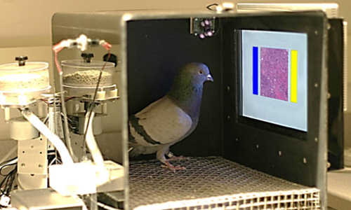
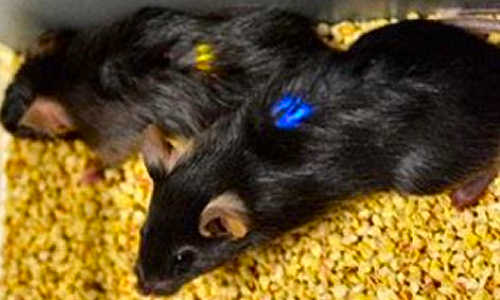
The pigeons’ training environment included a food pellet dispenser, a touch-sensitive screen which projected the medical image, as well as blue and yellow choice buttons on either side of the image. Pecks to those buttons and to the screen were automatically recorded. Photo Credit: Univ. Iowa/Wassermann Lab

“With some training and selective food reinforcement, pigeons do just as well as humans in categorizing digitized slides and mammograms of benign and malignant human breast tissue,” said Richard Levenson, professor of pathology and laboratory medicine at UC Davis Health System and lead author of the study.

“The pigeons were able to generalize what they had learned, so that when we showed them a completely new set of normal and cancerous digitized slides, they correctly identified them,” Levenson said. “Their accuracy, like that of humans, was modestly affected by the presence or absence of color in the images, as well as by degrees of image compression. The pigeons also learned to correctly identify cancer-relevant microcalcifications on mammograms, but they had a tougher time classifying suspicious masses on mammograms — a task that is extremely difficult, even for skilled human observers.”

The pigeons’ successes and difficulties provide a window into how physicians process visual cues present on slides and x-rays to diagnose and classify disease risk. This work also suggests that pigeons’ remarkable ability to discriminate between complex visual images could be put to good use as trained medical image observers, to help researchers explore image quality and the impact of color, contrast, brightness, and image compression artifacts on diagnostic performance.

The study appears online Nov. 18 in PLOS ONE.

Outstanding learners

Although a pigeon’s brain is no bigger than the tip of an index finger, it turns out that the neural pathways involved, including the basal ganglia and cortical-striatal synapses, operate in ways very similar to those at work in the human brain.

According to Edward Wasserman, professor of psychological and brain sciences at The University of Iowa, co-author of the study, the common pigeon (Columba livia) has a tremendous capacity to discriminate and categorize a wide range of objects and images.

“Research over the past 50 years has shown that pigeons can distinguish identities and emotional expressions on human faces, letters of the alphabet, misshapen pharmaceutical capsules, and even paintings by Monet vs. Picasso,” Wasserman said. “Their visual memory is equally impressive, with a proven recall of more than 1,800 images.”

When Levenson learned about Wasserman’s earlier research on the visual short-term memory capacities of pigeons and people, conducted with UC Davis Center for Mind and Brain Director Steven Luck, he wondered how pigeons would perform on pathology slides. And a new collaboration began.

Pigeons especially adept at discriminating breast cancer slides

For the study, each pigeon learned to discriminate cancerous from non-cancerous images and slides using traditional “operant conditioning,” a technique in which a bird was rewarded only when a correct selection was made; incorrect selections were not rewarded and prompted correction trials. Training with stained pathology slides included a large set of benign and cancerous samples from routine cases at UC Davis Medical Center.

Some birds, for example, first learned to recognize benign or malignant samples in full color at low magnification (4X) and then progressed to medium (10X) and high (20X) magnifications. They also were tested using monochrome samples to eliminate color and brightness as potential cues, as well as samples with different levels of image compression, a procedure commonly used to reduce the size of digital data sets.

To rule out the possibility that the birds were relying on rote memorization on the tests, brand-new samples were presented and food was dispensed regardless of whether the pigeons made a correct selection. And, indeed, the pigeons performed virtually as well on images that they had never been shown before, indicating that they had, in an extremely narrow sense, learned pathology.

“The birds were remarkably adept at discriminating between benign and malignant breast cancer slides at all magnifications, a task that can perplex inexperienced human observers, who typically require considerable training to attain mastery,” Levenson said. “Pigeons’ accuracy from day one of training at low magnification increased from 50 percent correct to nearly 85 percent correct at days 13 to 15.

Wasserman, who has conducted studies on pigeons for over 40 years, found the pigeons especially adept at discerning pathology slides.

“The pigeons learned to discriminate benign from cancerous slides as fast in this research as in any other study we’ve conducted on pigeons in our laboratory,” Wasserman said. “In fact, when we showed a cohort of four birds a set of uncompressed images, an approach known as “flock-sourcing,” the group’s accuracy level reached an amazing 99 percent correct, higher than that achieved by any of the four individual birds.”

Density on mammograms a challenge for pigeons

For the mammogram study, the birds were trained to detect images with and without microcalcifications and to discriminate the presence of malignancy in breast masses using a similar process. Their accuracy averaged 84 percent for images with microcalcifications that they had been trained upon, and 72 percent for novel images — a level of performance on par with human radiologists and radiology residents who were given the same cases to review.

The birds, however, had difficulty evaluating the malignant potential of breast masses (without microcalcifications) detected on mammograms, a task the authors acknowledge as “very challenging.” Human radiologists achieved an accuracy rate of about 80 percent when viewing images of the relatively subtle masses used in this study. But, the pigeons took many weeks — instead of days that they had needed to master the histopathology tasks — to learn to classify the breast masses in the mammogram training set. More strikingly, after the training phase, when they were finally shown novel, previously unseen images, the birds utterly failed to perform at a level better than chance.

“The data suggest that the birds were just memorizing the masses in the training set, and never learned how to key in on stellate margins and other features of the lesions that can correlate with malignancy,” Levenson said. “But, as this task reflects the difficulty even humans have, it indicates how pigeons may be faithful mimics of the strengths and weaknesses of humans in viewing medical images.”

Pigeons as human surrogates?

After years of education and training, physicians can sometimes struggle with the interpretation of microscope slides and mammograms. Levenson, a pathologist who studies artificial intelligence for image analysis and other applications in biology and medicine, believes there is considerable room for enhancing the process.

“While new technologies are constantly being designed to enhance image acquisition, processing, and display, these potential advances need to be validated using trained observers to monitor quality and reliability,” Levenson said. “This is a difficult, time-consuming, and expensive process that requires the recruitment of clinicians as subjects for these relatively mundane tasks.

“Pigeons’ sensitivity to diagnostically salient features in medical images suggest that they can provide reliable feedback on many variables at play in the production, manipulation, and viewing of these diagnostically crucial tools, and can assist researchers and engineers as they continue to innovate.”

Story Source:

The above post is reprinted from materials provided by University of Iowa.

22 Kasım 2015 Pazar

Electronic plants created

from
BIOENGINEER.ORG http://bioengineer.org/electronic-plants-created/

Researchers at Linköping University in Sweden have created analog and digital electronics circuits inside living plants. The group at the Laboratory of Organic Electronics (LOE), under the leadership of Professor Magnus Berggren, have used the vascular system of living roses to build key components of electronic circuits.

electronic plant

Augmenting plants with electronic functionality would make it possible to combine electric signals with the plant’s own chemical processes. Photo Credit: Laboratory of Organic Electronics

The article featured in the journal Science Advances demonstrates wires, digital logic, and even displays elements — fabricated inside the plants — that could develop new applications for organic electronics and new tools in plant science.

Plants are complex organisms that rely on the transport of ionic signals and hormones to perform necessary functions. However, plants operate on a much slower time scale making interacting with and studying plants difficult. Augmenting plants with electronic functionality would make it possible to combine electric signals with the plant’s own chemical processes. Controlling and interfacing with chemical pathways in plants could pave the way to photosynthesis-based fuel cells, sensors and growth regulators, and devices that modulate the internal functions of plants.

“Previously, we had no good tools for measuring the concentration of various molecules in living plants. Now we’ll be able to influence the concentration of the various substances in the plant that regulate growth and development. Here, I see great possibilities for learning more,” says Ove Nilsson, professor of plant reproduction biology and director of the Umeå Plant Science Center, as well as a co-author of the article.

The idea of putting electronics directly into trees for the paper industry originated in the 1990s while the LOE team at Linköping University was researching printed electronics on paper. Early efforts to introduce electronics in plants were attempted by Assistant Professor Daniel Simon, leader of the LOE’s bioelectronics team, and Professor Xavier Crispin, leader of the LOE’s solid-state device team, but a lack of funding from skeptical investors halted these projects.

Thanks to independent research money from the Knut and Alice Wallenberg Foundation in 2012, Professor Berggren was able to assemble a team of researchers to reboot the project. The team tried many attempts of introducing conductive polymers through rose stems. Only one polymer, called PEDOT-S, synthesized by Dr. Roger Gabrielsson, successfully assembled itself inside the xylem channels as conducting wires, while still allowing the transport of water and nutrients. Dr. Eleni Stavrinidou used the material to create long (10 cm) wires in the xylem channels of the rose. By combining the wires with the electrolyte that surrounds these channels she was able to create an electrochemical transistor, a transistor that converts ionic signals to electronic output. Using the xylem transistors she also demonstrated digital logic gate function.

Dr. Eliot Gomez used methods common in plant biology — vacuum infiltration — to infuse another PEDOT variant into the leaves. The infused polymer formed “pixels” of electrochemical cells partitioned by the veins. Applied voltage caused the polymer to interact with the ions in the leaf, subsequently changing the color of the PEDOT in a display-like device — functioning similarly to the roll-printed displays manufactured at Acreo Swedish ICT in Norrköping.

These results are early steps to merge the diverse fields of organic electronics and plant science. The aim is to develop applications for energy, environmental sustainability, and new ways of interacting with plants. Professor Berggren envisions the potential for an entirely new field of research:

“As far as we know, there are no previously published research results regarding electronics produced in plants. No one’s done this before,” he says.

Professor Berggren adds, “Now we can really start talking about ‘power plants’ — we can place sensors in plants and use the energy formed in the chlorophyll, produce green antennas, or produce new materials. Everything occurs naturally, and we use the plants’ own very advanced, unique systems.”

Story Source:

The above post is reprinted from materials provided by Linköping University.

21 Kasım 2015 Cumartesi

Nanocarriers may carry new hope for brain cancer therapy

from
BIOENGINEER.ORG http://bioengineer.org/nanocarriers-may-carry-new-hope-for-brain-cancer-therapy/

Glioblastoma multiforme, a cancer of the brain also known as “octopus tumors” because of the manner in which the cancer cells extend their tendrils into surrounding tissue, is virtually inoperable, resistant to therapies, and always fatal, usually within 15 months of onset. Each year, glioblastoma multiforme (GBM) kills approximately 15,000 people in the United States. One of the major obstacles to treatment is the blood brain barrier, the network of blood vessels that allows essential nutrients to enter the brain but blocks the passage of other substances. What is desperately needed is a means of effectively transporting therapeutic drugs through this barrier. A nanoscience expert at Lawrence Berkeley National Laboratory (Berkeley Lab) may have the solution.

nanopaarticle and cancer

At only 20 nanometers in size and featuring a unique hierarchical structure, 3HM nanocarriers meet all the size and stability requirements for effectively delivering therapeutic drugs to brain cancer tumors. Photo Credit: Ting Xu, Berkeley Lab

Ting Xu, a polymer scientist with Berkeley Lab’s Materials Sciences Division who specializes in self-assembling bio/nano hybrid materials, has developed a new family of nanocarriers formed from the self-assembly of amphiphilic peptides and polymers. Called “3HM” for coiled-coil 3-helix micelles, these new nanocarriers meet all the size and stability requirements for effectively delivering a therapeutic drug to GBM tumors. Amphiphiles are chemical compounds that feature both hydrophilic (water-loving) and lipophilic (fat-loving) properties. Micelles are spherical aggregates of amphiphiles.

In a recent collaboration between Xu, Katherine Ferrara at the University of California (UC) Davis, and John Forsayeth and Krystof Bankiewicz of UC San Francisco, 3HM nanocarriers were tested on GBM tumors in rats. Using the radioactive form of copper (copper-64) in combination with positron emission tomography (PET) and magnetic resonance imaging (MRI), the collaboration demonstrated that 3HM can cross the blood brain barrier and accumulate inside GBM tumors at nearly double the concentration rate of current FDA-approved nanocarriers.

“Our 3HM nanocarriers show very good attributes for the treatment of brain cancers in terms of long circulation, deep tumor penetration and low accumulation in off-target organs such as the liver and spleen,” says Xu, who also holds a joint appointment with the UC Berkeley’s Departments of Materials Sciences and Engineering, and Chemistry. “The fact that 3HM is able to cross the blood brain barrier of GBM-bearing rats and selectively accumulate within tumor tissue, opens the possibility of treating GBM via intravenous drug administration rather than invasive measures. While there is still a lot to learn about why 3HM is able to do what it does, so far all the results have been very positive.”

Glial cells provide physical and chemical support for neurons. Approximately 90-percent of all the cells in the brain are glial cells which, unlike neurons, undergo a cycle of birth, differentiation, and mitosis. Undergoing this cycle makes glial cells vulnerable to becoming cancerous. When they do, as the name “multiforme” suggests, they can take on different shapes, which often makes detection difficult until the tumors are dangerously large. The multiple shapes of a cancerous glial cell also make it difficult to identify and locate all of the cell’s tendrils. Removal or destruction of the main tumor mass while leaving these tendrils intact is ineffective therapy: like the mythical Hydra, the tendrils will sprout new tumors.

Although there are FDA approved therapeutic drugs for the treatment of GBM, these treatments have had little impact on patient survival rate because the blood brain barrier has limited the accumulation of therapeutics within the brain. Typically, GBM therapeutics are ferried across the blood brain barrier in special liposomes that are approximately 110 nanometers in size. The 3HM nanocarriers developed by Xu and her group are only about 20 nanometers in size. Their smaller size and unique hierarchical structure afforded the 3HM nanocarriers much greater access to rat GBM tumors than 110-nanometer liposomes in the tests carried out by Xu and her colleagues.

“3HM is a product of basic research at the interface of materials science and biology,” Xu says. “When I first started at Berkeley, I explored hybrid nanomaterials based on proteins, peptides and polymers as a new family of biomaterials. During the process of understanding the hierarchical assembly of amphiphilic peptide-polymer conjugates, my group and I noticed some unusual behavior of these micelles, especially their unusual kinetic stability in the 20 nanometer size range. We looked into critical needs for nanocarriers with these attributes and identified the treatment of GBM cancer as a potential application.”

Copper-64 was used to label both 3HM and liposome nanocarriers for systematic PET and MRI studies to find out how a nanocarrier’s size might affect the pharmacokinetics and biodistribution in rats with GBM tumors. The results not only confirmed the effectiveness of 3HM as GBM delivery vessels, they also suggest that PET and MRI imaging of nanoparticle distribution and tumor kinetics can be used to improve the future design of nanoparticles for GBM treatment.

“I thought our 3HM hybrid materials could bring new therapeutic opportunities for GBM but I did not expect it to happen so quickly,” says Xu, who has been awarded a patent for the 3HM technology.

Story Source:

The above post is reprinted from materials provided by DOE/Lawrence Berkeley National Laboratory.

20 Kasım 2015 Cuma

Functional vocal cord tissue grown in lab

from
BIOENGINEER.ORG http://bioengineer.org/functional-vocal-cord-tissue-grown-in-lab/

University of Wisconsin-Madison scientists have succeeded in growing functional vocal cord tissue in the laboratory, a major step toward restoring a voice to people who have lost their vocal cords to cancer surgery or other injuries.

nathan welharn

Dr. Nathan Welham, a UW speech-language pathologist, and colleagues from several disciplines were able to bioengineer vocal cord tissue able to transmit sound, they reported in a study published in the journal Science Translational Medicine.

About 20 million Americans suffer from voice impairments, and many have damage to the vocal cord mucosae, the specialized tissues that vibrate as air moves over them, giving rise to voice. While injections of collagen and other materials can help some in the short term, Welham says not much can be done for people who have had larger areas of their vocal cords damaged or removed.

“Voice is a pretty amazing thing, yet we don’t give it much thought until something goes wrong,” says Welham, an associate professor of surgery in the UW School of Medicine and Public Health. “Our vocal cords are made up of special tissue that has to be flexible enough to vibrate, yet strong enough to bang together hundreds of times per second. It’s an exquisite system and a hard thing to replicate.”

Welham and colleagues began with vocal cord tissue from a cadaver and four patients who had their larynxes removed but did not have cancer. They isolated, purified and grew the cells from the mucosa, then applied them to a 3-D collagen scaffold, similar to a system used to grow artificial skin in the laboratory.

In about two weeks, the cells grew together to form a tissue with a pliable but strong connective tissue beneath, and layered epithelial cells on top. Proteomic analysis showed the cells produced many of the same proteins as normal vocal cord cells. Physical testing showed that the epithelial cells had also begun to form an immature basement membrane, which helps create a barrier against pathogens and irritants in the airway.

Welham says the lab-grown tissue “felt like vocal cord tissue,” and materials testing showed that it had qualities of viscosity and elasticity similar to normal tissue.

To see if it could transmit sound, the researchers transplanted the bioengineered tissue onto one side of larynges that had been removed from cadaver dogs. The larynges were attached to artificial windpipes and warm, humidified air was blown through them. Not only did the tissue produce sound, but high-speed digital imaging showed the engineered mucosa vibrating like the native tissue on the opposing side. Acoustic analysis also showed the two types of tissue had similar sound characteristics.

Finally, the researchers wanted to see if the tissue would be rejected or accepted by mice that had been engineered to have human immune systems. The tissue grew and was not rejected, performing equally well in mice that had the larynx-cell donor’s immune system (created via a blood donation from the larynx-cell donors) and mice with different human immune systems.

“It seems like the engineered vocal cord tissue may be like cornea tissue in that it is immunoprivileged, meaning that it doesn’t set off a host immune reaction,” Welham says, adding that earlier studies had also suggested this.

In one way, the tissue was not as good as the real thing: Its fiber structure was less complex than adult vocal cords, but the authors said this was not surprising because human vocal cords continue to develop for at least 13 years after birth.

Welham says vocal cord tissue that is free of cancer is a rare commodity, so clinical applications will either require banking and expansion of human cells, or the use of stem cells derived from bone marrow or other tissues. Stem cells could be primed to differentiate into vocal cord cells by exposing them to vibration and tensile forces in a “laryngeal bioreactor.” Such work is being pursued by other laboratories, including at Wisconsin.

Clinical applications are still years away, but Welham says this proof-of-principle study is a “robust benchmark” along the route to replacement vocal cord tissue. Moving this promising work forward requires more testing of safety and long-term function.

Story Source:

The above post is reprinted from materials provided by University of Wisconsin-Madison.

Neuroscientists reveal how the brain can enhance connections

from
BIOENGINEER.ORG http://bioengineer.org/neuroscientists-reveal-how-the-brain-can-enhance-connections/

When the brain forms memories or learns a new task, it encodes the new information by tuning connections between neurons. MIT neuroscientists have discovered a novel mechanism that contributes to the strengthening of these connections, also called synapses.

neuron

Neuron connections (stock image). At each synapse, a presynaptic neuron sends chemical signals to one or more postsynaptic receiving cells. In most previous studies of how these connections evolve, scientists have focused on the role of the postsynaptic neurons. However, an MIT team has found that presynaptic neurons also influence connection strength.

At each synapse, a presynaptic neuron sends chemical signals to one or more postsynaptic receiving cells. In most previous studies of how these connections evolve, scientists have focused on the role of the postsynaptic neurons. However, the MIT team has found that presynaptic neurons also influence connection strength.

“This mechanism that we’ve uncovered on the presynaptic side adds to a toolkit that we have for understanding how synapses can change,” says Troy Littleton, a professor in the departments of Biology and Brain and Cognitive Sciences at MIT, a member of MIT’s Picower Institute for Learning and Memory, and the senior author of the study, which appears in the Nov. 18 issue of Neuron.

Learning more about how synapses change their connections could help scientists better understand neurodevelopmental disorders such as autism, since many of the genetic alterations linked to autism are found in genes that code for synaptic proteins.

Richard Cho, a research scientist at the Picower Institute, is the paper’s lead author.

Rewiring the brain

One of the biggest questions in the field of neuroscience is how the brain rewires itself in response to changing behavioral conditions — an ability known as plasticity. This is particularly important during early development but continues throughout life as the brain learns and forms new memories.

Over the past 30 years, scientists have found that strong input to a postsynaptic cell causes it to traffic more receptors for neurotransmitters to its surface, amplifying the signal it receives from the presynaptic cell. This phenomenon, known as long-term potentiation (LTP), occurs following persistent, high-frequency stimulation of the synapse. Long-term depression (LTD), a weakening of the postsynaptic response caused by very low-frequency stimulation, can occur when these receptors are removed.

Scientists have focused less on the presynaptic neuron’s role in plasticity, in part because it is more difficult to study, Littleton says.

His lab has spent several years working out the mechanism for how presynaptic cells release neurotransmitter in response to spikes of electrical activity known as action potentials. When the presynaptic neuron registers an influx of calcium ions, carrying the electrical surge of the action potential, vesicles that store neurotransmitters fuse to the cell’s membrane and spill their contents outside the cell, where they bind to receptors on the postsynaptic neuron.

The presynaptic neuron also releases neurotransmitter in the absence of action potentials, in a process called spontaneous release. These ‘minis’ have previously been thought to represent noise occurring in the brain. However, Littleton and Cho found that minis could be regulated to drive synaptic structural plasticity.

To investigate how synapses are strengthened, Littleton and Cho studied a type of synapse known as neuromuscular junctions, in fruit flies. The researchers stimulated the presynaptic neurons with a rapid series of action potentials over a short period of time. As expected, these cells released neurotransmitter synchronously with action potentials. However, to their surprise, the researchers found that mini events were greatly enhanced well after the electrical stimulation had ended.

“Every synapse in the brain is releasing these mini events, but people have largely ignored them because they only induce a very small amount of activity in the postsynaptic cell,” Littleton says. “When we gave a strong activity pulse to these neurons, these mini events, which are normally very low-frequency, suddenly ramped up and they stayed elevated for several minutes before going down.”

Synaptic growth

The enhancement of minis appears to provoke the postsynaptic neuron to release a signaling factor, still unidentified, that goes back to the presynaptic cell and activates an enzyme called PKA. This enzyme interacts with a vesicle protein called complexin, which normally acts as a brake, clamping vesicles to prevent release neurotransmitter until it’s needed. Stimulation by PKA modifies complexin so that it releases its grip on the neurotransmitter vesicles, producing mini events.

When these small packets of neurotransmitter are released at elevated rates, they help stimulate growth of new connections, known as boutons, between the presynaptic and postsynaptic neurons. This makes the postsynaptic neuron even more responsive to any future communication from the presynaptic neuron.

“Typically you have 70 or so of these boutons per cell, but if you stimulate the presynaptic cell you can grow new boutons very acutely. It will double the number of synapses that are formed,” Littleton says.

The researchers observed this process throughout the flies’ larval development, which lasts three to five days. However, Littleton and Cho demonstrated that acute changes in synaptic function could also lead to synaptic structural plasticity during development.

“Machinery in the presynaptic terminal can be modified in a very acute manner to drive certain forms of plasticity, which could be really important not only in development, but also in more mature states where synaptic changes can occur during behavioral processes like learning and memory,” Cho says.

Littleton’s lab is now trying to figure out more of the mechanistic details of how complexin controls vesicle release.

Story Source:

The above post is reprinted from materials provided by Massachusetts Institute of Technology.

19 Kasım 2015 Perşembe

Scientists turn tastes on and off by activating and silencing clusters of brain cells

from
BIOENGINEER.ORG http://bioengineer.org/scientists-turn-tastes-on-and-off-by-activating-and-silencing-clusters-of-brain-cells/

Most people probably think that we perceive the five basic tastes — sweet, sour, salty, bitter and umami (savory) — with our tongue, which then sends signals to our brain “telling” us what we’ve tasted. However, scientists have turned this idea on its head, demonstrating in mice the ability to change the way something tastes by manipulating groups of cells in the brain.

brain

The findings were published today in the online edition of Nature.

“Taste, the way you and I think of it, is ultimately in the brain,” said study leader Charles S. Zuker, PhD, professor of biochemistry and molecular biophysics and of neuroscience, a member of the Kavli Institute for Brain Science and the Mortimer B. Zuckerman Mind Brain Behavior Institute, and a Howard Hughes Medical Institute Investigator at Columbia University Medical Center (CUMC). “Dedicated taste receptors in the tongue detect sweet or bitter and so on, but it’s the brain that affords meaning to these chemicals.”

The primary aim of Dr. Zuker’s lab is to understand how the brain transforms detection of chemical stimuli into perception. Over the past decade or so, Dr. Zuker and his colleagues proved that there are dedicated receptors for each taste on the tongue, and that each class of receptor sends a specific signal to the brain. More recently, they demonstrated that each taste is sensed by unique sets of brain cells, located in separate locations in the brain’s cortex -generating a map of taste qualities in the brain.

The scientists used optogenetics, which allowed them to directly activate specific neurons with laser light. Yueqing Peng, a postdoctoral associate in Dr. Zuker’s lab, examined whether manipulating the neurons in these brain regions could evoke the perception of sweet or bitter, without the mouse actually tasting either. (Sweet and bitter tastes were chosen because they are most critical and recognizable tastes for humans and other animals. Sweet taste permits the identification of energy-rich nutrients, while bitter warns against the intake of potentially noxious chemicals).

“In this study, we wanted to know if specific regions in the brain really represent sweet and bitter. If they do, silencing these regions would prevent the animal from tasting sweet or bitter, no matter how much we gave them,” he said. “And if we activate these fields, they should taste bitter or sweet, even though they’re only getting plain water.”

This is exactly what the researchers observed. When scientists injected a substance into the mice to silence the sweet neurons, the animals could not reliably identify sweet. They could, however, still detect bitter. The animals regained their ability to taste sweet when the drug was flushed from the brain. Conversely, silencing the bitter neurons prevented the mice from recognizing bitter, but they could still taste sweet.

Remarkably, the researchers were also able to make the animals think they were tasting bitter or sweet, even when the animal was only drinking water. When the researchers activated the sweet neurons during drinking, they observed behavioral responses in the mice associated with sweet, such as impressively increased licking. In contrast, stimulating bitter neurons dramatically suppressed licking, and elicited classic taste-rejection responses, including the activation of gagging behavior. These results showed that by manipulating the brain centers representing sweet and bitter taste they could directly control an animal’s sensory perception and behavioral actions, says Peng.

The researchers also performed optogenetic tests on animals that had never tasted sweet or bitter chemicals, and showed that activation of the corresponding neurons triggered the appropriate behavioral response. “These experiments formally prove that the sense of taste is completely hardwired, independent of learning or experience, said Dr. Zuker, which is different from the olfactory system. Odors don’t carry innate meaning until you associate them with experiences. One smell could be great for you and horrible to me.” (As humans, of course, we can eventually learn to enjoy bitters and dislike sugar).

In a final set of experiments, animals were trained to report the identity of an orally applied sweet and bitter stimulus by performing a novel behavioral task, allowing the researchers to test what the animal is tasting. In the experiments, the mice tasted real bitter, sweet and salty chemicals at times, but at other times the researchers used the laser to activate the animals’ sweet or bitter cortical fields. The behavior of the mice did not differ between the real and virtual tastes, demonstrating that the light is mimicking the perception of bitter and sweet. “In other words, taste is all in the brain,” said Zuker.

The paper is titled, “Sweet and bitter taste in the brain of awake behaving animals.” The other contributors are Yueqing Peng (CUMC), Sarah Gillis-Smith (CUMC), Hao Jin (CUMC), Dimitri Tränkner (CUMC and Howard Hughes Medical Institute, Asburn, Va.), and Nicholas J. P. Ryba (National Institute of Dental and Craniofacial Research, National Institutes of Health, Bethesda, Md.).

The study was supported by grants from the National Institute of Drug Abuse (DA035025) and the Intramural Research Program of the National Institutes of Health and the National Institute of Dental and Craniofacial Research.

Story Source:

The above post is reprinted from materials provided by Columbia University Medical Center.

18 Kasım 2015 Çarşamba

Experimental drug targeting Alzheimer’s disease shows anti-aging effects

from
BIOENGINEER.ORG http://bioengineer.org/experimental-drug-targeting-alzheimers-disease-shows-anti-aging-effects/

Salk Institute researchers have found that an experimental drug candidate aimed at combating Alzheimer’s disease has a host of unexpected anti-aging effects in animals.

500

As mice age, those treated with J147 (right) showed improved physiology, memory and appearance that more closely resembled younger mice. Photo Credit: the Salk Institute for Biological Studies

The Salk team expanded upon their previous development of a drug candidate, called J147, which takes a different tack by targeting Alzheimer’s major risk factor–old age. In the new work, the team showed that the drug candidate worked well in a mouse model of aging not typically used in Alzheimer’s research. When these mice were treated with J147, they had better memory and cognition, healthier blood vessels in the brain and other improved physiological features, as detailed November 12, 2015 in the journal Aging.

“Initially, the impetus was to test this drug in a novel animal model that was more similar to 99 percent of Alzheimer’s cases,” says Antonio Currais, the lead author and a member of Professor David Schubert’s Cellular Neurobiology Laboratory at Salk. “We did not predict we’d see this sort of anti-aging effect, but J147 made old mice look like they were young, based upon a number of physiological parameters.”

Alzheimer’s disease is a progressive brain disorder, recently ranked as the third leading cause of death in the United States and affecting more than five million Americans. It is also the most common cause of dementia in older adults, according to the National Institutes of Health.

“While most drugs developed in the past 20 years target the amyloid plaque deposits in the brain (which are a hallmark of the disease), none have proven effective in the clinic,” says Schubert, senior author of the study.

Several years ago, Schubert and his colleagues began to approach the treatment of the disease from a new angle. Rather than target amyloid, the lab decided to zero in on the major risk factor for the disease–old age. Using cell-based screens against old age-associated brain toxicities, they synthesized J147.

Previously, the team found that J147 could prevent and even reverse memory loss and Alzheimer’s pathology in mice that have a version of the inherited form of Alzheimer’s, the most commonly used mouse model. However, this form of the disease comprises only about 1 percent of Alzheimer’s cases. For everyone else, old age is the primary risk factor, says Schubert. The team wanted to explore the effects of the drug candidate on a breed of mice that age rapidly and experience a version of dementia that more closely resembles the age-related human disorder.

In this latest work, the researchers used a comprehensive set of assays to measure the expression of all genes in the brain, as well as over 500 small molecules involved with metabolism in the brains and blood of three groups of the rapidly aging mice. The three groups of rapidly aging mice included one set that was young, one set that was old and one set that was old but fed J147 as they aged.

The old mice that received J147 performed better on memory and other tests for cognition and also displayed more robust motor movements. The mice treated with J147 also had fewer pathological signs of Alzheimer’s in their brains. Importantly, because of the large amount of data collected on the three groups of mice, it was possible to demonstrate that many aspects of gene expression and metabolism in the old mice fed J147 were very similar to those of the young animals. These included markers for increased energy metabolism, reduced brain inflammation and reduced levels of oxidized fatty acids in the brain.

Another notable effect was that J147 prevented the leakage of blood from the microvessels in the brains of old mice. “Damaged blood vessels are a common feature of aging in general, and in Alzheimer’s, it is frequently much worse,” says Currais.

Currais and Schubert note that while these studies represent a new and exciting approach to Alzheimer’s drug discovery and animal testing in the context of aging, the only way to demonstrate the clinical relevance of the work is to move J147 into human clinical trials for Alzheimer’s disease.

“If proven safe and effective for Alzheimer’s, the apparent anti-aging effect of J147 would be a welcome benefit,” adds Schubert. The team aims to begin human trials next year.

Other authors on the paper include Oswald Quehenberger of the University of California, San Diego; and Joshua Goldberg, Catherine Farrokhi, Max Chang, Marguerite Prior, Richard Dargusch, Daniel Daugherty and Pamela Maher of the Salk Institute.

This study was supported by the Salk Institute Pioneer Fund Postdoctoral Scholar Award and the Salk Nomis Fellowship Award, fellowships from the Hewitt Foundation and Bundy Foundation, and grants from the Burns Foundation and NIH.

Story Source:

The above post is reprinted from materials provided by Salk Institute.

A network of artificial neurons learns to use human language

from
BIOENGINEER.ORG http://bioengineer.org/a-network-of-artificial-neurons-learns-to-use-human-language/

A group of researchers from the University of Sassari (Italy) and the University of Plymouth (UK) has developed a cognitive model, made up of two million interconnected artificial neurons, able to learn to communicate using human language starting from a state of “tabula rasa,” only through communication with a human interlocutor. The model is called ANNABELL (Artificial Neural Network with Adaptive Behavior Exploited for Language Learning) and it is described in an article published in the international scientific journal PLOS ONE. This research sheds light on the neural processes that underlie the development of language.

language

An artificial system of nerons called ANNABELL has learned to pick up language by interacting with humans. Photo Credit: Aysezgicmeli/Shutterstock

How does our brain develop the ability to perform complex cognitive functions, such as those needed for language and reasoning? This is a question that certainly we are all asking ourselves, to which the researchers are not yet able to give a complete answer. We know that in the human brain there are about one hundred billion neurons that communicate by means of electrical signals. We learned a lot about the mechanisms of production and transmission of electrical signals among neurons. There are also experimental techniques, such as functional magnetic resonance imaging, which allow us to understand which parts of the brain are most active when we are involved in different cognitive activities. But a detailed knowledge of how a single neuron works and what are the functions of the various parts of the brain is not enough to give an answer to the initial question.

We might think that the brain works in a similar way to a computer: after all, even computers work through electrical signals. In fact, many researchers have proposed models based on the analogy brain-is-like-a-computer since the late ’60s. However, apart from the structural differences, there are profound differences between the brain and a computer, especially in learning and information processing mechanisms. Computers work through programs developed by human programmers. In these programs there are coded rules that the computer must follow in handling the information to perform a given task. However there is no evidence of the existence of such programs in our brain. In fact, today many researchers believed that our brain is able to develop higher cognitive skills simply by interacting with the environment, starting from very little innate knowledge. The ANNABELL model appears to confirm this perspective.

ANNABELL does not have pre-coded language knowledge; it learns only through communication with a human interlocutor, thanks to two fundamental mechanisms, which are also present in the biological brain: synaptic plasticity and neural gating. Synaptic plasticity is the ability of the connection between two neurons to increase its efficiency when the two neurons are often active simultaneously, or nearly simultaneously. This mechanism is essential for learning and for long-term memory. Neural gating mechanisms are based on the properties of certain neurons (called bistable neurons) to behave as switches that can be turned “on” or “off” by a control signal coming from other neurons. When turned on, the bistable neurons transmit the signal from a part of the brain to another, otherwise they block it. The model is able to learn, due to synaptic plasticity, to control the signals that open and close the neural gates, so as to control the flow of information among different areas.

The cognitive model has been validated using a database of about 1500 input sentences, based on literature on early language development, and has responded by producing a total of about 500 sentences in output, containing nouns, verbs, adjectives, pronouns, and other word classes, demonstrating the ability to express a wide range of capabilities in human language processing.

Story Source:

The above post is reprinted from materials provided by University of Sassari.

Most extensive face transplant to date

from
BIOENGINEER.ORG http://bioengineer.org/most-extensive-face-transplant-to-date/

NYU Langone Medical Center announced today the successful completion of the most extensive face transplant to date, setting new standards of care in this emerging field. Equally important, for the first time a face transplant has been performed on a first responder — a volunteer firefighter who suffered a full face and scalp burn in the line of duty.

health

Patrick Hardison pre-surgery, and again post-surgery on November 11, 2015 Credit: NYU Langone

The surgery — the first of its kind performed in New York State — began the morning of August 14, 2015 and concluded 26 hours later on the morning of August 15. It involved a team of more than 100 physicians, nurses, technical and support staff, led by Eduardo D. Rodriguez, MD, DDS, the Helen L. Kimmel Professor of Reconstructive Plastic Surgery and chair of the Hansjörg Wyss Department of Plastic Surgery. The team worked in two adjoining operating rooms — in one room, the donor’s face was procured (along with other donated organs), and in the other, the recipient’s face and scalp burn was removed and the transplant took place.

The recipient, volunteer firefighter Patrick Hardison, 41, of Senatobia, MS, was injured in September 2001 — ironically just days before the 9/11 attacks. Patrick entered a burning home on a rescue search, when its roof collapsed on him, leaving him with disfiguring burns across his entire face, head, neck and upper torso. He lost his eyelids, ears, lips, most of his nose as well as his hair including his eyebrows. After enduring more than 70 surgeries in Mississippi and elsewhere, Patrick was still unable to return to a normal life. He was brought to Dr. Rodriguez’s attention by a member of his church and fellow firefighter, who wrote to the doctor describing Patrick’s situation.

Highlights from the Surgery

Dr. Rodriguez — who previously performed a complex face transplant in 2012 — and his NYU Langone team transplanted not only the face, but also the entire scalp. Patrick’s surgery was pivotal in that the donor’s eyelids and the muscles that control blinking were transplanted–a significant milestone and a procedure that had not been previously performed on a seeing patient.

Among other milestones achieved in Patrick’s surgery:

Transplantation of the ears and ear canals
Transplantation of selective bony structures from the donor, including portions of the chin, cheeks and the entire nose
Advanced use of three-dimensional modeling, computerized modeling and 3D printed patient-specific cutting guides designed from the recipient’s and the donor’s CT scans to provide the most precise “snap-fit” of the skeleton
Precise placement of patient-specific metal plates and screws to ensure the proper contour and symmetry of the transplanted face
The transplantation of the donor’s eyelids and blinking mechanisms was particularly important to the surgery’s success, as Patrick was in danger of losing his sight and had been unable to perform independent daily tasks such as driving. Blinking enables the body to appropriately hydrate and clean the eyes to prevent infection and preserve vision. Earlier this year, Dr. Rodriguez and others published a study in the peer-reviewed journal, Plastic and Reconstructive Surgery, in which they detailed the importance of eyelid preservation and enhancement in facial transplantation.

Patrick’s Recovery

Within the final hours of surgery, signs of success were evident. Patrick’s new face, particularly his new lips and ears, were robust with color, indicating circulation had been restored. The hair on his scalp, as well as the beard on his face, began growing back immediately. He was able to use his new eyelids and blink on the third day of recovery, after the swelling began to diminish. He was sitting up in a chair within a week. And now, just three months removed from surgery, swelling has greatly subsided and he is quickly returning to the routines of daily life independently.

As part of his recovery, Patrick continues to go through extensive rehabilitative therapy, including:

Physical therapy to build his strength and stamina
Speech and swallowing therapy to further restore and enhance his ability to speak correctly using his new lips and to regain normal eating and swallowing abilities
Occupational therapy to re-learn daily tasks, such as shaving again for the first time in 14 years
Patrick, like all transplant patients, will need to remain on anti-rejection medication for the rest of his life to prevent transplant rejection. Patrick will also rely on his family and friends — particularly his fellow firefighters in Senatobia — to support him in his recovery and his transition back to his hometown after he is discharged from the hospital. He will also have regular monthly checkups with Dr. Rodriguez and the face transplant team.

About the Donor

With every successful transplant surgery, there is always a donor and a donor family that makes the altruistic gifts possible during one of the most difficult times in their lives. In Patrick’s case, his donor was David P. Rodebaugh, 26, an Ohio-born Brooklyn artist and bicycling enthusiast, who tragically died from injuries sustained in an accident. David’s career pursuits took him to New York, where he was advancing his training in cycling mechanics, design, and customization. He also won several cycling competitions, gaining a loyal following of fans and admirers in New York and across the country in the close-knit BMX cycling community. David also was a registered organ donor.

LiveOnNY, the organ recovery organization for the greater New York metropolitan area, approached David’s mother and informed her of David’s wishes to be an organ donor, explaining to her the importance of organ donation. They comforted David’s loved ones as they made the decision to donate David’s face, as well as his heart, liver, and kidneys to other recipients, and to research.

The implementation of the NYU Langone face transplant program required extensive collaboration with LiveOnNY, which began over a year ago — after Patrick was identified to receive the program’s first face transplant. Unlike other situations in which organs can be recovered and transported from distant hospitals, Dr. Rodriguez and his team needed to perform the recovery of David’s face in an operating room adjacent to the OR where Patrick’s transplant would take place. LiveOnNY also coordinated with Dr. Rodriguez and his team and other hospitals and transplant teams to procure David’s other donated organs.

Preparing for the Surgery

An important lessons learned was that with a skilled and experienced leader at the helm, a facial transplantation program can be created and — in Dr. Rodriguez’s case — re-created at a medical facility with the appropriate talent, resources and multi-disciplinary commitment to teamwork.

When Dr. Rodriguez joined the faculty of NYU Langone in November 2013 as chair of plastic surgery, one of his goals was to develop and launch a face transplant program. He assembled a team and educated them on the intricacies of facial transplantation. Most of these individuals were physicians, nurses and staff already at NYU Langone, representing numerous departments including plastic surgery, anesthesiology, clinical psychology, critical care medicine, emergency medicine, medical ethics, nursing, perioperative services, physical medicine and rehabilitation, psychiatry, radiology and social work.

They planned extensively to ensure appropriate systems were in place to respond immediately once a donor was identified. Preparations also included carefully executed surgical rehearsals over the ensuing months, including practice on cadavers.

Story Source:

The above post is reprinted from materials provided by NYU Langone Medical Center / New York University School of Medicine.

Repairing neurons with light

from
BIOENGINEER.ORG http://bioengineer.org/repairing-neurons-with-light/

The nervous system is built to last a lifetime, but diverse diseases or environmental insults can overpower the capacity of neurons to maintain function or to repair after trauma. A team led by Dr. Hernán López-Schier, head of the Research Unit Sensory Biology and Organogenesis at Helmholtz Zentrum München, now succeeded in promoting the repair of an injured neural circuit in zebrafish.

light

These are zebrafish neurons projecting to the brain (green). One neuron expresses a light-activatable enzyme (red). Scientist were able to stimulate the regeneration of injured neurons using optogenetics. Photo Credit: Helmholtz Zentrum München

Key for the researchers’ success was the messenger molecule cAMP, which is produced by an enzyme called adenylyl cyclase. For their experiment, the scientist used a special form of this enzyme which is inducible by blue light. Therefore, the scientists are able to specifically modulate the production of cAMP in cells expressing this enzyme by the use of blue light.*

The researchers used this system in zebrafish larvae** which had interrupted sensory lateralis nerves***. “However, when blue light was shone on severed nerves that expressed a photoactivatable adenylyl cyclase, their repair was dramatically increased,” remembers PhD student Yan Xiao who is the first author of the study. “While untreated nerve terminals only made synapses again in five percent of the cases, about 30% did after photostimulation.” In simple terms: the scientists were able to stimulate the repair of a neuronal circuit by elevating cAMP with blue light.

“Optogenetics have revolutionized neurobiology, since the method has already been used to modify for instance the electrical activity of neurons. However, our results show for the first time how the repair of a complex neural circuit in a whole animal can be promoted remotely by the use of light,” explains López-Schier.

But the head of the study thinks that this is only the beginning: “Our results are a first step. Now we would like to investigate, whether these results can be extrapolated beyond single neurons in zebrafish, to more complex neuronal circuits of higher animals.” The scientist could think of using this method for future therapeutic approaches for the treatment of neuropathies like those occurring in the wake of Diabetes and other diseases.

Further information

* Optogenetics: As the name indicates, this cutting-edge technology combines elements of Optics and Genetics. Scientists make use of proteins which are sensitive to certain wavelengths of light. These are brought into the target cells with certain genetic methods. The so treated cells then change their respective phenotype depending on the exposure to light.

** Larvae of zebrafish are particularly well suited for optogenetic approaches, since their skin in transparent/translucent. Thus, the light can reach the respective target cells easily.

*** These nerves normally communicate external sensory signals to the brain, but cannot normally repair after injury.

Story Source:

The above post is reprinted from materials provided by Helmholtz Zentrum München – German Research Center for Environmental Health.

Self-healing sensor brings ‘electronic skin’ closer to reality

from
BIOENGINEER.ORG http://bioengineer.org/self-healing-sensor-brings-electronic-skin-closer-to-reality/

Flexible sensors have been developed for use in consumer electronics, robotics, health care, and space flight. Future possible applications could include the creation of ‘electronic skin’ and prosthetic limbs that allow wearers to ‘feel’ changes in their environments.

skin

One problem with current flexible sensors, however, is that they can be easily scratched and otherwise damaged, potentially destroying their functionality. Researchers in the Department of Chemical Engineering at the Technion — Israel Institute of Technology in Haifa (Israel), who were inspired by the healing properties in human skin, have developed materials that can be integrated into flexible devices to “heal” incidental scratches or damaging cuts that might compromise device functionality. The advancement, using a new kind of synthetic polymer (a polymer is a large molecule composed of many repeated smaller molecules) has self-healing properties that mimic human skin, which means that e-skin “wounds” can quickly “heal” themselves in remarkably short time — less than a day.

A paper outlining the characteristics and applications of the unique, self-healing sensor has been published in the current issue of Advanced Materials.

“The vulnerability of flexible sensors used in real-world applications calls for the development of self-healing properties similar to how human skins heals,” said self-healing sensor co-developer Prof. Hossam Haick. “Accordingly, we have developed a complete, self-healing device in the form of a bendable and stretchable chemiresistor where every part — no matter where the device is cut or scratched — is self-healing.”

The new sensor is comprised of a self-healing substrate, high conductivity electrodes, and molecularly modified gold nanoparticles. “The gold particles on top of the substrate and between the self-healing electrodes are able to “heal” cracks that could completely disconnect electrical connectivity,” said Prof. Haick.

Once healed, the polymer substrate of the self-healing sensor demonstrates sensitivity to volatile organic compounds (VOCs), with detection capability down to tens of parts per billion. It also demonstrates superior healability at the extreme temperatures of -20 degrees C to 40 degrees C. This property, said the researchers, can extend applications of the self-healing sensor to areas of the world with extreme climates. From sub-freezing cold to equatorial heat, the self-healing sensor is environment-stable.

The healing polymer works quickest, said the researchers, when the temperature is between 0 degrees C and 10 degrees C, when moisture condenses and is then absorbed by the substrate. Condensation makes the substrate swell, allowing the polymer chains to begin to flow freely and, in effect, begin “healing.” Once healed, the nonbiological, chemiresistor still has high sensitivity to touch, pressure and strain, which the researchers tested in demanding stretching and bending tests.

Another unique feature is that the electrode resistance increases after healing and can survive 20 times or more cutting/healing cycles than prior to healing. Essentially, healing makes the self-healing sensor even stronger. The researchers noted in their paper that “the healing efficiency of this chemiresistor is so high that the sensor survived several cuttings at random positions.”

The researchers are currently experimenting with carbon-based self-healing composites and self-healing transistors.

“The self-healing sensor raises expectations that flexible devices might someday be self-administered, which increases their reliability,” explained co-developer Dr. Tan-Phat Huynh, also of the Technion, whose work focuses on the development of self-healing electronic skin. “One day, the self-healing sensor could serve as a platform for biosensors that monitor human health using electronic skin.”

Story Source:

The above post is reprinted from materials provided by American Technion Society.

17 Kasım 2015 Salı

Gene patterns that make our brains human

from
BIOENGINEER.ORG http://bioengineer.org/gene-patterns-that-make-our-brains-human/

The human brain may be the most complex piece of organized matter in the known universe, but Allen Institute researchers have begun to unravel the genetic code underlying its function.

brain

Researchers prepare tissue used in the Allen Human Brain Atlas. Photo Credit: Allen Institute for Brain Science

Research published this month in Nature Neuroscience identified a surprisingly small set of molecular patterns that dominate gene expression in the human brain and appear to be common to all individuals, providing key insights into the core of the genetic code that makes our brains distinctly human.

“So much research focuses on the variations between individuals, but we turned that question on its head to ask, what makes us similar?” says Ed Lein, Ph.D., Investigator at the Allen Institute for Brain Science. “What is the conserved element among all of us that must give rise to our unique cognitive abilities and human traits?”

Researchers used data from the publicly available Allen Human Brain Atlas to investigate how gene expression varies across hundreds of functionally distinct brain regions in six human brains. They began by ranking genes by the consistency of their expression patterns across individuals, and then analyzed the relationship of these genes to one another and to brain function and association with disease.

“Looking at the data from this unique vantage point enables us to study gene patterning that we all share,” says Mike Hawrylycz, Ph.D., Investigator at the Allen Institute for Brain Science. “We used the Allen Human Brain Atlas data to quantify how consistent the patterns of expression for various genes are across human brains, and to determine the importance of the most consistent and reproducible genes for brain function.”

Despite the anatomical complexity of the brain and the complexity of the human genome, most of the patterns of gene usage across all 20,000 genes could be characterized by just 32 expression patterns. While many of these patterns were similar in human and mouse, the dominant genetic model organism for biomedical research, many genes showed different patterns in human. Surprisingly, genes associated with neurons were most conserved across species, while those for the supporting glial cells showed larger differences.

The most highly stable genes–the genes that were most consistent across all brains–include those that are associated with diseases and disorders like autism and Alzheimer’s and include many existing drug targets. These patterns provide insights into what makes the human brain distinct and raise new opportunities to target therapeutics for treating disease.

The researchers also found that the pattern of gene expression in cerebral cortex is correlated with “functional connectivity” as revealed by neuroimaging data from the Human Connectome Project. “It is exciting to find a correlation between brain circuitry and gene expression by combining high quality data from these two large-scale projects,” says David Van Essen, Ph.D., professor at Washington University in St. Louis and a leader of the Human Connectome Project.

“The human brain is phenomenally complex, so it is quite surprising that a small number of patterns can explain most of the gene variability across the brain,” says Christof Koch, Ph.D., President and Chief Scientific Officer at the Allen Institute for Brain Science. “There could easily have been thousands of patterns, or none at all. This gives us an exciting way to look further at the functional activity that underlies the uniquely human brain.”

Story Source:

The above post is reprinted from materials provided by Allen Institute.

10 Kasım 2015 Salı

Researchers advance genome editing of blood stem cells

from
BIOENGINEER.ORG http://bioengineer.org/researchers-advance-genome-editing-of-blood-stem-cells/

Genome editing techniques for blood stem cells just got better, thanks to a team of researchers at USC and Sangamo BioSciences.

hiv

HIV (yellow) infecting a human immune cell. Photo Credit: Seth Pincus, Elizabeth Fischer and Austin Athman, National Institute of Allergy and Infectious Diseases, National Institutes of Health

In an upcoming study in Nature Biotechnology, co-first authors Colin M. Exline, PhD, from USC and Jianbin Wang, PhD, from Sangamo BioSciences describe a new, more efficient way to edit genes in blood-forming or “hematopoietic” stem and progenitor cells (HSPCs).

“Gene therapy using HSPCs has enormous potential for treating HIV and other diseases of the blood and immune systems,” said co-corresponding author Paula Cannon, PhD, professor of molecular microbiology and immunology, pediatrics, biochemistry and molecular biology, and stem cell biology and regenerative medicine at USC. “And using genome editing techniques now allows us to make very precise changes that could repair genetic mutations — the gene typos — that can cause disease.”

Despite the enormous potential of such targeted gene medicine to cure patients, getting genome editing to work has proven challenging in human HSPCs — especially in the most primitive, least differentiated cells with the greatest ability to become any blood cell type.

Cannon’s group, working with a team at Sangamo, has been using “genetic scissors” called zinc finger nucleases (ZFNs) to cut a cell’s DNA at a precise location or sequence. The cell normally uses a copy of the cut DNA sequence as a template to repair the DNA break. During this process, there is the opportunity to introduce new DNA sequences or to repair mutations, effectively fooling the cell into making a genetic edit.

To provide the cell with both the targeted nuclease and the new DNA template, scientists can use a variety of delivery vehicles or vectors, including viruses and a type of genetic material known as messenger RNA (mRNA).

In the study, the team discovered a highly effective way to deliver the DNA repair template using a specific type of viral vector, known as an adeno-associated virus (AAV) serotype 6, which can naturally enter HSPCs. At the same time, they found that delivering the ZFNs as short-lived mRNA molecules allowed the DNA cutting and repair process to occur without disrupting the HSPCs. By combining these two delivery methods, the scientists were able to insert a gene at a precise site in even the most primitive human HSPCs with unprecedented efficiency rates ranging from 15 to 40 percent.

The team then transplanted these genetically edited human HSPCs into immune-deficient mice, and found that the cells thrived and differentiated into many different blood cell types — all retaining the edits to their DNA.

“Our results provide a strategy for broadening the application of genome editing technologies in HSPCs,” said co-corresponding author Michael C. Holmes, PhD, vice president of research at Sangamo BioSciences. “This significantly advances our progress towards applying genome editing to the treatment of human diseases of the blood and immune systems.”

Story Source:

The above post is reprinted from materials provided by University of Southern California.

Implantable wireless devices trigger, and may block, pain signals

from
BIOENGINEER.ORG http://bioengineer.org/implantable-wireless-devices-trigger-and-may-block-pain-signals/

Building on wireless technology that has the potential to interfere with pain, scientists have developed flexible, implantable devices that can activate — and, in theory, block — pain signals in the body and spinal cord before those signals reach the brain.

implant

Implanted microLED devices light up, activating peripheral nerve cells in mice. The devices are being developed and studied by researchers at Washington University School of Medicine in St. Louis and the University of Illinois at Urbana-Champaign as a potential treatment for pain that does not respond to other therapies. Photo Credit: Gereau lab/Washington University

The researchers, at Washington University School of Medicine in St. Louis and the University of Illinois at Urbana-Champaign, said the implants one day may be used in different parts of the body to fight pain that doesn’t respond to other therapies.

“Our eventual goal is to use this technology to treat pain in very specific locations by providing a kind of ‘switch’ to turn off the pain signals long before they reach the brain,” said co-senior investigator Robert W. Gereau IV, PhD, the Dr. Seymour and Rose T. Brown Professor of Anesthesiology and director of the Washington University Pain Center.

The study is published online Nov. 9 in the journal Nature Biotechnology.

Because the devices are soft and stretchable, they can be implanted into parts of the body that move, Gereau explained. The devices previously developed by the scientists had to be anchored to bone.

“But when we’re studying neurons in the spinal cord or in other areas outside of the central nervous system, we need stretchable implants that don’t require anchoring,” he said.

The new devices are held in place with sutures. Like the previous models, they contain microLED lights that can activate specific nerve cells. Gereau said he hopes to use the implants to blunt pain signals in patients who have pain that cannot be managed with standard therapies.

The researchers experimented with mice that were genetically engineered to have light-sensitive proteins on some of their nerve cells. To demonstrate that the implants could influence the pain pathway in nerve cells, the researchers activated a pain response with light. When the mice walked through a specific area in a maze, the implanted devices lit up and caused the mice to feel discomfort. Upon leaving that part of the maze, the devices turned off, and the discomfort dissipated. As a result, the animals quickly learned to avoid that part of the maze.

The experiment would have been very difficult with older optogenetic devices, which are tethered to a power source and can inhibit the movement of the mice.

Because the new, smaller, devices are flexible and can be held in place with sutures, they also may have potential uses in or around the bladder, stomach, intestines, heart or other organs, according to co-principal investigator John A. Rogers, PhD, professor of materials science and engineering at the University of Illinois.

“They provide unique, biocompatible platforms for wireless delivery of light to virtually any targeted organ in the body,” he said.

Rogers and Gereau designed the implants with an eye toward manufacturing processes that would allow for mass production so the devices could be available to other researchers. Gereau, Rogers and Michael R. Bruchas, PhD, associate professor of anesthesiology at Washington University, have launched a company called NeuroLux to aid in that goal.

Story Source:

The above post is reprinted from materials provided by Washington University School of Medicine.

9 Kasım 2015 Pazartesi

Tissue engineers recruit cells to make their own strong matrix

from
BIOENGINEER.ORG http://bioengineer.org/tissue-engineers-recruit-cells-to-make-their-own-strong-matrix/

Extracellular matrix is the material that gives tissues their strength and stretch. It’s been hard to make well in the lab, but a Brown University team reports new success. The key was creating a culture environment that guided cells to make ECM themselves.

tissue engineering

Fluorescent labeling shows extra cellular matrix (green) aligned with the directions specified by shaped tissue culture molds. Photo Credit: Jacquelyn Schell/Brown University

Imitation may be the sincerest form of flattery but the best way to make something is often to co-opt the original process and make it work for you. In a sense, that’s how scientists at Brown University accomplished a new advance in tissue engineering.

In the journal Biomaterials, the team reports culturing cells to make extracellular matrix (ECM) of two types and five different alignments with the strength found in natural tissue and without using any artificial chemicals that could make it incompatible to implant.

ECM is the fibrous material between cells in tissues like skin, cartilage, or tendon that gives them their strength, stretchiness, squishiness, and other mechanical properties. To help patients heal wounds and injuries, engineers and physicians have strived to make ECM in the lab that’s aligned as well as it is when cells make it in the body. So far, though, they’ve struggled to recreate ECM. Using artificial materials provides strength, but those don’t interact well with the body. Attempts to extract and build upon natural ECM have yielded material that’s too weak to reimplant.

The Brown team tried a different approach to making both collagen, which is strong, and elastin, which is stretchy, with different alignments of their fibers. They cultured ECM-making cells in specially designed molds that promoted the cells to make their own natural but precisely guided ECM.

“What we hypothesized is that the cells are making it the same way they do in the body, because we’re starting them in a more natural environment,” said lead author Jacquelyn Schell, assistant professor (research) of molecular pharmacology, physiology and biotechnology. “We’re not adding exogenous materials.”

The strategy built on the insight that when cells clump together and grow in culture, they pull on each other and communicate as they would in the body, Schell said. The molds therefore were made from agarose so that cells wouldn’t stick to the sides or bottom. Instead they huddled together.

To guide ECM growth in particular alignments, the researchers used molds with very specific shapes, often constrained by pegs the cells had to grow around. For instance, to make a rod with collagen fibers aligned along its length (like a tendon) they cultured chondrocyte cells in a dog bone-shaped mold with loops on either end. To make a skin-like “trampoline” of elastin, where the ECM fibers run in all directions, they cultured fibroblast cells to grow in an open area suspended at the center of a honeycomb shape.

“The placement of the pegs that this group of cells wraps itself around and then exerts force on each other is what dictates their alignment and the direction of the ECM they are going to synthesize,” said senior author Jeffrey Morgan, professor of medical science and engineering and co-director of Brown’s Center for Biomedical Engineering. “That’s a new ability to control the cells’ synthesis of extracellular matrix.”

After the researchers grew various forms of ECM, they did some stress testing. They took the dog bone-shaped tissues to the lab of Christian Franck, assistant professor of engineering, and together made precise measurements of the tissue strength under the force of being pulled apart. The measurements confirmed the self-assembled tissue was about as strong as that found in some of the body’s tissues, such as skin, cartilage or blood vessels.

The team’s next goal is to identify a prospective clinical application, Morgan said. The lab will pursue the needed testing to see if this new way of growing ECM can help future patients.

In addition to Schell, Morgan, and Franck, the paper’s other authors are Benjamin Wilks, Mohak Patel, Vijaya Chalivendra, Xuan Cao, and Vivek Shenoy.

The Department of Defense (grant: W81XWH-10-1-0643_ and the National Science Foundation (grants: 1129172, CBET-1428092) funded the study.

Story Source:

The above post is reprinted from materials provided by Brown university.

7 Kasım 2015 Cumartesi

Breakthrough: Redefining How Blood Is Made

from
BIOENGINEER.ORG http://bioengineer.org/breakthrough-redefining-how-blood-is-made/

Stem-cell scientists led by Dr. John Dick have discovered a completely new view of how human blood is made, upending conventional dogma from the 1960s.

John Dick

Dr. John Dick, Senior Scientist at the Princess Margaret Cancer Centre. Photo Credit: UHN

The findings, published online in the journal Science, prove “that the whole classic ‘textbook’ view we thought we knew doesn’t actually even exist,” says principal investigator John Dick, Senior Scientist at Princess Margaret Cancer Centre, University Health Network (UHN), and Professor in the Department of Molecular Genetics, University of Toronto.

“Instead, through a series of experiments we have been able to finally resolve how different kinds of blood cells form quickly from the stem cell — the most potent blood cell in the system — and not further downstream as has been traditionally thought,” says Dr. Dick, who holds a Canada Research Chair in Stem Cell Biology and is also Director of the Cancer Stem Cell Program at the Ontario Institute for Cancer Research. He talks about the research at www.youtu.be/D08FMKDppVQ .

The research also topples the textbook view that the blood development system is stable once formed. Not so, says Dr. Dick. “Our findings show that the blood system is two-tiered and changes between early human development and adulthood.”

Co-authors Dr. Faiyaz Notta and Dr. Sasan Zandi from the Dick lab write that in redefining the architecture of blood development, the research team mapped the lineage potential of nearly 3,000 single cells from 33 different cell populations of stem and progenitor cells obtained from human blood samples taken at various life stages and ages.

For people with blood disorders and diseases, the potential clinical utility of the findings is significant, unlocking a distinct route to personalizing therapy.

Dr. Dick says: “Our discovery means we will be able to understand far better a wide variety of human blood disorders and diseases — from anemia, where there are not enough blood cells, to leukemia, where there are too many blood cells. Think of it as moving from the old world of black-and-white television into the new world of high definition.”

There are also promising implications for advancing the global quest in regenerative medicine to manufacture mature cell types such as platelets or red blood cells by engineering cells (a process known as inducing pluripotent stem cells), says Dr. Dick, who collaborates closely with Dr. Gordon Keller, Director of UHN’s McEwen Centre for Regenerative Medicine.

“By combining the Keller team’s ability to optimize induced pluripotent stem cells with our newly identified progenitors that give rise only to platelets and red blood cells, we will be able develop better methods to generate these mature cells,” he says.

Currently, human donors are the sole source of platelets — which cannot be stored or frozen — for transfusions needed by many thousands of patients with cancer and other debilitating disorders.

Today’s discovery builds on Dr. Dick’s breakthrough research in 2011, also published in Science, when the team isolated a human blood stem cell in its purest form — as a single stem cell capable of regenerating the entire blood system.

“Four years ago, when we isolated the pure stem cell, we realized we had also uncovered populations of stem-cell like ‘daughter’ cells that we thought at the time were other types of stem cells,” says Dr. Dick.

“When we burrowed further to study these ‘daughters’, we discovered they were actually already mature blood lineages. In other words, lineages that had broken off almost immediately from the stem cell compartment and had not developed downstream through the slow, gradual ‘textbook’ process.

“So in human blood formation, everything begins with the stem cell, which is the executive decision-maker quickly driving the process that replenishes blood at a daily rate that exceeds 300 billion cells.”

For 25 years, Dr. Dick’s research has focused on understanding the cellular processes that underlie how normal blood stem cells work to regenerate human blood after transplantation and how blood development goes wrong when leukemia arises. His research follows on the original 1961 discovery of the blood stem cell by Princess Margaret Cancer Centre scientists Dr. James Till and the late Dr. Ernest McCulloch, which formed the basis of all current stem-cell research.

Story Source:

The above post is reprinted from materials provided by University Health Network (UHN).